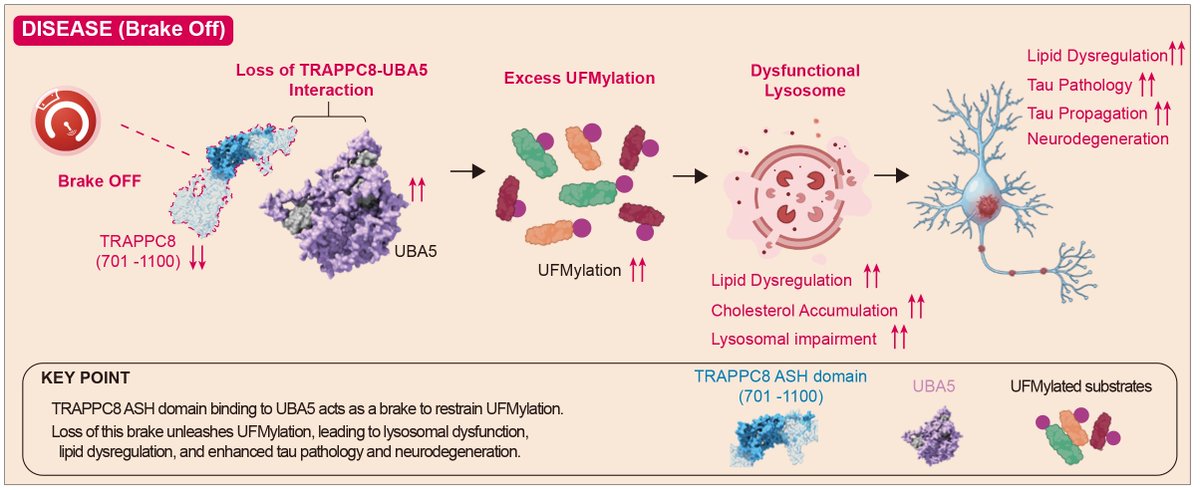
We identified TRAPPC8 as a brake on UFMylation via direct binding of UBA5. Loss of this brake disrupts lysosomal function and drives tau propagation. Restoration rescues this effect in vivo—revealing it as a potential therapeutic target for tauopathies.
https://t.co/DEsmfUmJeK
New in Nature! 🐭 Lövestam et al. injects tau filaments from the brains of AD and CBD patients into WT mice and demonstrates propagation through prion-like templated seeding that results in disease-specific pathologic phenotypes and seed specific folding.
https://t.co/38bKnW0RHh
New in Nature Methods! 🔬🧪Joly et al. profiled intact tau molecules one at a time. Across cell models and brain tissue, they found 130 forms of full-length tau and certain phosphorylation sites that tend to show up together on the same molecule. https://t.co/recuyThn3F
New in Nature 🧠! Hao et al., showed tauopathy mice lacking cDC1s or antigen cross-presentation have reduced brain infiltration of CD8+ T cells, glial response, and neurodegeneration; but no change in tau phosphorylation or aggregation. Read here:
https://t.co/gfG8Pfpn0K
Konrad-Vicario et al. developed STARFISH to spatially resolve endogenous mRNA translation in neurons, finding Mapt mRNA as exclusively translated in dendrites and degraded by the neuroproteasome, failure of which leads to accumulation of mislocalized tau aggregates. Amazing work!
Congratulations to Dr. @naguib_sarah, postdoctoral researcher in the Gan Lab, on receiving an NIH K99 Pathway to Independence Award! 🎉
This prestigious award recognizes her research achievements and potential to become an independent investigator.
Congrats, Sarah! 🔬🧠
🎉 Huge congratulations to our postdoc, Wenhui Qu, on receiving an NIH K99/R00 Pathway to Independence Award! A fantastic recognition of her exciting research and an important step toward research independence. We’re incredibly proud—well deserved! 👏
🎉New in Neuron! We identified that microglia-derived soluble DLK1 links aging to hypomyelination. In mouse and human iPSC models, telomere shortening drives microglial senescence, ↑sDLK1, and impaired oligodendrocyte maturation and neuronal activity. https://t.co/iNR0jS1xJa
A new 2026 Neuron paper shows that tau directly activates mitochondrial RET via complex I, driving a feed-forward cycle. These findings align with our Cell 2022 and Cell 2024 studies, supporting tau–mitochondria coupling as a central driver of tauopathy. https://t.co/KCrk9scjTv
Tau interactome maps synaptic and mitochondrial processes associated with neurodegeneration. https://t.co/Nx7fYq3BXc. Human iPSC 4R tauopathy model uncovers modifiers of tau propagation. https://t.co/UGufKqYQlR.
Our new study is out today in @NatureNeuro! Stressed neurons release DNA, activating cGAS–type I interferon signaling and driving inflammation, seizures and mortality in Dravet syndrome. Blocking cGAS disrupts this immune cascade and improves outcomes. https://t.co/1AJ8iNTEk8
From @Nature : The aging human brain is more dynamic than we thought. Somatic mutations revealed marrow-derived cells in every human brain studied, sometimes forming a large share of microglia, Clonal hematopoiesis was also linked to a lower risk of AD. https://t.co/mzah3qDhq8
Proud of Dr. @naguib_sarah for her new co-first author paper in @MolNeuro in collaboration with
@BlurtonJonesLab
& the Paz lab. Transplanted iPSC-derived microglia in progranulin KO mice restores pathology, circuitry, & behavior https://t.co/NhtRQMa7Ty