#EchoFirst@ase360#Cardiotwitter#oxorntips Remember the “guide” to 2D TEE IMAGING is just a guide. Example: we are always told that the commissural view is P3__A2__P1 but in this case of flail P2, the P2 segment “breaks” the plane and appears above A2. PLEASE CLICK ON IMAGE
1st day of A.R.C.A. meeting. Great opportunity of learning and meeting with amazing colleagues. Thanks for the opportunity to @Matte_Cameli@CardiouniSiena And the organization of the event
9/11
Vasoactive substances released by carcinoid tumors can damage the heart valves, leading to stenosis and/or regurgitation. Left-sided valves are spared in most cases because the lungs inactivate the vasoactive substances before they reach the left side of the heart.
@ekgpress Sinus rhythm, ca 68 bpm, pr and qrs and qt are normal, the st segment looks "stiff" in d2, downs loping in III and aVF, with reciprocal in aVL. Hyper acute T waves in antero lateral leads with st elevation
For those not familiar with the Ashman Phenomenon — CLICK on this LINK for my 8:00 minute Audio Pearl on this topic = https://t.co/r3TOKcds90 — :)
Detailed explanation of a relevant case to illustrate this phenomena on Blog #288 — https://t.co/6IIspqkqt2 — :)
Thanks to Professor Ken Grauer for the elegant discussion he has made about the tracing I have sent to him. An honor, and always a pleasure to read his thoughts
Rhythm from an older woman awaiting surgery. Hemodynamically stable. Not responding to several antiarrhythmic drugs. — Runs of VT? — YOUR thoughts on this case? — Details on Blog #288 — https://t.co/6IIspqkqt2 — #ECG - #ECG_Ken_Grauer - #EKG — :)
@ekgpress Narrow complex fairly regular tachicardia with a 150~ ventricular response. 2:1 A flutter until proven otherwise (V1 shows pointed p waves halfway R-R, typical A flutter)
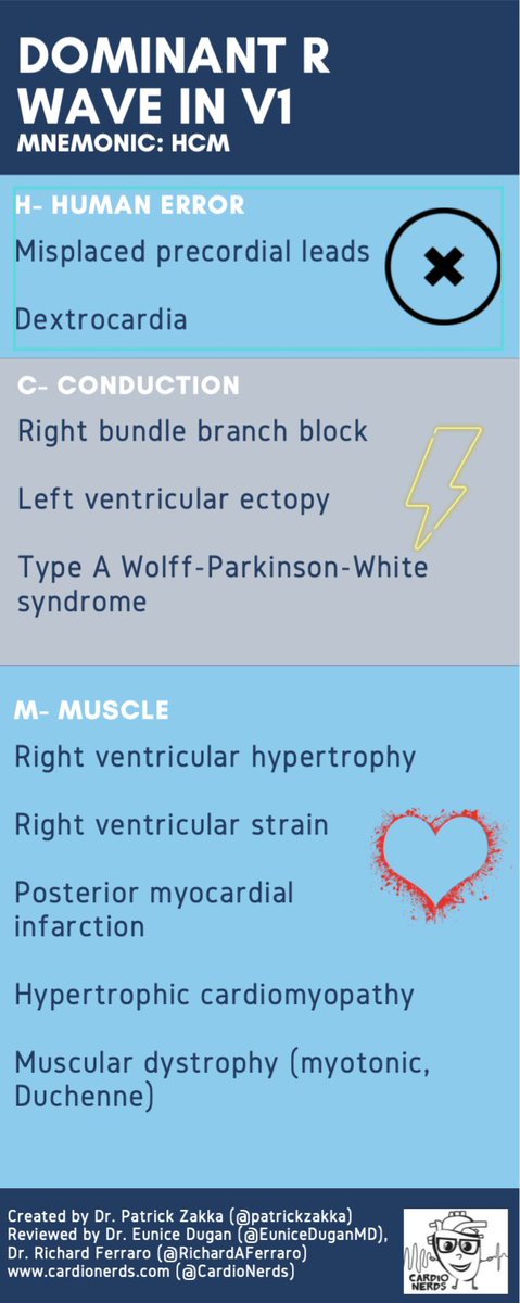
We can think of 3 broad categories to start with, using the mnemonic HCM (hint: this is also one of the diagnoses!) This stands for:
1⃣ Human Error ❌
2⃣ Conduction and ⚡️
3⃣ Muscle or myocardium. 💓
Check out this summary table on tall R waves in V1!