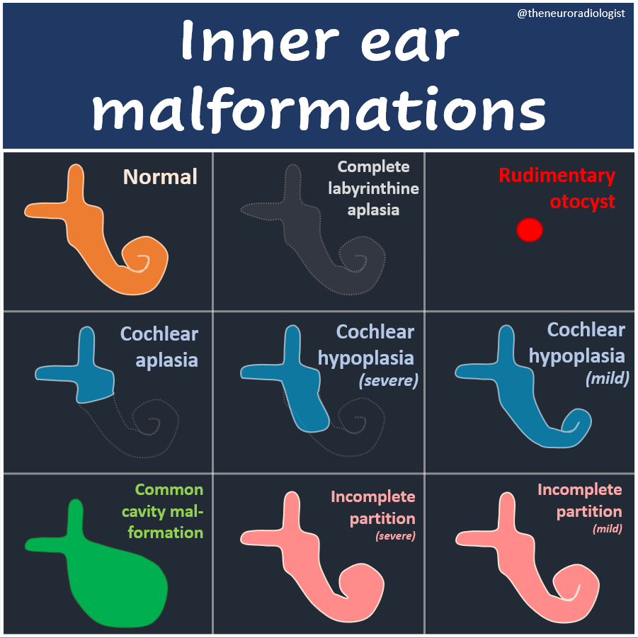
Inner ear malformations made easy (a non-exhaustive overview focussing on the cochlea; many of these can be further subdivided, but you've got to start somewhere). #radres#radiology#MedEd#FOAMed#FOAMrad#RadEd
A unique case of intraventricular fetus-in-fetu in a 1-year-old girl with motor delay and macrocephaly. Imaging shows a mass with vertebral column, femur, and tibia, distinguishing it from a teratoma.
https://t.co/NNeWvFuk3Q
Tip of the day 💡: HIE (hypoxic ischemic encephalopathy) is a 'clinical diagnosis', not a radiological one. We only look at injury on imaging if any. This one does not show any- but does not mean he didn't suffer from HIE. This weekend, know your limitations!
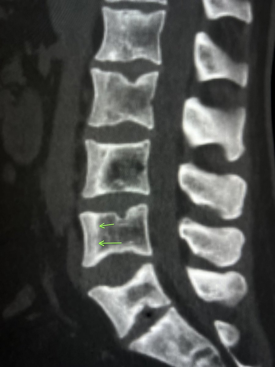
Patchy sclerosis of the vertebral body with a “ruler straight” margin suggests bone infarction.
In a patient with sickle cell disease.
— today’s reporting list
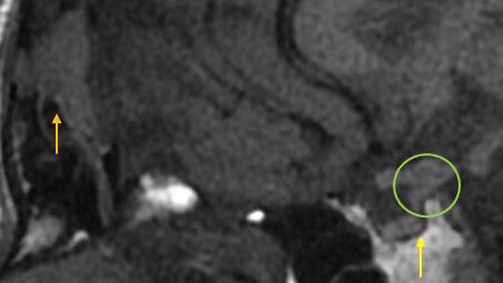
Langerhans Cell Histiocytosis (LCH). Three in a row:
🟠 lytic skull mass
🟢 pituitary stalk thickening
🟡 absent posterior pituitary T1 hyperintensity
Put the pieces of the puzzle together.
Easily missed injuries: Lisfranc fracture dislocation
Increased C1-M2 distance on an AP foot radiograph- > 3 MM- is a red-flag. Ask for CT or MRI.
I prefer CT in all suspicious cases. MRI only after negative CT.
—on-call wisdom
How durable is your knowledge of dural lesions?
How’s your knowledge base of the skull base?
Know the difference between petroclival & petrous face? Middle fossa & middle sphenoid wing? Clinoid & clivus?
Here is a map of the names of skull base meningiomas. How many do you know?
▶️Recognizing a lesion is only the first step. Communication is the next step.
▶️If you can’t accurately communicate where a lesion is located, then you have only done have your job.
➡️When it comes to skull base meningiomas, high risk lesions are the medial lesions:
🔸Clinoid
🔸Cavernous sinus
🔸Tuberculum sellae
🔸Medial/incisural tentorium
🔸Clivus
🚨I think of a cone coming up from the foramen magnum encompassing these lesions that I call the “cone of risk”!
Hopefully, this anatomy of skull base dural lesions with stay with you for the duration!
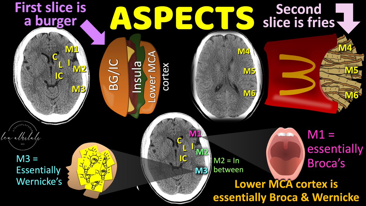
Can’t remember all the aspects of...well, ASPECTS?
Need a stroke of genius to recall this stroke score?
Knowing which regions to count is a very important aspect of ASPECTS!
All you need to remember is burger & fries to calculate the ASPECT score!
Here's how you can do it!
➡️You start at the level of the basal ganglia or “ganglionic” level
▶️Here, you have:
🔸3 structures of the basal ganglia/internal capsule medially
🔸Insula in the middle
🔸Lower MCA cortex on the outside--separated into 3 sections.
🚨Remember the insula is INSULAted—so it is sandwiched between the basal ganglia/internal capsule & MCA cortex like a burger!
▶️Here’s how to remember the 3 MCA cortex sections:
🔸M1 is essentially Broca’s area. W/Broca’s you can only get one word out at a time, so it’s M1.
🔸Wernickes’s rhymes w/three, so it’s M3.
🔸M2 is just in between.
➡️Next section is higher, above the basal ganglia. You have 3 MCA cortical region here sticking out like fries
➡️MCA cortices stick out like french fries corresponding to same MCA cortex regions from the ganglionic level, just higher up!
So now you know the burger and fries of the ASPECTS score--a fitting meal for those hungry to learn about stroke imaging!