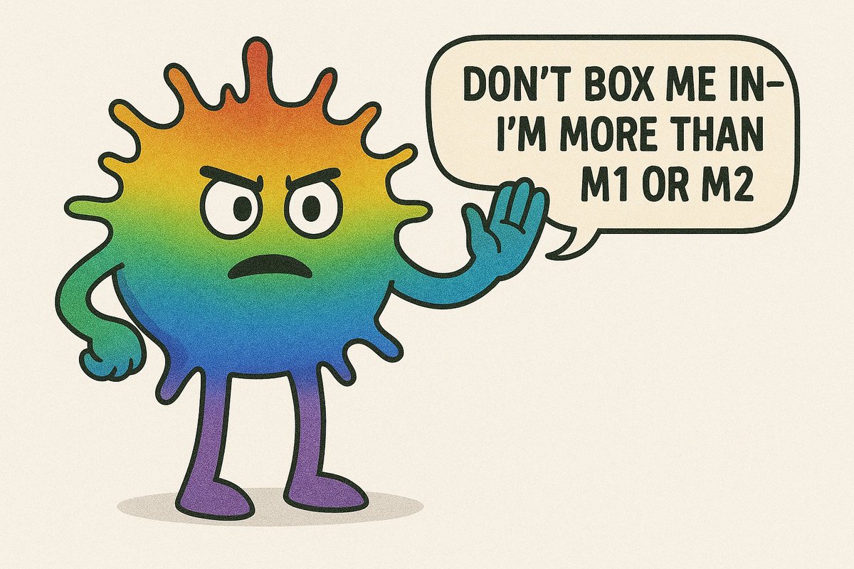
PSA: Stop calling macrophages in tissue M1 or M2. These are not states that exist in biology. The only use of M1 is a macrophage cultured ex vivo with LPS and IFNg; and for M2: IL-4/13/10. Macrophages in tissues are highly complex and diverse and do not resemble either of the aforementioned M1/M2 states. M1/M2 language causes confusion and sets the field back. Refer to your macrophages by the molecules they express and the cytokines they make.
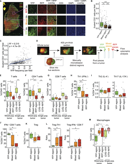
Our story made the cover of @JExpMed! So excited to share our work showing how tumor cells profoundly shape their local immune microenvironment @MihoTanakaMT
In a recent Science #Immunology Review, researchers discuss ways in which genetic abnormalities enable #cancer cells to evade #antitumor immunity and resist treatment. https://t.co/CrW5eEAywh #DayOfImmunology
Is there anything more fun than starting your talk with a walk-up song?! Thank you to the brilliant 3rd year Immunobiology PhD students at Yale who organized the '12th Yale Immunobiology Student Symposium' today.
A new Review suggests that genomic changes in #cancer cells can shape antitumor immune cells and facilitate #immunogenomic cancer evolution, which could inform future strategies for precision medicine.
Learn more in Science #Immunology: https://t.co/CrW5eEAywh
How does tumor heterogeneity impair the immune response to cancer? @MihoTanakaMT, @melissaqreeves@UUtah et al. show tumor cells shape their microenvironment on a highly local level, using CX3CL1 to create spatial pockets of immunosuppression. https://t.co/5ihRk2FMEY
Just published in Nature (Fed 2025), Tyler Miller, MD, PhD et al. have made a major conceptual leap in characterizing glioma-associated myeloid cells--not by their lineage but by their functional states dictated by local microenvironmental cues (Bradley Bernstein lab at Dana-Farber Cancer Institute).
In this study, Miller et al. pinpoint four key "activity programs" in glioma-associated myeloid cells: systemic inflammatory, microglial inflammatory, complement immunosuppressive, and scavenger immunosuppressive. Remarkably, any of the myeloid subsets (microglia, macrophages, monocytes, and dendritic cells) can adopt these programs in response to local cues like hypoxia, IL-1B, TGFb, or steroids.
Notably, the "scavenger immunosuppressive" programs correlate strongly with worse clinical outcomes, and ex vivo reprogramming data reveal transcriptional regulators (AP-1, p300/CBP) with the potential to convert an immunosuppressive environment into an inflammatory, T cell-supportive one.
1/Excited to share our latest work published in @CD_AACR. Led with #MiyaHugaboom, #KellyStreet, we show that the presence of tertiary lymphoid structures and exhausted tissue-resident T cells determines clinical response to PD-1 blockade in #RCC patients: https://t.co/Apw81TaY0Q
Gasdermin D pore-laden pyroptotic vesicles transplant these pores on neighboring cell surfaces and propagate cell death to bystander cells @CellCellPress@RathinamVA
https://t.co/MR0s9dg2i5
Online now: Antigen presentation by tumor-associated macrophages drives T cells from a progenitor exhaustion state to terminal exhaustion https://t.co/fnnRUAvbBg
In the latest issue! Multiparameter imaging reveals clinically relevant cancer cell-stroma interaction dynamics in head and neck cancer https://t.co/J2AwhDcisj