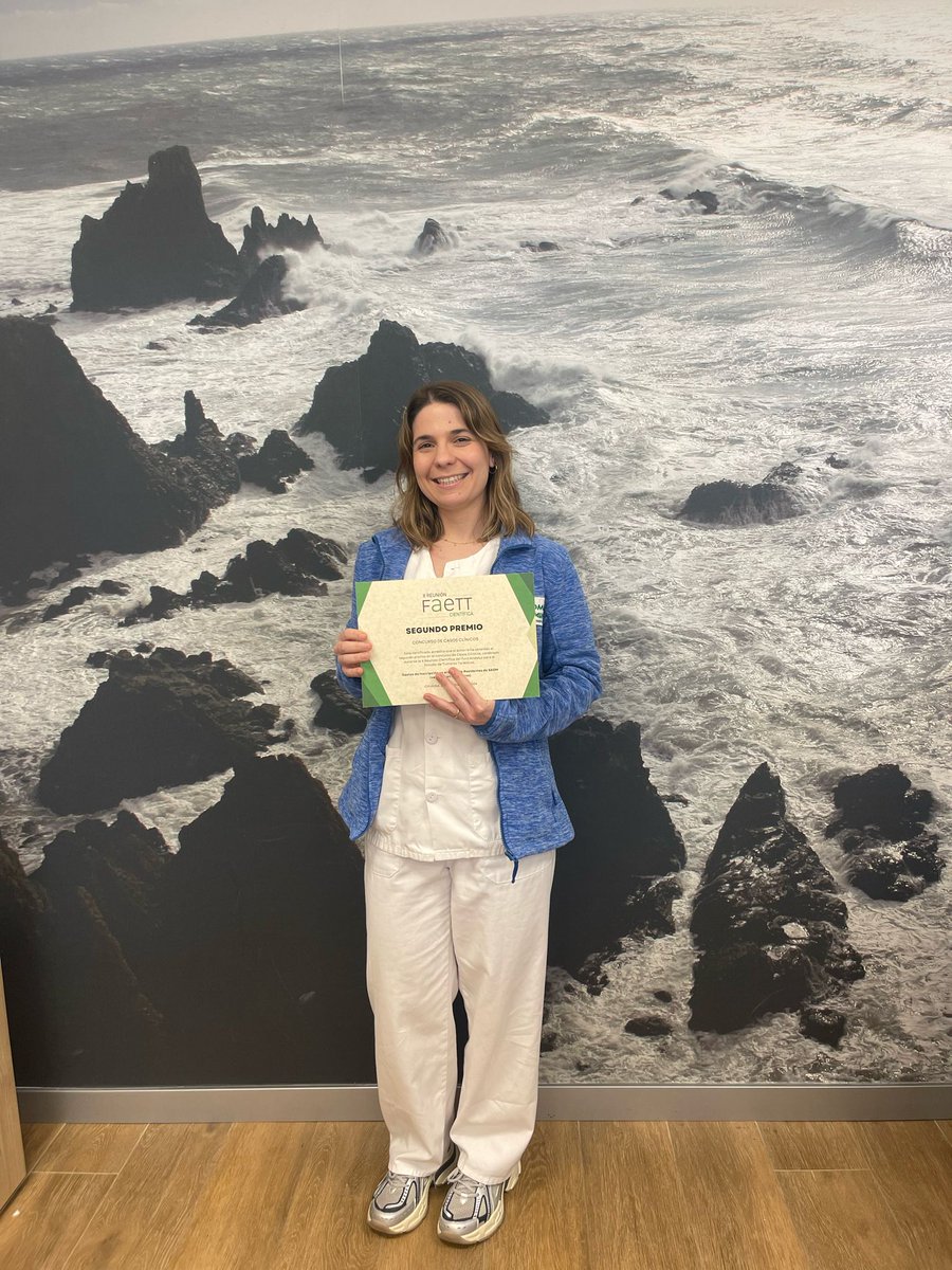
Es un orgullo para nuestra UGC #Anatomía_patológica y para el @TorrecardenasHU la obtención del segundo premio, en el concurso de casos clinicos, celebrado en la X reunión científica del #Faett por nuestra residente @MJPath Felicidades 👏
Gracias al trabajo incansable por parte del Dr. Velasco Albendea, sale a la luz nuestra humilde visión y experiencia sobre cryoebus🫁🔬❄️
Intentaremos pronto informar de los 60 pacientes que ya se han beneficiado de esta técnica en el 🏥 @TorrecardenasHU
https://t.co/CaVlCmHqe3
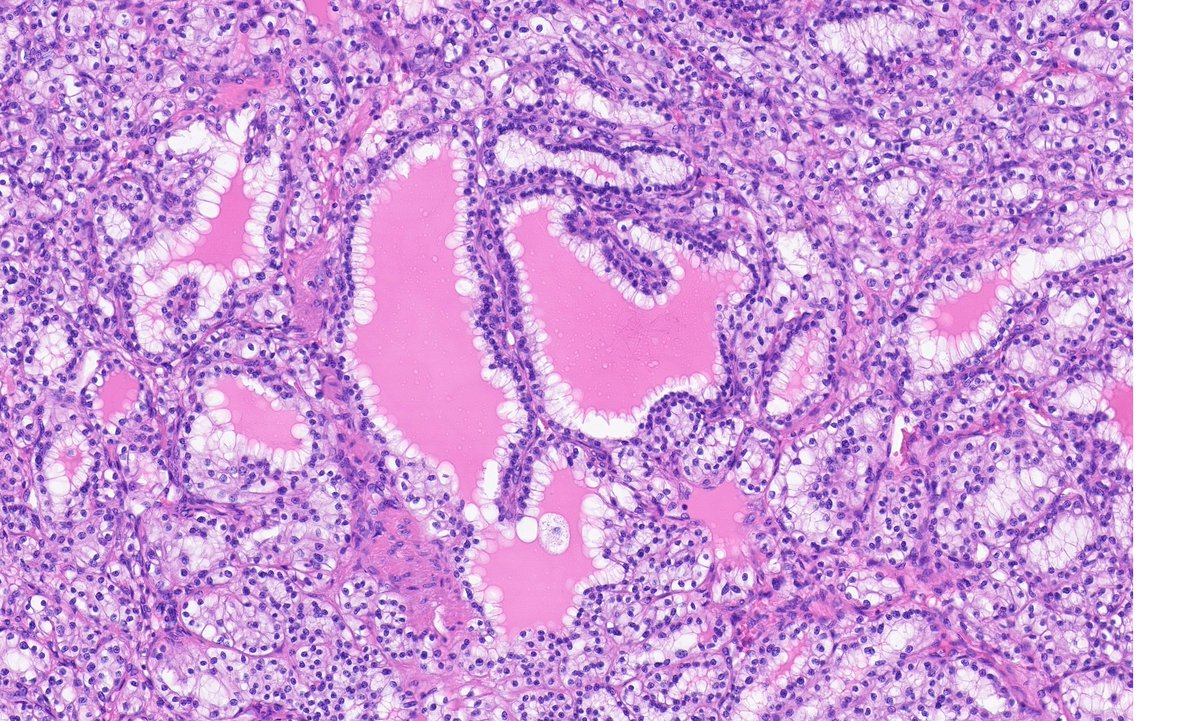
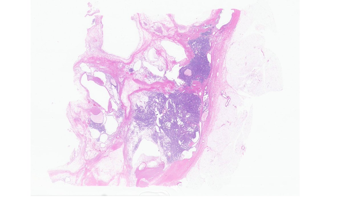
Need some help. 64yo woman with a solid-cystic encapsulated renal mass limited to kidney. Tubular and nested-cord-like architecture with clear cells basally/centrally located - not luminal (G2 max). Here some representative pics. IHQ on thread #GUpath#PathTwitter
Ayuda patólogos generales y digestivos! Solo 2 min. Podeis colaborar en esta encuesta sobre el tallado óptimo de los ganglios en piezas de ca. colorrectal. Vuestras respuestas serán muy útiles para comprobar diferencias regionales. https://t.co/v8XDrpP7nB
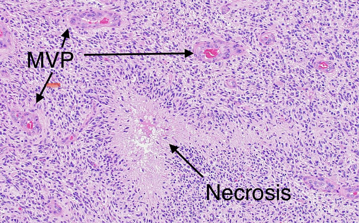
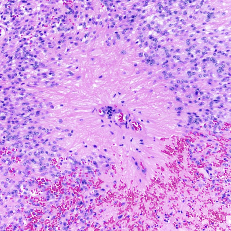
#GlioblastomaAwarenessDay#Neuropathologists look at tissue under a microscope to diagnose #GBM so that patients can receive appropriate treatment. Features we look for include pseudopalisading necrosis and microvascular proliferation.
Vamos a iniciar una serie de casos clínicos dirigidos tanto a estudiantes de Grado de #AnatomíaPatológica como a residentes.
Mujer de 50 años de edad, con HTA e imagen 👇 (bibliografía al final).
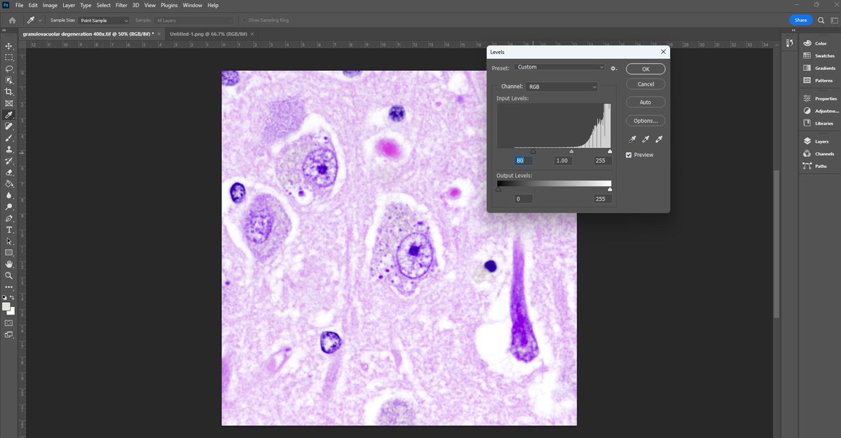
How to make photomicrographs more vibrant:
1. open a picture in photoshop
2. CTRL+M
3. select the white eyedropper and click it on a pure white area of the image
4. click "OK"
5. CTRL+L
6. adjust the left and/or right markers (not the center)
7. click "OK"
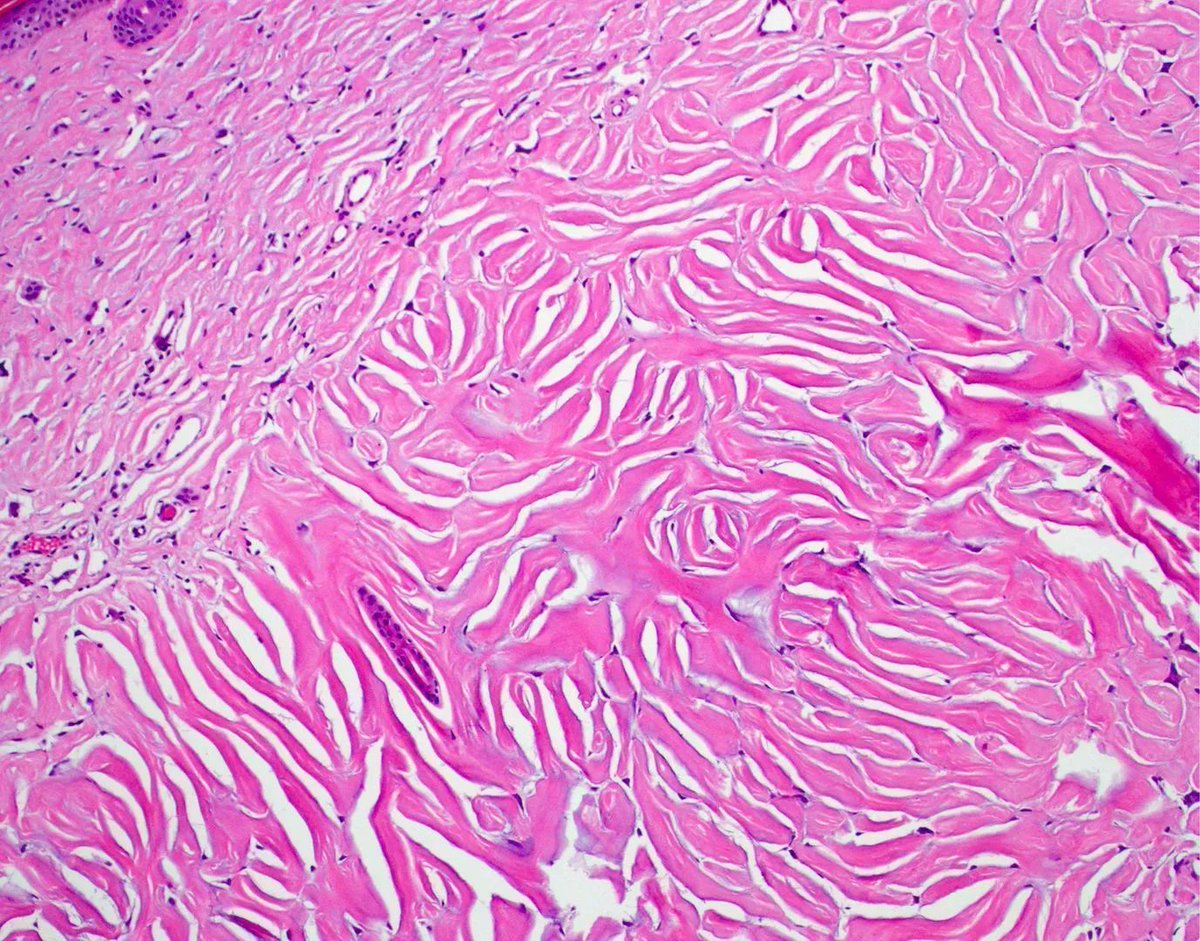
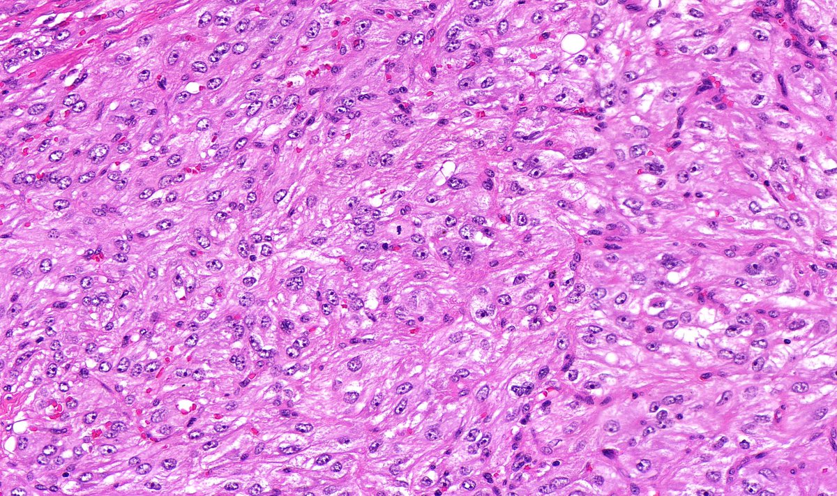
Sacral mass in an adult. Diagnosis?
WSI digital slide: https://t.co/RxlYHO1czv
Answer & video: https://t.co/8luDC0VfDl
Another great example: https://t.co/fgv6TxmhAm
#neuropath#BSTpath#pathologists#pathology#pathTwitter
The JHH Atlas of Colon Pathology is out! This searchable atlas features numerous color images, including inflammatory and neoplastic disorders. Each image is annotated with a diagnosis, caption, and list of diagnostic features. #GIpath@LizMontgomeryMD