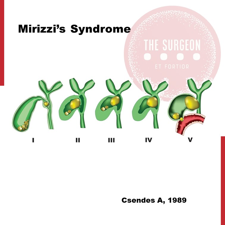
Easy to miss #
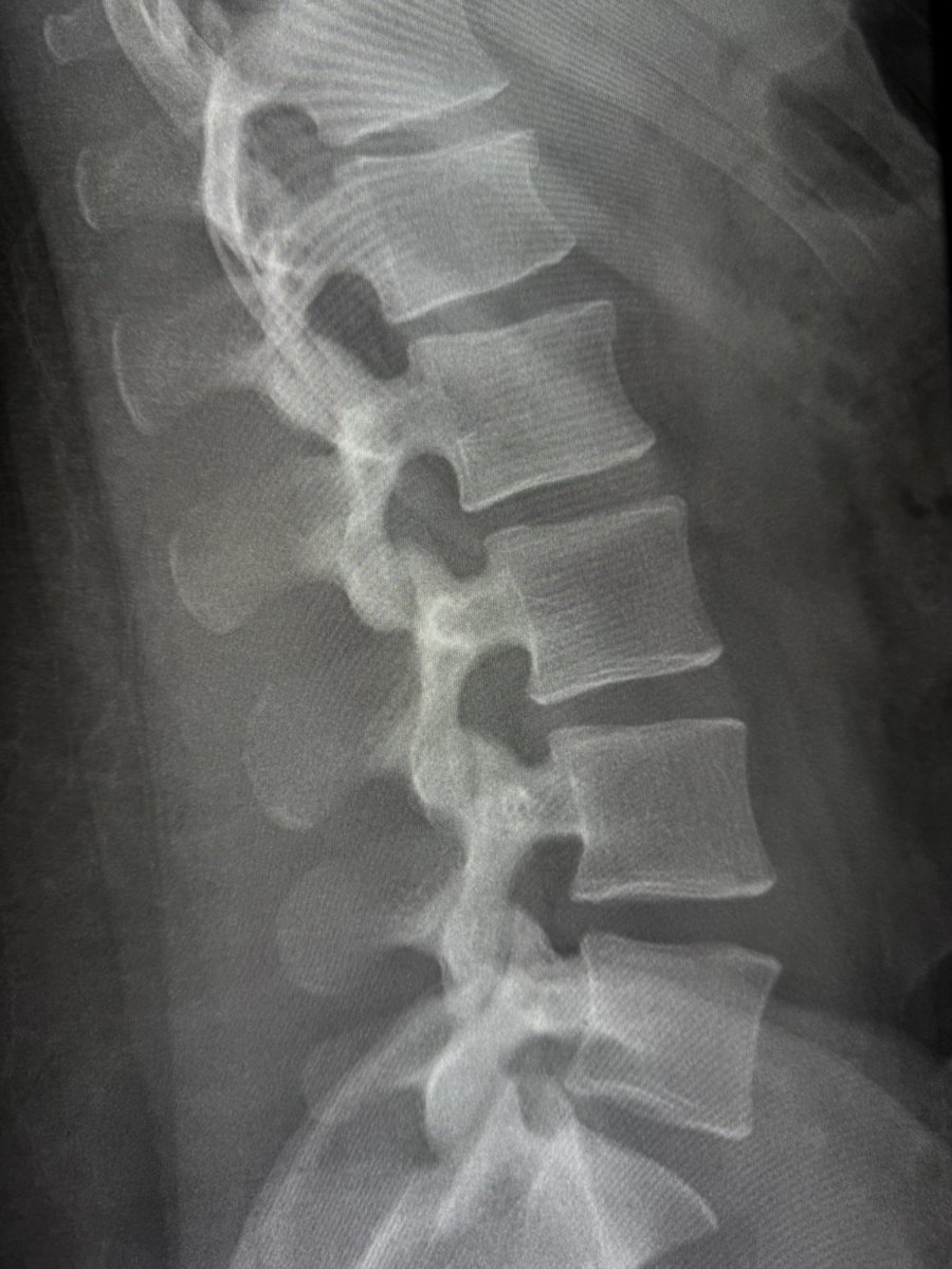
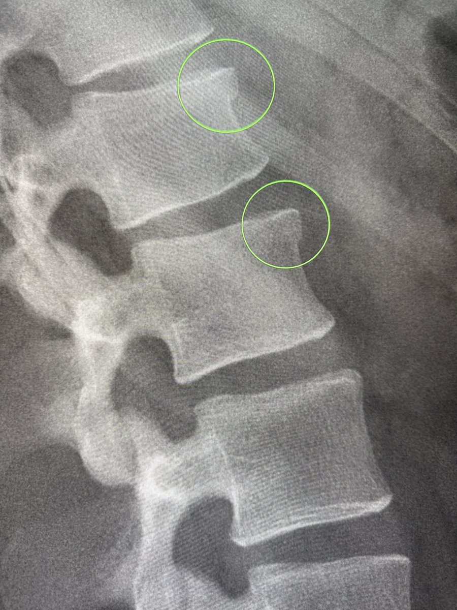
Acute compression fractures of L1 and L2 with anterior cortical step-off—one of the most specific signs of an acute vertebral fracture. Always look for the step-off.
All fractures on radiographs will go for CT irrespective of how subtle or mild they look.
—On-call tips: my reporting list
#GoodNews dal mondo #AlumniSSC!
Complimenti alla socia Sara Romano per l'importante risultato clinico raggiunto presso l'ospedale Buccheri La Ferla di #Palermo permettendo a un bambino di continuare la terapia grazie al #BatTest.
La news completa è qui👇🏻 https://t.co/LB8Hii8MK4
The circle of Willis is often incomplete. But what kind of incomplete is associated with poor stroke outcomes. Get to know more about this specific anatomic characterization for your next Code Stroke patient at https://t.co/KyLW9bYPUP @UCStrokeTeam@TheAJNR
Procalcitonin(PCT) Guideline for ED.
https://t.co/SJw9XHs0Kx
This guideline offers procalcitonin (PCT) recommendations for emergency departments to optimize antibiotic use and infection management. For lower respiratory infections, use antibiotics if PCT >0.25 ng/mL. In COPD exacerbations or febrile patients, PCT alone is insufficient. For acute pancreatitis, antibiotics are advised if PCT >1.0 ng/mL. In sepsis diagnosis, combine PCT with SIRS or qSOFA for accuracy. Post-gastrointestinal surgery, PCT may yield false positives but remains reliable after cardiac surgery or burns. For immunocompromised patients (e.g., neutropenia, lupus), PCT has low sensitivity. In bone/joint infections, PCT confirms but doesn’t exclude infections. For dialysis patients, PCT detects bacterial infections but is less effective in excluding peritonitis. In liver cirrhosis, PCT aids in diagnosing bacterial infections and peritonitis. For organ transplant patients, PCT is useful post-solid organ transplant but not post-stem cell transplant. Combining PCT with respiratory virus testing helps differentiate viral from bacterial infections, reducing unnecessary antibiotics. Overall, PCT is valuable but should be used alongside clinical judgment for optimal decision-making.
#GoodNews dal mondo #AlumniSSC! Complimenti al socio @plvdac, tra i vincitori del bando Multiround Telethon, che vedrà finanziato per due anni il suo progetto di ricerca sulla sindrome da deficit di glut1.
Complimenti e in bocca al lupo da parte di tutti noi, Marco 🎉👏🏻
My attending once said, “If you don’t want to miss something, you have to know where to look for it!”
MCA aneurysms may occur at many branch points along the way, but overwhelming occur at the bifurcation (or trifurcation)
Interestingly, the right MCA is more prone to distal aneurysms, while the left MCA is more prone to proximal branch aneurysms
So look hard at the bifurcation bc you miss 100% of the aneurysms you don’t look for!
Research Article
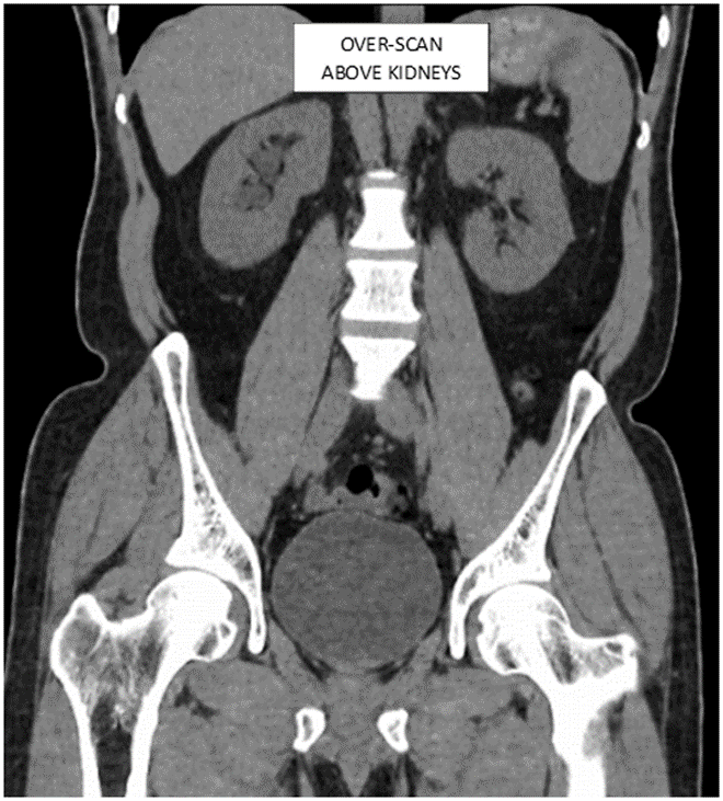
Striving for excellence: multicentre quality improvement project to optimize CT KUB technique in renal colic patients
https://t.co/tf1JQXcbTh
#Radiology
Worried that hydrocephalus is hiding in plain sight?
Normal pressure hydrocephalus (NPH) can mimic volume loss
How can you tell the difference?
This month’s @theAJNR SCANtastic gives you want you need to know
https://t.co/twxJl6438G
Many findings can be seen with NPH—you may use 1 or more. But do you know how well your sign performs?
Here are how common NPH signs perform:
Evans index
SENS: 100
SPEC: 74
Cingulate sulcus sign:
SENS: 63
SPEC: 61
Callosal angle:
SENS: 49
SPEC: 78
Disproportionately enlarged subarachnoid spaces (DESH):
SENS: 94
SPEC: 41
Anteroposterior diameter of the lateral ventricle index:
SENS: 98
SPEC: 100
For the SINPHONI trial, a combination of Evans index > 0.3 & DESH had a 70-80% PPV for shunt responsiveness.
In this month’s @theAJNR, Leary et al decided not to pick & choose what to use & instead used an AI algorithm based on T2 & FLAIR images. They found they could get >80% sensivity & >80% specificity. This could be used as a screening tool in NPH!
Now you know the signs of NPH & how good they may or may not be. Hopefully, you can now set the bar high when it comes to hydrocephalus!
Review
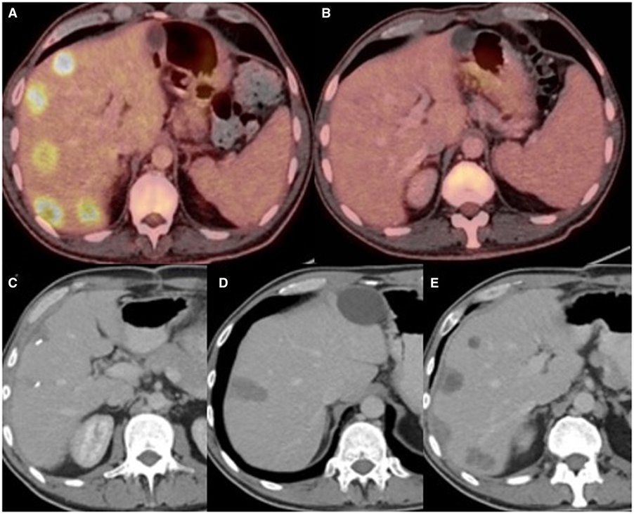
Preoperative imaging of colorectal liver metastases: what the radiologist and the multidisciplinary team need to know
https://t.co/2AJul4fdPD #Radiology
Cross-sectional patterns of spinal cord involvement in various causes of myelopathy.
Source: Kranz, P. G., & Amrhein, T. J. (2018). Imaging Approach to Myelopathy. Radiologic Clinics of North America. doi:10.1016/j.rcl.2018.09.006 (https://t.co/lAqWdUrDbZ)