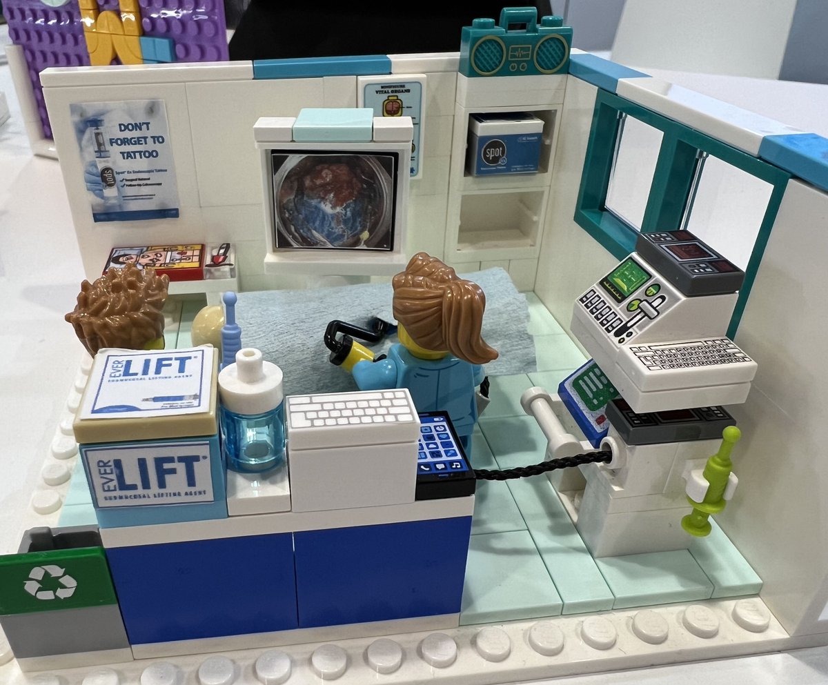
Should expensive endoscopy accessories be thrown out once they have reached their expiry date?
Sustainability data presented at #ESGEDays2026 shows that these devices remain sterile and functional even after expiry.
The iron supplements were supposed to FIX his anemia.
Instead, they were causing it.
A 60-year-old man with worsening anemia despite oral iron. EGD revealed the answer.
Here's what every endoscopist should know about iron pill gastritis:
EGD showed diffuse gastritis and a focally enlarged gastric fold on the greater curvature of the proximal stomach.
Biopsies? Iron-pill gastritis.
Iron deposits brown-black crystalline hemosiderin into the mucosa — essentially a chemical burn.
Key diagnostic features:
→ Focally enlarged, erythematous gastric fold
→ Greater curvature of stomach body (most common location)
→ Tiny blood clots or active oozing
→ Erosions, ulceration, or diffuse gastritis pattern
→ Biopsy shows hemosiderin deposits in mucosa
Why it matters:
Iron pill gastritis causes occult GI bleeding — worsening the very anemia you're treating.
It's frequently overlooked and missed.
Next time you see a focally enlarged gastric fold with blood clots → think iron pill gastritis.
By @theendoscopist | Bapaye, Lebel & Mönkemüller
#GIEndoscopy #Gastroenterology #EndoscopyTips #MedTwitter #GITwitter
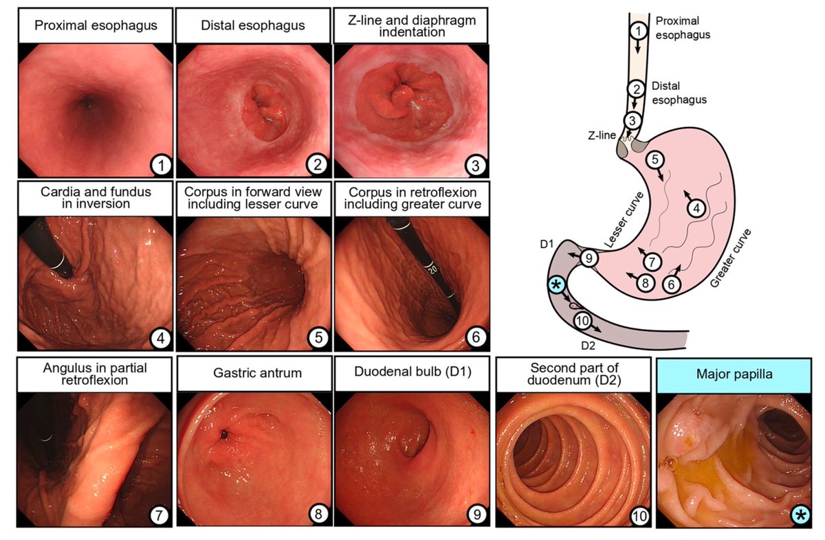
Classification systems for upper GI endoscopy #GITwitter
From @my_ueg “mistakes in gastroscopy and how to avoid them” - well worth a read!
https://t.co/DpvRYwOoLP
🇸🇦 Get ready for a groundbreaking medical event in Riyadh! 🌟Join us at the 9th International Conference on Live Endoscopy in Saudi Arabia, taking place January 29–31, 2026. Experience cutting-edge procedures, expert panels,
🔗 Register now at https://t.co/FtvkSGgZVm
Excellent Endoscopy Nurses ⭐️ are the cornerstones for any successful endoscopy unit!
Luckily we had our 4th Annual Endoscopy Nurse Skills Program with more than 150 Nurses from the whole Kingdom! 👏🏼
Many thanks to all Speakers & Participants
@SGA_KSA More to come! 💪🏼
#ENSP
👉Menetrier's disease (also called hypertrophic gastropathy)
👉Diagnosis
Clinical Features:
Epigastric pain
Nausea and vomiting
Weight loss
Peripheral edema (due to hypoalbuminemia)
Anemia (due to mucosal bleeding or poor nutrition)
Laboratory Findings:
Hypoalbuminemia (low serum albumin)
Hypochlorhydria or achlorhydria (due to loss of parietal cells)
Imaging and Endoscopy:
Upper GI endoscopy shows thickened, rugal folds, especially in the body and fundus of the stomach (sparing the antrum)
Biopsies reveal:
Foveolar hyperplasia (mucus cell hyperplasia)
Glandular atrophy
Decreased parietal and chief cells
Minimal inflammation
CT or MRI may show enlarged gastric folds
Histopathology (Gold Standard):
Large, tortuous gastric rugae
Hyperplasia of surface mucous (foveolar) cells
Loss of acid-secreting cells
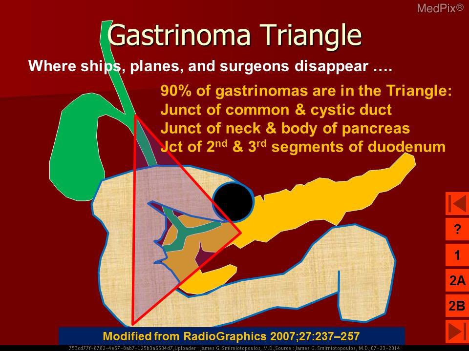
This is the gastrinoma (or Passaro’s) triangle where 90% of gastrinomas arise. Gastrinomas are a type of neuroendocrine tumour which can secrete gastrin, leading to peptic ulcers underlying Zollinger-Ellison syndrome. Gastrinomas can occur de novo but 25% occurs as part of MEN-1 syndrome (pancreatic, parathyroid and pituitary malignancies). Consider this triangle when hunting for the primary.
📸: Wikipedia