Cardiac Conduction Axis—From the AV Node to the Bundle Branches⚡️💡
👉The AV node continues as the penetrating bundle through the central fibrous body and membranous septum before becoming the branching bundle.
👉The branching bundle gives rise to the LBB, while the RBB emerges onto the RV septal surface posterior to the muscle of Lancisi.
Successful TAVI implantation relies on much more than accurate valve positioning, it requires an understanding of the relationship between the prosthesis and the cardiac conduction system.
@TAVRBot#cardioed#cardiotwitter
CardioNugget™ 🦀
The Crab View (Suprasternal Coronal View)
📍 How to obtain
Start in the suprasternal notch
From the long-axis aortic arch ("candy cane") view, rotate ~90° into the coronal plane and make slight tilt adjustments until the LA and pulmonary veins come into view.
👀 What you'll see
❤️ Left atrium = Crab's body
🦵 4 pulmonary veins = Crab's legs
🎯 Why it matters
✅ Confirms all pulmonary veins drain into the LA
✅ Detects Total anomalous pulmonary venous return and partial anomalous pulmonary venous return
✅ Evaluates pulmonary venous obstruction/stenosis
#CardioNugget™ #EchoBoards #CongenitalHeartDisease #ASE #CardioTwitter
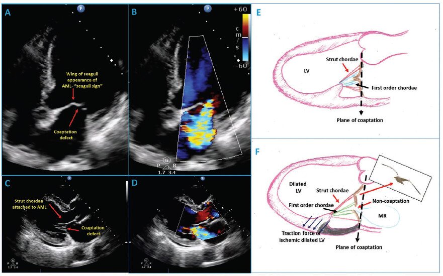
The Seagull Sign of Anterior Mitral Leaflet
🩺 What is the Seagull Sign?
In echo, the mitral valve seagull sign refers to a distinctive wing-like distortion or "kink" in the middle of the anterior mitral valve leaflet during systole, mimicking the wings of a flying seagull.
Assessing LV diastolic function is one of the most nuanced tasks in echocardiography, overlapping terminology, load dependence, and discordant parameters make it genuinely hard to do consistently at the bedside.
This review in #EHJCVI offers a pragmatic, tier-based framework built around the 2025 ASE recommendations, using a probabilistic approach that integrates clinical context, echo findings, advanced imaging, and emerging AI to sharpen both diagnosis and prognosis.
Kudos to Dr @argulian , who led this work with exceptional clarity and vision. Beyond being one of the very best imaging cardiologists in the world, Edgar is a wonderful clinician and exceptional human being. It is a privilege to learn from and collaborate with him.
And what an honour to share authorship with two giants of the field, @SNagueh and @OttoSmiseth , whose work has shaped how all of us think about diastology.
Don’t miss it ➡️ https://t.co/Gt32R2DzqA
#Echocardiography #Cardiology #DiastolicFunction #HeartFailure #CardiacImaging #EHJCVI
📌 Mitral Regurgitation: Signal Density & Contour on CW Doppler
Continuous-wave (CW) Doppler assessment of the mitral regurgitation (MR) jet provides important clues about MR severity.
Follow this thread 👇
Ejercicio & prueba de esfuerzo en el Sx de Brugada. 🏃♂️🫀⚡️
🔹 El momento clave no es el ejercicio, sino la recuperación. Solo ~1% de los pacientes desarrolla patrón tipo 1 durante el esfuerzo, mientras que hasta 25.5% lo manifiesta en la fase de recuperación, cuando ocurre el rebote vagal tras el retiro del estímulo simpático. 🤔🏃♂️⏱️
🔹 La recuperación temprana (primeros 2-3 minutos) es la ventana más importante. Es aquí donde pueden aparecer el patrón tipo 1 de Brugada, elevación del ST/J-point, extrasístoles ventriculares, TV o FV en pacientes de alto riesgo. 🧐⚠️⚡️
🔹 Se asociaron con mayor riesgo de eventos arrítmicos: Aumento del ST durante la recuperación, extrasístoles ventriculares entre 1.5 y 3 minutos de recuperación, recuperación rápida de la FC (marcador de hiperactividad vagal), prolongación de la pendiente ascendente de la onda S en ejercicio máximo. 👨⚕️📝
📄🆓️⤵️ State-Of-The-Art Review 2026 @JACCJournals 👌🏻💯
https://t.co/pKL8FJz2HZ
https://t.co/4PnV0aMMfn
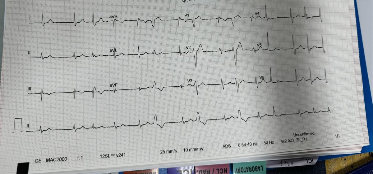
🧵Excellent EKG! #EP_Fellow called to help managing AF with RVR. 40-year-old structurally normal heart.
✅ What is the antiarrhythmic of choice?
✅ What about Flecainide / Propafenone?
✅ What unique⚡️abnormality present in this EKG?
#Epeeps
Baseline ECG👇
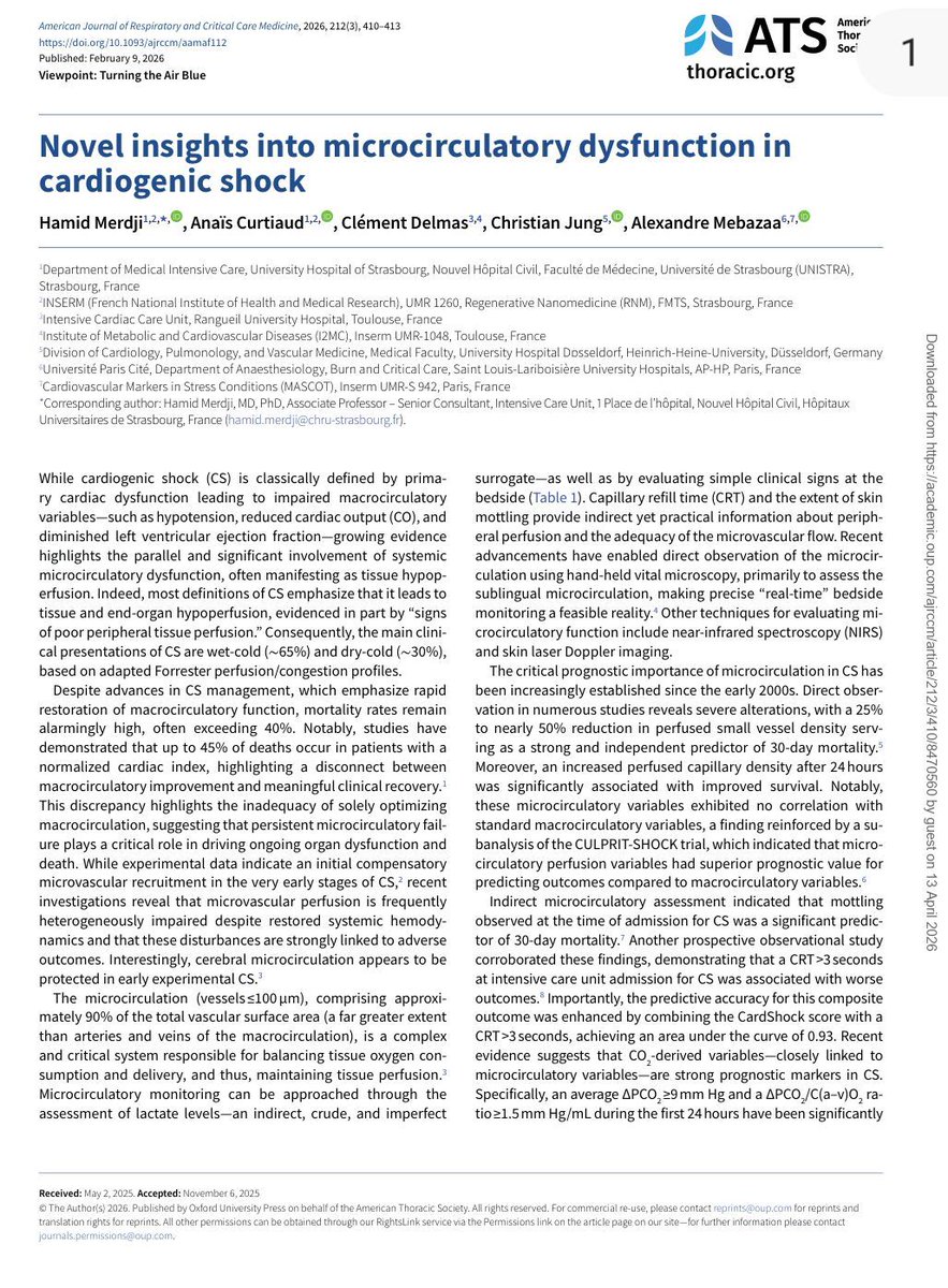
🫀In cardiogenic shock, we still focus heavily on MACROcirculation:
📉 blood pressure
📉 cardiac output
📉 LVEF
📉 vasopressor dose
But what if the real battle is happening deeper?
🩸 At the microcirculatory level.
This excellent ATS viewpoint highlights one of the most important evolving concepts in shock physiology:
⚠️ normalization of macrocirculation does not necessarily mean restoration of tissue perfusion.
Despite advances in cardiogenic shock management, mortality remains >40%.
Even more striking, up to 45% of deaths occur in patients with normalized cardiac index.
That disconnect may be explained by persistent:
🩸 microvascular dysfunction
🩸 impaired capillary perfusion
🩸 endothelial dysfunction
🩸 tissue hypoxia despite “acceptable” hemodynamics
The review reinforces that: Microcirculation is not a passive bystander.
It may be a central driver of:
• organ dysfunction
• lactate persistence
• shock progression
• mortality
Particularly interesting is the emphasis on simple bedside tools.
We often think microcirculation requires advanced devices, yet:
📌 capillary refill time (CRT)
📌 mottling
📌 ΔPCO₂
📌 lactate trends
still carry strong prognostic value.
A CRT >3 seconds at ICU admission was associated with worse outcomes, and combining CRT with the CardShock score achieved an impressive AUC of 0.93 for outcome prediction.
The article also reviews modern technologies:
🔬 handheld vital microscopy
🔬 sublingual microcirculation imaging
🔬 NIRS
🔬 laser Doppler assessment
bringing “real time” bedside microcirculatory monitoring closer to clinical practice.
One of the strongest physiological messages:
⚠️ Shock is not only about flow. It is about effective tissue level oxygen delivery.
The review beautifully summarizes the four major mechanisms of microvascular dysfunction:
• heterogeneity
• hemodilution
• congestion
• edema
Particularly relevant for intensivists:
📌 venous congestion itself may worsen microvascular flow
📌 elevated filling pressures impair driving pressure
📌 edema increases oxygen diffusion distance
This is highly relevant in:
• advanced heart failure
• VA ECMO
• mixed shock states
• fluid overloaded patients
Another important takeaway: Persistent microcirculatory dysfunction after VA ECMO initiation was associated with increased mortality, even when macrocirculation improved.
Perhaps the key message of this paper is:
🩸 Microcirculation should no longer be considered a secondary endpoint in cardiogenic shock.
It may become one of the most important physiological targets of the next decade.
📖 Merdji H, American Journal of Respiratory and Critical Care Medicine. 2026, 212(3), 410–413 https://t.co/81Zp3aj274.
WHAT?
Hb 4 g/dL… AND HR IS 78??
I often ask fellows:
“What heart rate (HR) would you expect in a patient with profound chronic anemia?”
Most say:
“High.”
But over the years, I’ve realized that many patients with severe chronic anemia have surprisingly normal resting HRs.
🚨 ECG Breakdown
Axis:- Marked left axis deviation
Broad QRS with complete LBBB
Deep QS/rS in V1–V3
Broad tall R waves in I, aVL, V5–V6
Secondary discordant ST-T changes consistent with LBBB
Mild lateral strain-like changes in I & aVL
Negative Sgarbossa criteria
Impression:- Complete LBBB with LAD and expected secondary repolarization abnormalities.
Possible underlying LVH/hypertensive heart disease. No ECG evidence of acute STEMI.
#MedX #ECG
Coronary Artery Dominance: A Key Concept in Angiography
Dominance is defined by which artery gives rise to the posterior descending artery (PDA):
• Right dominance – PDA from RCA
• Left dominance – PDA from LCx
• Balanced – PDA from both RCA & LCx
Prevalence:
• Right dominant – ~85%
• Left dominant – ~8%
• Balanced – ~7%
Understanding dominance is crucial in assessing myocardial perfusion, planning revascularization, and anticipating outcomes in coronary artery disease.
Image:
Top – Right dominance
Middle – Left dominance
Bottom – Balanced dominance
(Courtesy Dr. Annapoorna Kini, Mount Sinai, NY)
Ref: Braunwald’s Heart Disease, 12th Ed.
🚨 ECG breakdown
🔻STE in aVL > Lead I
🔻Reciprocal STD in inferior leads (II, III, aVF)
🔻Significant STE across V2–V6
🔻Positive/tall R waves present in anterior leads
🔻1st degree AV block seen ( PR interval is prolonged)
🔻NSIVCD present
Overall impression :
Extensive anterior–anterolateral STEMI, most likely due to proximal LAD occlusion involving a large myocardial territory. Reciprocal inferior changes strengthen the diagnosis of an acute occlusive MI with intramural injury.
(QOH- Culprit vessel picture diagram included). Immediate PCI is life saving.
#MedX
Are all MRAs equal? Short answer: no. 🫀
#HeartFailure26 | Anne-Christine Ruwald
Harrington JL et al. JACC Heart Fail. 2025;13(10):102637
Steroidal MRAs (spironolactone, eplerenone) vs Non-steroidal MRAs (finerenone, balcinrenone, aparenone, esaxerenone)
Key differences:
🔵 Steroidal MRAs:
• Higher binding in kidneys vs heart
• More off-target binding → increased side effects (gynecomastia, hyperkalemia)
• Clinical data: benefit in HFrEF ✅ | Role in HFpEF unclear ❓
🟠 Non-steroidal MRAs:
• More even distribution between heart and kidneys
• More potent MR antagonism
• Clinical data: Role in HFrEF unclear ❓ | Benefit in HFpEF ✅
The pharmacological differences are not trivial — tissue distribution, specificity, potency, and tolerability all differ.
Fascinating inversion: sMRAs work in HFrEF, ns-MRAs work in HFpEF. Same target, different patients, different drugs.
The MRA class is not one-size-fits-all.
#MRA #Finerenone #HFpEF #HFrEF #HeartFailure #Cardiology #ESC #HeartFailure26
Diuretic resistance is not a therapeutic dead end → it’s a signal that we need a smarter strategy.”
The perfect closing slide from Dr. Ana Belen Mendez Fernandez at #HeartFailure26 👏
5-step framework for smarter decongestion:
1️⃣ Recognize the problem
Multifactorial, common, predictable — requires a structured approach
2️⃣ Objective assessment
Weight, ultrasound, BNP, renal function, volume status, urine output
3️⃣ Optimize loop diuretics
Adequate dose & frequency, IV route, check absorption
4️⃣ Sequential nephron blockade
Target different nephron segments, overcome compensatory mechanisms
5️⃣ Treat the drivers
Venous congestion, low cardiac output, neurohormonal activation
And the line that should be on every HF ward wall:
“Decongestion is not just symptomatic relief. It changes outcomes.”
#CardioTwitter #HeartFailure #Cardiology #Decongestion #Diuretics