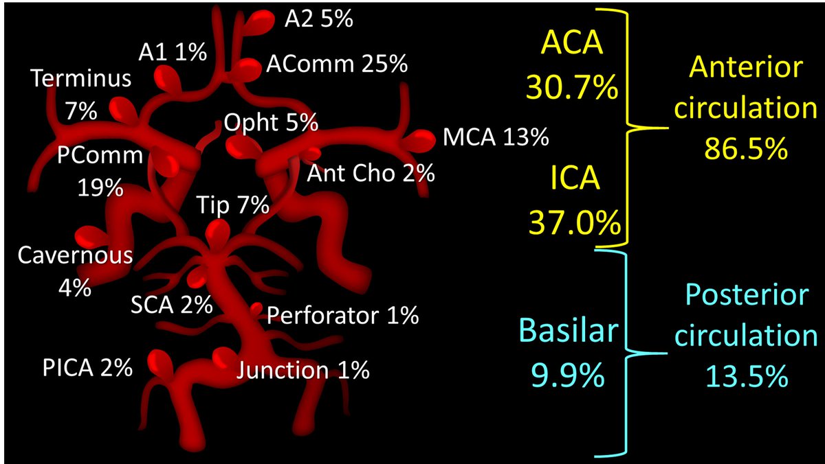
Saccular aneurysms account for approximately 90% of lesions and typically form at arterial bifurcations in the internal carotid artery, the anterior and posterior communicating arteries, and the middle cerebral artery. In the posterior circulation, they commonly occur at the basilar artery bifurcation and cerebellar artery branch points (seen in figure). Less common types of unruptured intracranial aneurysms (UIAs) include fusiform aneurysms involving elongated segments of the artery, mycotic aneurysms associated with infections, and dissecting aneurysms resulting from arterial injury. Up to 20% of patients with UIAs have multiple intracranial aneurysms. The formation of aneurysms is thought to result from degeneration of the internal elastic lamina, endothelial dysfunction, and hemodynamic stress; inflammation, which leads to instability of the vascular wall, plays a crucial role in both their formation and rupture.
Learn more in the new Clinical Practice article, “Unruptured Intracranial Aneurysms,” by Christopher S. Ogilvy, MD (@chrisogilvyMD), from @BIDMChealth and @harvardmed: https://t.co/lAIar9rnAx
#Neurology #Cardiology
Anyone can see the bright spot of diffusion imaging!
What sets you apart is your ability to tell them where it is—what vascular territory
Here are the vascular territories you need to know:
* Posterior Inferior Cerebellar Artery (PICA)
Soft under surface of the cerebellum
* Superior Cerebellar Artery (SCA)
The SCA territory is the top of the cerebellum (SCA is the SCAlp of the cerebellum)
* Basilar perforators
Make a line in the pons like a penetrating injury. So perforator infarcts look like perforating injuries
* Anterior Choroidal artery
This supplies the posterior limb of the internal capsule & corticospinal tract. Remember COR-roidal is COR-ticospinal
* Lenticulostriate arteries
Infarct territory looks like a tadpole
* Heubner's artery: infarct is the caudate head.
Remember "who called" which sounds like "heu caud" so heubner takes out the caudate
* Anterior cerebral artery (ACA)
Medial, anterior surface of the cerebral hemispheres
* Middle cerebral artery (MCA)
Lateral surface of the cerebral hemispheres
* Posterior cerebral artery (PCA)
Medial, posterior surface of the cerebral hemispheres
Hopefully now when it comes to the vascular territories, you will always be right when it's bright!!
My attending once said, “If you don’t want to miss something, you have to know where look for it!”
Intracranial aneurysms may occur at many locations, but overwhelming at the ACOM & PCOMM. Look hard at these regions!
Remember, you miss 100% of aneurysms you don't look for!
1/I always say you can tell a bad read on a spine MR if it doesn’t talk about the lateral recess
What will I think when I see your read? What do you say about lateral recesses?
Here’s a #tweetorial on lateral recess #anatomy & grading stenosis
#medtwitter#neurorad#spine
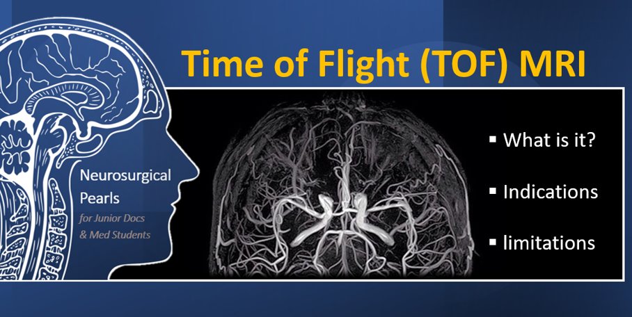
1/ Your patient is suspected of having cerebral aneurysm(s), needs an angiogram, but is allergic to Iodine/Gadolinium contrast? Patient needs an angiogram but their GFR is shocking?!
Time to learn about TOF MRI 🧠#NSurgPearls
Hello #MedTwitter, I am pleased to announce the launch of a series of educational posts, aimed at #JDocs & #medstudents.
Online #MedEd has been a passion of mine and as a JDoc myself, I learn best by sharing and discussing what I learn from my mentors and patients #Neurosurgery
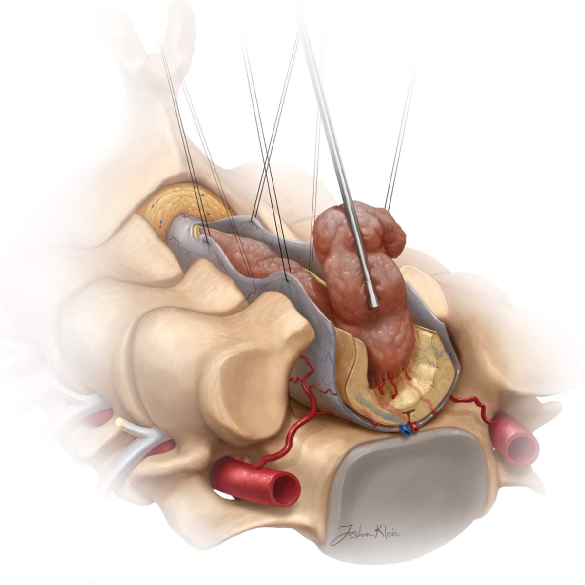
The most common intramedually spinal cord tumors are ependymomas, astrocytomas, and hemangioblastomas. In order of importance: preservation of function, tumor debulking, and gross total resection are the goals of surgery for these challenging entities.
#MedTwitter#Neurosurgery
Daily #Neurosurgery Quiz Question 316:
How many layers does the cerebral cortex have? How many layers does the cerebellar cortex have?
Comment with your answers!