The model of gene expression taught in school is highly misleading!
Transcription factors are proteins that bind to DNA and then help repress, or activate, the expression of genes. Cells have hundreds of different types of transcription factors, each tuned to regulate different genes based on short snippets of DNA located near those genes.
The basic model, taught in school, says that these transcription factor proteins float around the cell and, when they bump into a DNA sequence, either latch onto it strongly (CORRECT SITE!) or fall off quickly (WRONG SITE) and keep searching. All the other DNA in a cell is basically abstracted away as unimportant or irrelevant; mere background noise.
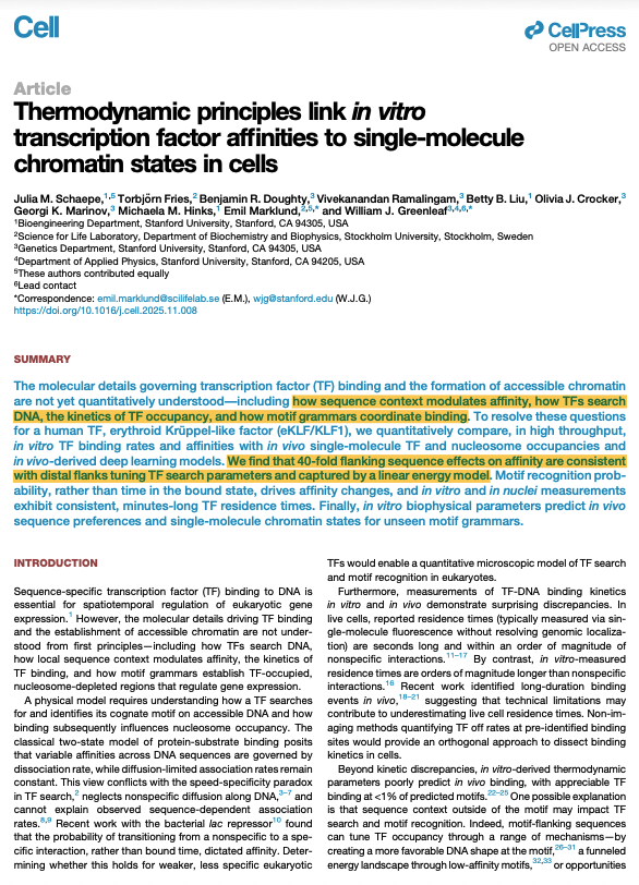
But again, this model is naive! And a new paper, published in Cell, beautifully shows how the sequences SURROUNDING a transcription factor's binding site also matter a great deal.
This won't be surprising to many biologists, as "cracks" in the standard two-state model began emerging decades(?) ago. Biologists have tagged transcription factors with fluorescent tags and then watched them move around living cells. And they have noticed that when transcription factors land in a "wrong" location in the genome, they skip or hop to a nearby location and repeat this until finally connecting with the "correct" sequence. So in other words, there are actually three states that a transcription factor can exist in: free-floating, "searching", or "bound."
(More technically, transcription factors first do a 3D search, then latch onto DNA and do a 1D search to find the correct location.)
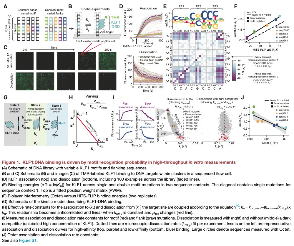
For this new paper, though, scientists exhaustively quantified *how* the sequences flanking a transcription factor binding site influence the search of the protein.
They did a huge in vitro experiment, wherein they placed a specific transcription factor with a known binding site, called KLF1, in a huge library of 11,812 different DNA sequences. These sequences had mutated "core" binding sites and variations in the flanking sequences. They also prepared negative controls. Then, these researchers measured the binding kinetics of KLF1 with each sequence to understand which bases in the flanking sites impact the 1D search.
What they found is that KLF1 has a basically flat disocciation rate from its core sequence, but that the PROBABILITY that it finds this sequence depends a lot on the surrounding context. Even mutations located dozens of bases away from the core site matter a lot, either pushing KLF1 to "hop" faster to find the site, or "trapping" KLF1 and slowing down its search. These flanking sequences can cause up to a 40-fold variation in the affinity of a transcription factor for its target site!
This is just one small part of the paper, though, so I encourage anyone interested to read the whole thing. It is challenging throughout.
This 1998 paper is, without question, one of the most beautiful in the history of biology.
It answers two questions:
First, how does a potassium channel let in K+ ions while excluding Na+ ions? And second, how does it funnel 100 million of those ions through each second?
These questions are interesting not only because potassium channels are so deeply involved in our brain's electrical signals, but also because K+ and Na+ both carry a positive charge and have similar sizes! A potassium ion has a Pauling radius (a measure of how far its electron cloud extends from the nucleus) of 1.33 Angstroms, compared to 0.95 for the sodium ion. So a potassium ion is slightly larger, but both ions carry exactly the same charge! And yet, despite these similarities, the potassium channel is “at least 10,000 times more permeant” to K+ than Na+.
How did evolution sculpt such an exquisitely-tuned machine?
To find out, scientists crystallized potassium channel proteins and solved its structure using X-ray crystallography. From this structure, a few things became immediately clear: First, the potassium channel’s interior measures 12 Angstroms long. And second, the channel's interior is lined with oxygens.
Normally, in a cell, ions are surrounded by water molecules. They must shed these waters to pass through the pore, but that's energetically expensive to do! The oxygens inside the channel are positioned at PERFECT locations to make this totally feasible; the K+ ions shed their waters and grab onto the oxygens instead.
The structure also revealed why Na+ cannot pass through. Because it is slightly smaller, Na+ ions cannot form contacts with all the oxygen atoms at once. The geometry is slightly off, giving potassium a decisive advantage.
Now onto the second question. Namely, how does this channel allow 100 million K+ ions to pass each second? That is very quick, considering these ions get "held" by their contacts with oxygen, presumably slowing them down a great deal.
Again, the scientists turned to structure. They again crystallized the potassium channel protein. But this time, they soaked those crystals in a liquid containing rubidium (Rb⁺) and cesium (Cs⁺). These ions behave like potassium but scatter X-rays more strongly, because they are heavier. Thus, they show up more brightly on the X-ray diffraction data.
When the scientists compared electron density maps with and without Rb⁺ or Cs⁺, they could literally see peaks where the ions bound inside the channel.
From this structure, they discovered that TWO K+ ions sit inside of the channel at once, separated by precisely 7.5 Angstroms. This distance is close enough that the ions "feel" each other’s electric repulsion (like charges repel!), but not so close that they destabilize the protein.
This repulsion is used by the channel as a feature, rather than bug! When a third K⁺ ion comes in from the top, the electrostatic “push” kicks the ions forward through the filter. In other words, instead of ions having to crawl through the pore one at a time, the channel uses their mutual repulsion to keep the flow moving; 100 million ions per second.
Beautiful paper. A classic in using 3D structures to reveal biophysical mechanisms.
Today, we’re announcing the first major discovery made by our AI Scientist with the lab in the loop: a promising new treatment for dry AMD, a major cause of blindness.
Our agents generated the hypotheses, designed the experiments, analyzed the data, iterated, even made figures for the paper. The resulting manuscript is a first-of-a-kind in the natural sciences, in which everything that needed to be done to write the paper was done by AI agents, apart from actually conducting the physical experiments in the lab and writing the final manuscript. We are also introducing Robin, the first multi-agent system that fully automates the in-silico components of scientific discovery, which made this discovery. This is the first time that we are aware of that hypothesis generation, experimentation, and data analysis have been joined up in closed loop, and is the beginning of a massive acceleration in the pace of scientific discovery that will be driven by these agents. We will be open-sourcing the code and data next week.
Robin is a multi-agent system that uses Crow, Falcon, and Finch, the agents on our platform, to generate novel hypotheses, plan experiments, and analyze data. We asked Robin to find a new treatment for dry age-related macular degeneration. Robin considered the disease mechanisms associated with dry AMD, proposed a specific experimental assay that could be used to evaluate hypotheses in the wet lab, and proposed specific molecules we could test in that assay. We tested the molecules and gave it the resulting data, which it analyzed before proposing more experiments. In the end, it identified Ripasudil, a Rho Kinase inhibitor (ROCK inhibitor) that is approved in Japan for several other diseases, which seems very promising as potential treatment for dry AMD. It also identified specific molecular mechanisms that might underlie the effects of Ripasudil in RPE cells, from an RNA sequencing experiment it proposed. To be clear, no one has proposed using ROCK inhibitors to treat dry AMD in the literature before, as far as we can find, and I think it would have been very difficult for us to come up with this hypothesis without the agents. We have also run the proposed treatment by several experts in AMD, who confirm that it is interesting and novel. Moreover, this project was fast: with Robin in hand, the entire project took about 10 weeks, which is way shorter than it would have taken if we had been doing all of the in-silico components ourselves.
Important caveats: We are real biologists at FutureHouse, so I want to be clear that although the discovery here is exciting, we are not claiming that we have cured dry AMD. Fully validating this hypothesis as a treatment for dry AMD will take human trials, which will take much longer. Also, this discovery is cool, but it is not yet a "move 37"-style discovery. At the current rate of progress, I'm sure we will get to that level soon.
Congratulations to the team. Congratulations in particular to Robin, which generated the hypotheses, proposed the experiments, analyzed the data and generated the figures. And major congratulations also to the human team, which built Robin: @MichaelaThinks, @agreeb66, @benjamin0chang, @ludomitch, Mo Razzak, Kiki Szostkiewicz, and Angela Yiu.
Interdisciplinary immunologists at all levels WANTED!
Faculty recruitment at Institute for Immunology, Tsinghua University, Beijing.
PLEASE SPREAD THE WORD!
The Kappel Lab is opening soon at UCLA and we’re hiring Staff Scientists to help lead our wet-lab projects & to assist with lab management! We have open positions for candidates with a PhD & for recent college grads. Learn more and apply: https://t.co/CoupN3mPHm
Please RT!
We're seeking a chemical (or molecular) biologist, predoc or postdoc, passionate about developing peptide and protein therapeutics! 🧪🧬 Be part of groundbreaking research at @ChemSynBioGroup and @IQSbarcelona . If interested in the offer, apply now!
Biggest medical discoveries of the week (🧵)
1/8
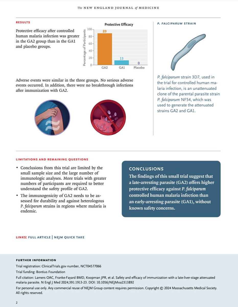
A malaria vaccine - attenuated P. falciparum arrested in the late liver stage - provided 89% protection in a trial of 9 people
This could improve the modest, short-lived protection of current vaccines
https://t.co/E5by1sbgXK
We have two open PhD positions in the lab! One together with @LandauMeytal on viral amyloids and one on phase separation of viral replication compartments! See https://t.co/SuuTVmzMvh for viral amyloids, and PM me for the second!
We are hiring multiple assistant professors in microbiology. Come and join our awesome research environment at Penn State.
Apply online at: https://t.co/MO1KArw6Dw
Please Retweet
I am excited to announce that the Tang lab (https://t.co/w6z4HWIf7N) at UT Arlington is recruiting 1-2 fully funded graduate students. Tang lab's general research focuses on microbiology and innate immunology. If you're interested, please don't hesitate to reach out and join us!