✨Take a look at this stunning confocal image of a mouse duodenum intestinal organoid!
💡This organoid was grown in Matrigel, fixed after the first passage, and captured using the Nikon AX/R confocal microscope
🔬Katarina Tokarcikova, Ales Pavlik, Lubomira Gresakova, Petr Slama
🗺️Did you know? Nikon's Microscope Imaging Software NIS-Elements is trusted by scientists worldwide and has been cited in over 30,000 peer-reviewed publications!
🔗Explore how Nikon microscopes and NIS-Elements are driving innovation: https://t.co/UX73UaKEQU
#NIS20
🌟 Discover how 3D imaging advances intestinal model research!
🔬 Using air-liquid interface culture & the Nikon AX/AX R confocal with NSPARC, researchers captured detailed 3D views of intestinal cells. No special prep needed!
📎Learn more: https://t.co/I8aUCezZpD
If we want #AI to think like humans, then we need to understand how humans think. 🤔
Studying humans is tough, so @Laurier researcher Nicolas Rouleau (@DrNRouleau) is creating miniaturized brains in petri dishes.
Learn more about his inspired research: https://t.co/2O3WI6IbIp
#WesternU PhD candidate Sabrina Staples stared at a small silicone chip for months, trying to coax cells into doing something remarkable. It paid off.
She's the lead author on a study that improves understanding of blood vessel growth. @SchulichMedDent https://t.co/zi7rXxk4uR
📣 Our #SuperResolution#NSPARC detector is compatible with the AX R MP #Multiphoton#Microscopy system!
🔬 Below is a comparison of a neuron in a cleared mouse brain imaged using the AX R MP with non-descanned detector (NDD) and NSPARC.
🔗 More info: https://t.co/jUSEeS7nhV
📣 Announcing the launch of our AX NIR with NSPARC!
🔬 Combining the #NIR option for our AX / AX R #Confocal#Microscope with #SuperResolution imaging using the #NSPARC detector makes it possible to image deeper in 3D models with high resolution: https://t.co/GzqadKPiRo
🤔 What if you could simultaneously improve the sensitivity and resolving power of your confocal?
🔬 The #SuperResolution#NSPARC detector does just that, using #ImageScanningMicroscopy to heighten the performance of Nikon AX / AX R #Confocal systems: https://t.co/MLuZ1XllMh
📣 #USCAP2025 starts this weekend!
🔍 Find us at booth 700 starting Monday, 3/24 to check out our #Microscopy solutions for the #ClinicalLaboratory.
#NikonBioImagingLabUSA members will be on-hand to discuss our #CRO services.
🔗 Click for more details: https://t.co/PgNPXphrs4
Announcing limited-time pricing on select AX / AX R #Confocal#Microscopy systems with #SuperResolution#NSPARC detector (US & Canada only) 🔬
Special pricing extends to upgrading existing AX / AX R systems with a new NSPARC!
Submit an inquiry here: https://t.co/eaYIN3BZ6f
Catch us starting Thursday, Nov. 7th at the American Society of Cytopathology meeting #ASCyto24 – booth 207!
We’ll have some of our leading #Microscope solutions for #Pathology on display - click for more info: https://t.co/PjmmcXFf9G
#Microscopy#PathTwitter@cytopathology
🎉 Announcing the winners of our 50th anniversary #NikonSmallWorld photomicrography competition!
1st place goes to Dr. Bruno Cisterna of the Dr. Eric Vitriol lab @chillinwithpfn1 for his image of mouse brain tumor cells 🧠 🔬
See the winning entries: https://t.co/0DfUQpJjLo
On TVO's Secrets of the Forest, Laurier researcher Nirosha Murugan (@msahsorin) teaches everything you want to know about slime mold. She and 10-year-old host Taviss Edwards watch this strange organism solve a maze all on its own!
Watch the full episode: https://t.co/QyftdciakE
In 3rd place of our Microscopy Competition, Yue Su (@Moran_MirabalRG ) takes the prize for her impressive confocal image of a cellulose aerogel, showing its 3D architecture and connecting pores. The aerogel is made of cellulose nanocrystals covalently labelled with Cy5.
Hold on to your hats! The winners of the 2024 #NikonSmallWorldInMotion competition have finally been revealed, and they're sure to blow you away. View the full video gallery here: https://t.co/sQ5ahDQMC8
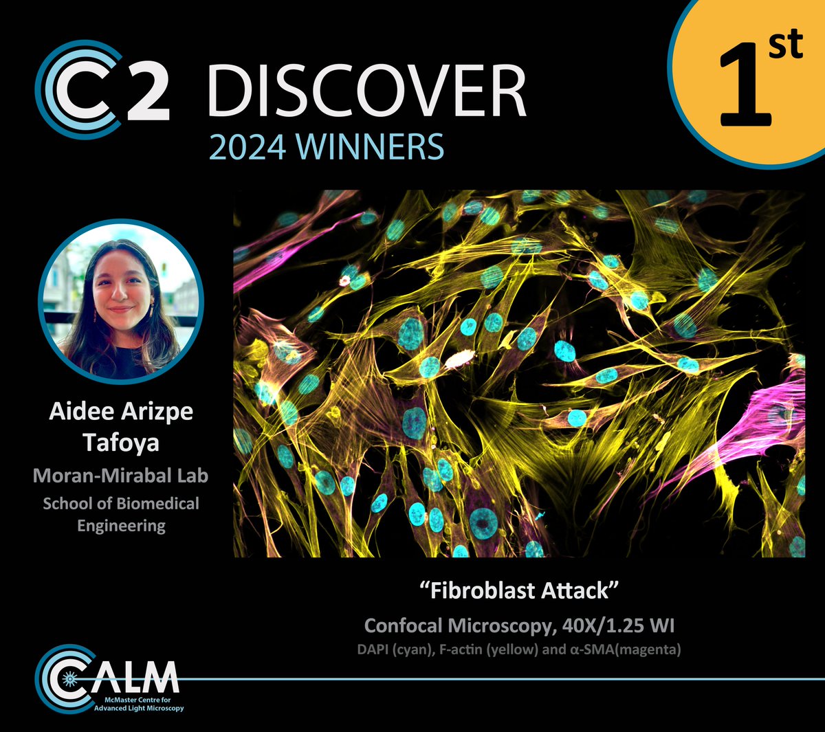
Aidee Arizpe Tafoya wins 1st place @aideearizpe@Moran_MirabalRG with a beautiful image of human lung fibroblasts that have been cultured on a hydrogel tissue model, where the expression of F-actin (yellow) and α-SMA (magenta) suggests their differentiation into myofibroblasts.
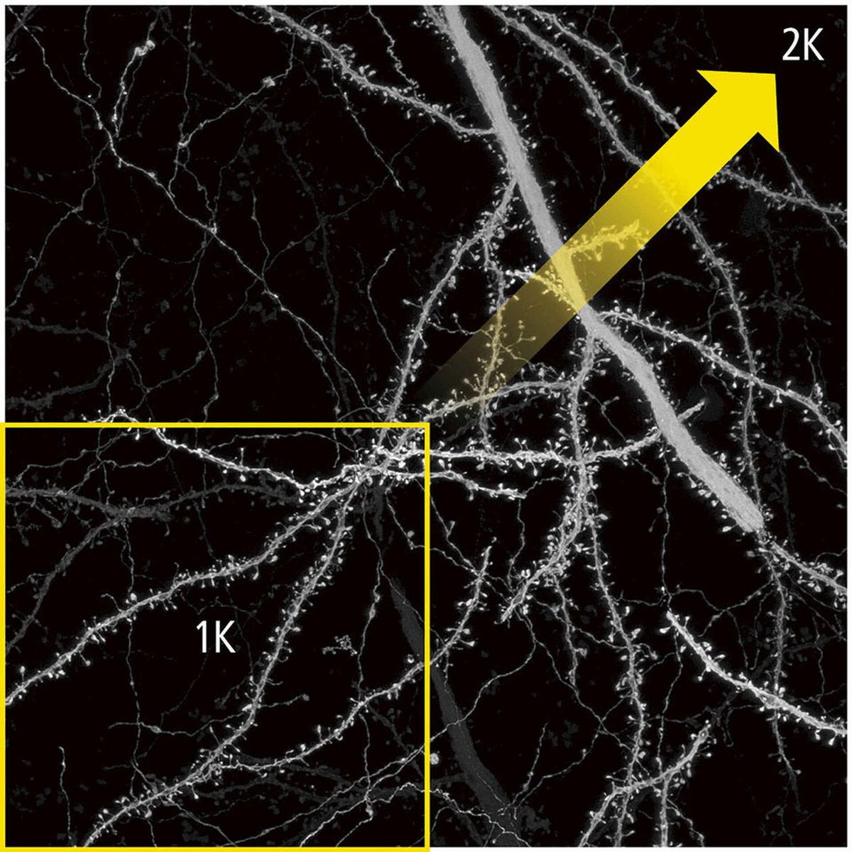
New option for resonant scanning at 2048 x 2048-pixel resolution with the #NSPARC#ImageScanningMicroscopy detector equipped on AX R #Confocal#Microscopy systems!
Realize fast #SuperResolution imaging across a 4X greater field of view. Learn more: https://t.co/fZzILvH2X3
Did you know the Nikon AX R MP #Multiphoton#Microscopy system is capable of third harmonic generation #THG#Imaging?
This #AppNote shows how THG imaging with the AX R MP enables #LabelFree detection of hemoglobin - used as a marker for red blood cells: https://t.co/I1DpvGl8uW
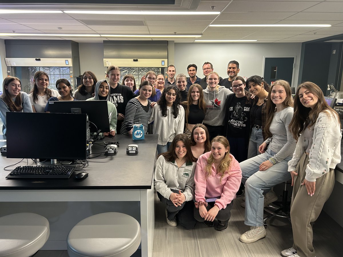
Earlier this week we had the great pleasure of receiving students from @MMRobinsonHS for some fun microscopy activities! We looked at all sorts of samples that ranged from individual cells and their organelles, to tissue sections and whole organisms.