Be honest!
Do you just say “thalamic infarct” and hope no one asks questions?
Or can you specify the thalamic vascular territories?
Do you know why the whole thalamus doesn’t infarct when you have a PCA infarct?
Answer: It’s supplied both by the PCA & PCOMM (ICA)!
Here are the thalamic vascular territories you NEED to know and HOW to remember them.
Tuberothalamic From the PCOMM
Supplies the anterior thalamus
I call it the Top-o-the-thalamic to remember its at the top
Paramedian
Branch of the P1
ParaMEDIAN is MEDIAL, so it supplies the medial aspect
Posterior choroidal
Supplied by the posterior choroidal artery
Posterior choroidal is POSTERIOR—it supplies the back of the thalamus
Thalamogeniculate
Branch of the P2
Geniculate is by the GENU of the internal capsule, so it is lateral by the internal capsule
Now you know the thalamic vascular territories & you won’t have lie when it comes to these guys!
Sowers et al. report a patient in whom axonal degeneration on MRI appeared unusually late at 6 months poststroke, underscoring the importance of longitudinal imaging to improve understanding of the natural history and evolution of stroke. https://t.co/dhN2LmX4eP
#NeurologyRF
Daily Pearl(s):
IgG4-Related Disease (IgG4-RD)
IgG4-RD is a systemic immune-mediated fibroinflammatory condition; classic sites are the pancreas, salivary or lacrimal glands, and retroperitoneum, but essentially any organ can be involved.
Consider IgG4-RD in the differential for cryptogenic stroke, especially with multifocal or atypical large-vessel vasculopathy in patients lacking traditional atherosclerotic risk factors.
In patients with vascular stenosis that progresses despite standard antiplatelet or anticoagulant therapy, consider an underlying inflammatory vasculitis.
A serum IgG4 >135 mg/dL is consistent with the diagnosis but is neither fully sensitive nor specific, so an elevated level is a clue rather than proof.
Diagnosis requires biopsy, since other inflammatory and malignant conditions can raise IgG4 levels; histology shows a lymphoplasmacytic infiltrate, storiform fibrosis, and obliterative phlebitis with an IgG4:IgG ratio >40%.
PET is especially useful for identifying biopsy sites, with salivary glands and superficial lymph nodes being accessible and high-yield.
Glucocorticoids are first-line and often produce rapid responses, with rituximab or MMF for maintenance or steroid-refractory disease; immunosuppression can not only halt progression but also reverse stenosis.
Source: Progressive Internal Carotid Artery Occlusion in IgG4-Related Disease (Annals of IM)
You might think it’s a seizure… but the truth is: after a group A streptococcal (GAS) throat infection, the immune response can misfire—cross‑reactive antibodies can disrupt the basal ganglia, leading to involuntary, dance‑like movements..😳😳
What is the diagnosis and treatment ?!
Alpha-Synuclein Seed Amplification Assay in CSF, Skin, and Submandibular Gland From Incidental Lewy Body Disease and Parkinson Disease https://t.co/2EBqfqT7SF
#ParkinsonDisease
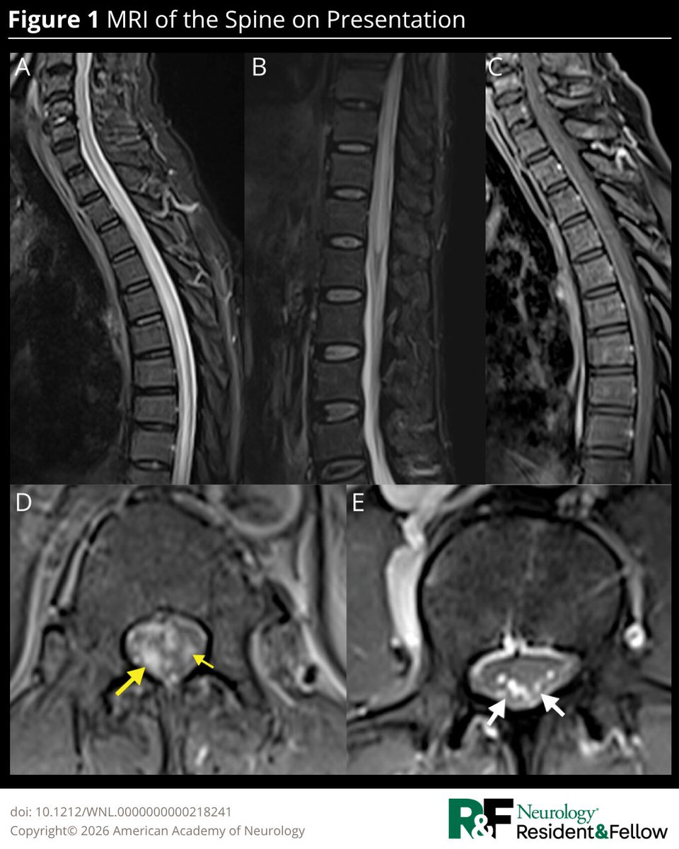
A 54-year-old farmer presented with a 5-day history of progressively worsening right leg weakness, truncal dysesthesia, and 1 day of painless urinary retention, without systemic symptoms. Read the full Teaching #NeuroImage case: https://t.co/pXsvEaTpSd
#NeurologyRF
It is July, I’m staffing the PN clinic and once again, it is essential to emphasize to the new PN/NM fellows the critical role of the clinical phenotype in diagnosing peripheral neuropathies.
NCS/EMG are valuable, but the neurological exam is the most important (as it should be in neurology but many have forgotten).
This is the neuropathy clinical phenotype classification I find the most helpful:
1-Isolated small fiber neuropathy
2-Length-dependent peripheral neuropathy (or distal symmetric polyneuropathy)
3-Multiple mononeuropathies
4-Mononeuropathy
5-Asymmetric neuropathy
6-Polyradiculoneuropathy
7-Plexopathy (brachial or lumbosacral, many times a radiculoplexus neuropathy)
8-Sensory neuronopathy
9-Motor neuronopathy
I always tell my trainees they don’t have to memorize the causes of each phenotype. If you define the syndrome correctly, you can ask a Chatbot what are the most likely causes. If you add the time course (tempo), presence of autonomic dysfunction, presence of systemic features (weight loss, rashes, fever, night sweats, or anasarca), comordities and response to immunotherapy, the chatbot will provide you an expert level discussion and differential.
1- Isolated Small Fiber Neuropathy
-Pure small fiber neuropathy.
-Only temperature and pinprick dysfunction on exam.
-Ankle reflexes must be normal in patients < 60 yo.
-NCS must be normal for age.
2- Length-dependent peripheral neuropathy (or distal symmetric polyneuropathy)
-Sensory predominant neuropathy, can also have motor involvement, distal predominant, worsens in an ascending length-dependent fashion: foot->leg-> hands/knee->elbow/anterior abdomen.
3- Multiple Mononeuropathies
Involvement of two or more non-contiguous (separate) motor, sensory or sensorimotor peripheral nerves.
4- Mononeuropathy
Involvement of a single sensory, motor or sensorimotor peripheral nerve.
5-Asymmetric neuropathy
It’s the pattern of overlapping multiple mononeuropathies.
Length-dependent polyneuropathy (distal predominant) involving bilateral extremities in an asymmetric fashion demonstrated by at least one grade of motor power difference by the Medical Research Council strength scale, more than 50% difference in sensory testing and involved individual nerves could not be identified separately.
6-Polyradiculoneuropathy
Proximal and distal weakness and sensory loss in the four limbs.
Can be distal>proximal; distal=proximal or distal<proximal.
7- Plexopathy
-Proximal and distal weakness in a single limb with decreased/absent reflexes and sensation loss (can be only proximal or only distal in brachial plexopathies; and of course can be bilateral).
- On exam can look like a radiculopathy or multiple radiculopathies.
-Plexopathies usually have more dense sensation loss than radiculopathies and may cause alodynea (pain caused by non-painful stimulus) which is rarer in radiculopathies.
8- Sensory Neuronopathy or Ganglionopathy
-The cell body of the sensory neurons are affected, usually asymmetric and affecting distal and proximal limbs, patchy, can affect the face or whole body, must have sensory ataxia and preserved strength.
- Can be accompanied by pseudoathetosis.
9- Motor neuronopathy
-Pure motor syndromes.
-The body of the motor neurons are affected.
-Usually asymmetric and distal predominant, accompanied by atrophy, fasciculations, decreased/absent reflexes (can also have upper motor neuron signs) and normal sensory exam.
Important update in neurology! 🧠✨ A comprehensive review on the current landscape of Multiple Sclerosis (MS) has just been published in NEJM.
Key takeaways: 👇
✅ MS is a chronic autoimmune disorder of the CNS, marked by inflammation, demyelination and neurodegeneration.
✅ Its etiology is complex, involving genetic susceptibility, Epstein–Barr virus, and immune responses centered on B cells, T cells and microglia.
✅ CD20-targeting therapies have greatly improved outcomes in relapsing forms, but major unmet needs remain: preventing progression and promoting myelin repair. 🛠️
A deeper understanding of neurodegeneration will be crucial to improving long-term outcomes and quality of life. 🧬💊
🔗👇🏻 https://t.co/GWD43Q2k8X
#MultipleSclerosis #Neurology #Neuroimmunology #Science #Medicine #Healthcare #MSResearch #NEJM
🩺Clinical & neuroimaging 🧠 clues to differentiate 🧩autoimmune encephalitis & its mimics🔎
Current opinions in Neurology 📖
10.1097/WCO.0000000000001489.
Many lumbar punctures are interpreted incorrectly.
Not because the CSF report is wrong.
Because the serum glucose was never looked at.
Every time I see a CSF report, the first question I ask is not...
What is the CSF glucose?
It is...
What was the serum glucose at the time of lumbar puncture?
Here's why.
CSF glucose is not an independent value.
It comes from the blood.
Normally,
CSF glucose is about 50–80% of serum glucose
(usually around 60%).
That means the absolute CSF glucose is far less important than the
CSF : Serum glucose ratio.
A normal ratio is 0.5–0.8.
A ratio <0.4 should immediately make you think of impaired glucose transport or increased glucose consumption within the CSF.
Now let's think like a clinician.
Scenario 1
Serum glucose: 100 mg/dL
CSF glucose: 30 mg/dL
Ratio = 0.3
Now hypoglycorrhachia is real.
Think of bacterial, tuberculous or fungal meningitis, or leptomeningeal malignancy.
Then correlate with CSF cells, protein, lactate, Gram stain, culture and PCR.
Scenario 2
Serum glucose: 200 mg/dL
CSF glucose: 70 mg/dL
Many people say,
CSF glucose is normal.
It isn't.
The ratio is only 0.35.
The patient may still have pathological CSF glucose.
This is one of the commonest interpretation errors during lumbar puncture.
Scenario 3
Serum glucose: 60 mg/dL
CSF glucose: 36 mg/dL
Ratio = 0.6
Both values are low.
The ratio is normal.
The CSF is simply reflecting systemic hypoglycaemia.
Not every low CSF glucose means meningitis.
Another practical pearl.
Draw the serum glucose immediately before, or at the time of lumbar puncture.
Why?
Because serum glucose can change rapidly after meals, insulin or IV dextrose.
CSF glucose changes much more slowly and reflects blood glucose over the preceding few hours.
If serum is sampled much earlier or much later, the ratio may become misleading.
My bedside protocol is simple.
• Draw serum glucose with every lumbar puncture.
• Send both samples together.
• Calculate the CSF/serum ratio.
• Never interpret CSF glucose in isolation.
• Always correlate with the clinical picture and the remaining CSF profile.
One number tells you very little.
The relationship between two numbers tells you the diagnosis.
#NeuroX #MedTwitter #FOAMed #LumbarPuncture #Meningitis #NeurologyTeaching
The image details Autonomic Hyperreflexia (AH)—also known as autonomic dysreflexia—which is a life-threatening, uninhibited sympathetic response to noxious stimuli below the level of injury in patients with a chronic spinal cord injury at T6 or above.
https://t.co/Lc7OlWwnvb
Big step forward for #MOGAD treatment!🚨
Our multicenter @JAMANeuro study shows that IL-6 receptor blockade (90% in Tocilizumab) markedly reduces relapses, achieving relapse-free outcomes comparable to high-dose IVIG (≥1g/kg/month).
If satralizumab gains approval for MOGAD, these data support #Tocilizumab as a widely available, lower-cost option thanks to biosimilars!
A great example of collaboration across the Americas🌎
#Neurology #MOG #NeuroTwitter
Olivary hypertrophy is an exceptional consequence of brainstem insults. Usually occurs after a latency period of few months. @atabisneuro et al reported a case of bilateral olivary degeneration following Scrub typhus rhombencephalitis @neuro_ian Link: 10.4103/aian.aian_931_23
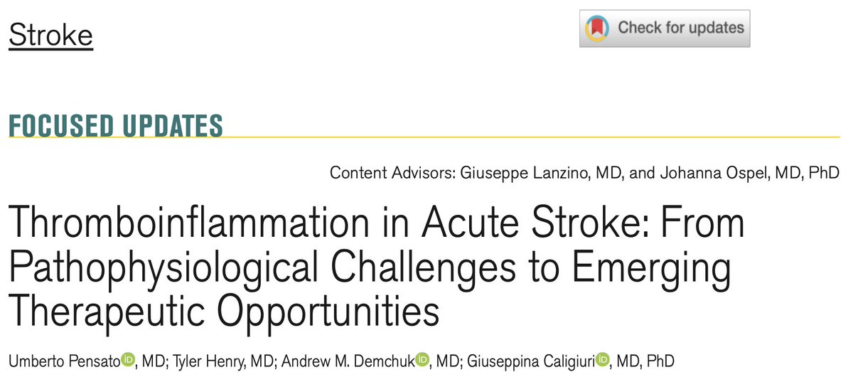
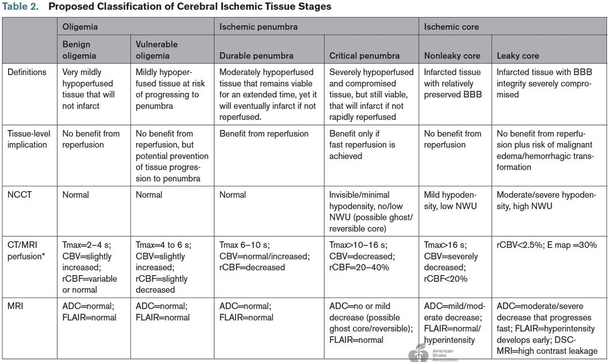
Thromboinflammation in Acute Stroke: From Pathophysiological Challenges to Emerging Therapeutic Opportunities | Stroke @StrokeAHA_ASA@AHAScience https://t.co/IDjWwnoZ24
Great @NEJM review;
#Neurology should take note
Inflammatory myopathies are moving beyond antibody status to a pathophysiology-based understanding👇🏽
Inclusion Body → CD8+ T cells
Necrotizing or antisynthetase → pathogenic antibodies
Dermatomyositis → acquired interferonopathy
Fascinating paper on Clinical Approach to 🧠 Encephalitis
by Prof Sarosh Irani & his team..@TheLancet
💡Teaching point: Identify Clues & patterns to differentiate Autoimmune vs Infectious causes of Encephalitis.
DOI: 10.1016/S0140-6736(26)00363-6