New publication: Our study shows that GFAP protein cleavage and complement C3 activation within astrocytes occur during epileptogenesis, highlighting potential markers of astrocyte stress and disease progression.
https://t.co/i4zS6ZDnzw
Fifteen years after microglia were first implicated in synaptic pruning, this new study now implicates B cells originating from the skull bone marrow in synaptic pruning. So much for the "immune privileged" brain 😉 Cool findings from @soyonhonglab's group https://t.co/6qMwFXRcTY
A new 2026 Neuron paper shows that tau directly activates mitochondrial RET via complex I, driving a feed-forward cycle. These findings align with our Cell 2022 and Cell 2024 studies, supporting tau–mitochondria coupling as a central driver of tauopathy. https://t.co/KCrk9scjTv
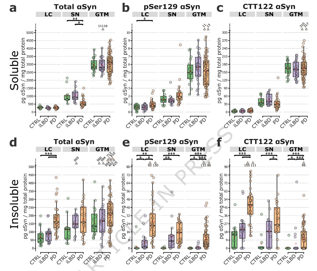
Quantitative biochemical profiling in post-mortem human brains demonstrates a link between pSer129-enriched α-synuclein deposition & GCase deficiency across the #Parkinsons spectrum, supporting lysosomal/GCase-enhancing therapies regardless of GBA1 status
https://t.co/bZEpWcZR3b
For decades, researchers thought tau tangles were the problem in Alzheimer's. A new Stanford study shows tau kills neurons before it even tangles, through a mitochondrial mechanism no one predicted.
The protein tau has become one of the strongest predictors of Alzheimer's disease. When tau shows up in cerebrospinal fluid or blood, cognitive decline usually follows. Inside the brains of Alzheimer's patients, tau forms neurofibrillary tangles, long filaments that accumulate inside dying neurons.
The working theory has been that tau either damages microtubules, the structural scaffolding inside neurons, or that the tangles themselves are toxic. But this Stanford-UCSF study reveals something different. Tau enters mitochondria and forces them to run backward, generating toxic free radicals that trigger neurodegeneration.
Neurons have especially high energy demands, which is why they're packed with mitochondria.
The process that generates ATP is called the electron transport chain. Think of it as an assembly line where electrons flow forward through a series of protein complexes, ultimately producing ATP and a small amount of reactive oxygen species as a byproduct.
Reverse electron transport, or RET, happens when this process runs backward. Instead of electrons flowing forward to make ATP, they flow in reverse through Complex I, the first station in the chain.
This generates massive amounts of ROS, reactive oxygen species, without producing any energy. Think of ROS as exhaust fumes from a malfunctioning engine. A healthy mitochondria produces a small amount as a normal byproduct, like a well-tuned car. But when the engine runs backward, it floods the system with toxic exhaust while consuming fuel instead of generating power.
Key findings from the study:*
• Tau protein enters mitochondria and binds directly to NDUFS3, a subunit of Complex I in the electron transport chain
• This interaction activates reverse electron transport, producing excessive reactive oxygen species while reducing the NAD+/NADH ratio
• Tau phosphorylation, the chemical modification linked to disease, enhances this effect in a dose-dependent manner
• The ROS produced by RET further drives tau hyperphosphorylation, creating a self-perpetuating cycle
• Removing tau completely eliminated stress-induced RET in fruit flies, mice, and human neurons derived from stem cells
• Blocking RET, either genetically or pharmacologically, protected against tau-induced neurodegeneration across all three species
The mechanism works like this. Normal tau can enter mitochondria, but it doesn't cause much damage. When tau becomes hyperphosphorylated, those chemical modifications make it stickier. It binds more tightly to NDUFS3 and activates RET more strongly.
The RET produces ROS, which damages cellular components and triggers more tau phosphorylation. That phosphorylated tau enters more mitochondria, activates more RET, produces more ROS, and the cycle accelerates.
This explains why mitochondrial dysfunction appears so consistently across tauopathies. It's not just Alzheimer's disease. Frontotemporal dementia, progressive supranuclear palsy, and potentially Parkinson's and Huntington's diseases all involve tau pathology and mitochondrial decline.
The study tested this mechanism across three different model systems. In Drosophila fruit flies expressing human tau, blocking RET prevented neurodegeneration. In mice engineered to develop tau pathology, the same interventions worked. In human iPSC-derived neurons carrying disease-causing tau mutations, blocking RET restored mitochondrial function.
The consistency across species and cell types suggests this is a fundamental mechanism, not an artifact of any single experimental system.
What makes this particularly actionable is that RET can be blocked. The researchers used both genetic approaches, knocking down specific Complex I subunits that enable reverse flow, and pharmacological compounds that inhibit RET without disrupting forward electron transport.
The therapeutic implication is that you don't necessarily need to clear tau tangles or prevent tau phosphorylation. Blocking the downstream mitochondrial damage might be sufficient to prevent neurodegeneration.
This also reframes what tau actually does under normal conditions. It's not just a microtubule stabilizer. Tau appears to regulate RET as part of its normal function, possibly as a stress response mechanism. Under acute stress, activating RET might serve a protective signaling role.
The problem emerges when this becomes chronic. Aging, metabolic stress, and tau phosphorylation shift the system from acute adaptive response to sustained pathological state. The mitochondria get stuck running backward, neurons lose their energy supply while simultaneously being poisoned by ROS.
The NAD+/NADH ratio matters here because it reflects cellular redox state. High NAD+ relative to NADH indicates healthy metabolism and oxidative capacity. When RET runs continuously, it consumes NAD+ and produces NADH, collapsing this ratio and impairing every NAD-dependent process in the cell.
Sirtuins, PARP enzymes, and DNA repair machinery all depend on NAD+ availability. Mitochondrial biogenesis requires NAD+. When tau-induced RET depletes the NAD+ pool, it doesn't just damage existing mitochondria. It prevents the cell from making new ones to replace the damaged units.
This study is still a preprint, not yet peer-reviewed, but the mechanistic detail, cross-species validation, and therapeutic proof-of-concept are compelling. The question is whether RET inhibitors can be developed that are safe and brain-penetrant enough for human use.
The bigger question is timing. At what point in disease progression does tau enter mitochondria and activate RET? If this happens early, before tangles form and before cognitive symptoms appear, then targeting RET could be preventive.
If tau pathology is already advanced by the time RET activation occurs, the therapeutic window narrows. The accumulating evidence suggests mitochondrial dysfunction appears early in Alzheimer's disease, potentially decades before diagnosis.
Our approach for untangling brain-wide interactions is out today in @NeuroCellPress!
Massive congrats to @mattperich for leading this incredible effort since 2020, and a huge thank you to all of our collaborators.
https://t.co/aIjzMDpQnE
A reproducible three-dimensional model of human brain tissue to investigate physiological and disease-associated microglia phenotypes | Nature Neuroscience https://t.co/oMVxXWm5TH
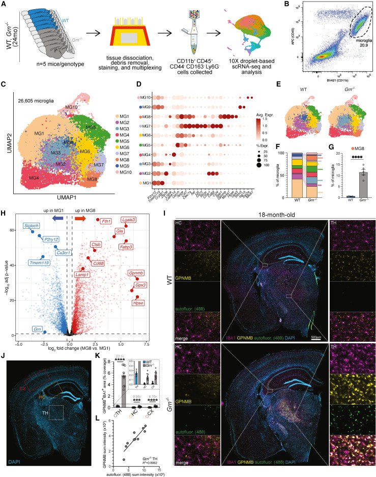
Lysosomal dysfunction is sufficient to drive epigenetic, transcriptional, and functional remodeling of microglia and macrophages, including in the context of progranulin deficiency @ImmunityCP
https://t.co/CDKR6fzmzc @LeonTejwani@Denalitx
Lysosomal dysfunction drives a transcriptional and epigenetic signature found in disease-associated microglia in neurodegenerative diseases: @ImmunityCP https://t.co/AOV5YSAqH1
Guo et al. show that microglial exosomes mediate α-synuclein transmission from microglia to neurons in vitro and in vivo, suggesting potential as a therapeutic target in #Parkinson's disease. https://t.co/M3Ve2h3WOh
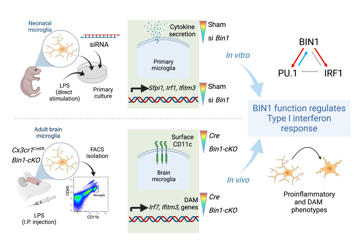
Excited to share our recent publication in @MolNeuro – BIN1 is a key regulator of proinflammatory and neurodegeneration-related activation in microglia
#microglia#neuroinflammation#Alzheimer’s disease