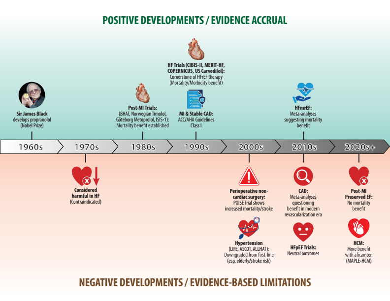
Sharing our state-of-the-art review on beta blockers just published in @JACCJournals! Thanks @rcstarling@DLBHATTMD@WilfriedMullens@AndreaRussoEP@MAbdeljawadmd for your expertise!
Key points:
1. Beta-blockers should no longer be prescribed by reflex. The modern question is: Which beta-blocker, for which patient, for which indication, and for how long? We propose an indication- and phenotype-based approach.
2. Beta-blockers do more than slow the heart rate. Blocking cardiac β1 receptors reduces heart rate, contractility and oxygen demand, while blocking β1 receptors in the kidney’s juxtaglomerular apparatus suppresses renin release.
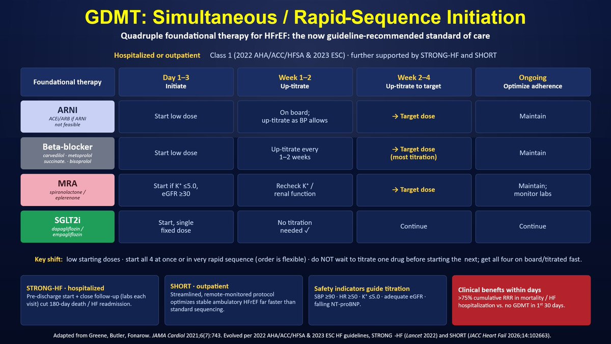
🫀💊 HF care has flipped
🚫 No more slow, one-drug-at-a-time titration over months
The new standard = all 4 pillars FAST 🚀 (Greene/Butler/Fonarow)
🔹ARNI 🔹β-blocker 🔹MRA 🔹SGLT2i
🏥 STRONG-HF (inpatient) + 🏡 SHORT (outpatient):
🎯 all 4 drugs ASAP
📉 >75% ⬇️ ☠️/HF🏨
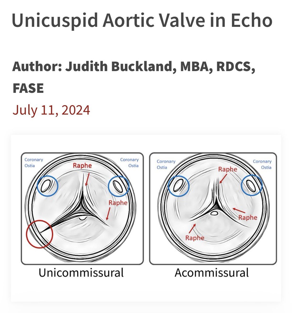
Valve imaging in TEE starts with the correct orientation. Keep in mind that during tricuspid valve surgery, the valve is typically oriented in the SURGEON’S view. This orientation differs slightly from that used for TEE-guided structural heart interventions #echofirst
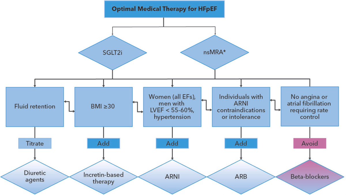
This is a treatment algorithm for optimal therapy in HFpEF
Under SGLT2i:
Fluid retention → titrate diuretic agent
BMI ≥30 → add incretin-based therapy (e.g., GLP-1 agonists)
Women (any EF), men with LVEF <55–60%, or hypertension → add ARNI
https://t.co/LtDakOyxO1
🫀📡 The stethoscope is evolving… and it’s now an ultrasound probe.
A new review highlights how Point-of-Care Ultrasound (POCUS) is transforming bedside cardiopulmonary diagnosis 👇
✨ Key insights:
⚡ POCUS = real-time, bedside, physiology-driven imaging
🫁 Lung ultrasound (LUS) → detects congestion better than CXR & exam
🩸 VExUS score → quantifies systemic venous congestion
❤️ FoCUS + LVOT-VTI → assess structure, function & cardiac output
🔄 Enables dynamic monitoring and therapy guidance
💡 As shown in the graphical abstract (page 2):
👉 HF, STEMI, shock → one tool, multimodal assessment
👉 From congestion ➝ to perfusion ➝ to haemodynamics
🚨 Clinical message:
Traditional exam = limited sensitivity
POCUS = immediate + actionable information
Not a replacement… but a powerful extension of clinical reasoning
🎯 Big take-home:
From “suspecting haemodynamics”
➡️ to
“seeing haemodynamics at the bedside”
📄 Read the full paper:
👉
🔗 DOI: 10.1093/ehjimp/qyaf147
#Cardiology #POCUS #Echocardiography #HeartFailure #CriticalCare #Ultrasound #CardioTwitter #MedTech #BedsideMedicine #DigitalHealth 🩺📊
In #echofirst it is pivotal to be aware of the settings of the machine to ensure that our results can be trusted
In national & international meetings I do miss this in echo based talks almost always
@EACVIPresident@escardio
My Conflict of Image Quality
Frequency
•2D Imaging: 1.4/2.8 MHz
•2D Color: 2.3 MHz
Frame Rate
•2D Imaging: ~50 FPS
•2D Color: 25–30 FPS
•3D (LV EF): 20–25 FPS
•3D Valve : 20–30 FPS
•3D Color: 20–25 FPS
Image quality requires balancing frequency, frame rate, spatial resolution, and temporal resolution. Optimizing one parameter often comes at the expense of another.
@TrackYourHeart Nice overview. With 3D TEE, you can display all three standard views simultaneously. If you don’t have a 3D probe, you can systematically assess all three mitral valve scallops in 2D TEE by advancing and withdrawing the TEE probe #echofirst
Current #guidelines recommend the mitral inflow/LVOT VTI ratio as an easy-to-measure semiquantitative index for isolated primary MR (>1.4 severe, <1 mild). But how robust is the evidence behind it? Let's take a closer look at this widely cited parameter. #EchoFirst Part 1
🔥 New in @JACCJournals#HF — SMR Trajectories & GDMT in HFrEF
🔑
❤️ 32% had significant SMR at baseline
📉 GDMT optimization → dropped to 21%
✅ 57.5% of significant SMR improved
⚠️ Persistent SMR → 60% ↑ mortality
💡 GDMT first -TEER if SMR persist
https://t.co/ecK1z02gdE
O diagnóstico de Carnell é que o sistema de saúde foi construído para reagir a doenças, não para preveni-las. Cada etapa, da consulta à farmácia, foi desenhada para o paciente que já está doente. Não para o que ainda pode não ficar.
🫀 New multicentre international study on HCM with apical aneurysms
A large cohort of 510 patients with hypertrophic cardiomyopathy (HCM) and left ventricular apical aneurysms has provided important insights into risk stratification and clinical management.
🔹 Key findings:
• The annual rate of life-threatening ventricular arrhythmic events was 2.1% per year.
• Risk increased progressively with aneurysm size:
<10 mm: 0.2%/year
10–19 mm: 2.3%/year
20–39 mm: 3.0%/year
≥40 mm: 8.2%/year
• Aneurysm size independently predicted ventricular arrhythmias, even after adjustment for traditional risk markers and ESC risk scores.
• Apical thrombus or thromboembolic events occurred in 13% of patients and were also strongly associated with larger aneurysm size.
• Small aneurysms (<10 mm) were associated with a relatively low arrhythmic risk, whereas aneurysms ≥10 mm identified a substantially higher-risk population.
📌 Clinical implications:
This study supports the concept that apical aneurysms are not all equal. Aneurysm size appears to be a key determinant of both arrhythmic and thromboembolic risk, potentially influencing decisions regarding ICD implantation, anticoagulation, and follow-up strategies.
#HCM #HypertrophicCardiomyopathy #Cardiology #CMR #CardiacImaging #SuddenCardiacDeath #Electrophysiology #JACCAdvances #RiskStratification
Think spatially #echofirst
The leaflet orientation of the mechanical mitral valve prosthesis on 3D echocardiography can be predicted from the 2D TEE image.
@CASivaram1
Ventricular Septal Defects (VSDs) Explained!
The ventricular septum is divided into four key regions: inlet, trabecular, outlet, and membranous. A VSD is an abnormal opening in this septum, allowing blood to shunt between the LV & RV.
Types of VSDs & Their Locations:
⚪ Perimembranous (pmVSD): Below the aortic valve, near the membranous septum
🔴 Muscular (mVSD): Located in the trabecular septum; can be multiple (Swiss-cheese VSD)
🟢 Inlet (iVSD): Near the AV valves, often linked to endocardial cushion defects
🔵 Supracristal (scVSD): Just below the pulmonic valve, may lead to aortic regurgitation
How Do We Diagnose VSDs?
Echocardiography plays a key role! Different echo views help identify VSDs:
🔺 Parasternal long-axis: Best for pmVSD & mVSD
🔺 Parasternal short-axis: Locates pmVSD (10 o’clock) & scVSD (1 o’clock)
🔺 Apical 4-chamber: Ideal for detecting inlet & apical muscular VSDs
Why does this matter?
VSD size, location, and associated defects determine hemodynamic impact & management! Some close spontaneously, while others require surgical or percutaneous closure.
Understanding VSDs = Better diagnosis & treatment!
💡TIPS and TRICKS
2D/3D TOE : a mobile echodense mass (red arrow) within the transverse sinus of the pericardium (blue arrow, 3D Flexi-light)
•DIAGNOSIS: fat within the transverse sinus. NO left atrial appendage thrombosis! #echofirst
•Do not forget pericardial sinuses! --> https://t.co/9qgOjiyCym
--------------------------------------------
Do not miss our next course MICS Echo Lab & Live Surgery 6-7 June in Rome - Ospedale San Carlo di Nancy.
💎 Arrhythmic MVP is characterized by ventricular arrhythmias and sudden arrhythmic death.
This infographic 👇 highlights the 🔑 clinical, ECG, echocardiographic, and CMR markers associated with ⬆️arrhythmic risk in MVP.
🎯 Risk stratification requires looking beyond the valve.
#ASEchoJC #EchoFirst #CardioX #MVP @ASE360 #EPeeps