Visual survey of surgical pathology with more than 14,000 high-quality images of benign and malignant neoplasms & related entities. Created by: @DharamRamnani
Please consider giving your pathology images a forever home on WebPathology. Now you can load them directly from the site using Submit Images form. Give it a try. https://t.co/bh8mbMrhQw: A Collection of Surgical Pathology Images #PathTwitter#PathX
I refrain from posting anything non-pathology related on this account....however, I couldn't resist sharing this with you. If you are ever in Richmond, Virginia, pay them a visit...their single malts and old-fashioned are to die for.....
Update in #HemePath - Intranodal Palisaded Myofibroblastoma; 15 images; check it out on the website under Hematopathology ➡️Lymph Node➡️Spindle & Vascular Proliferations #pathresidents#pathology
Rosai-Dorfman disease in skin -somebody please call the police - the emperipolice! An S100 protein stain highlights the nuclei (arrow) and cytoplasm of abnormal histiocytes but not the engulfed inflammatory cells in the cytoplasm (emperipolesis - engulfment without destruction).
I’m updating mediastinum section and looking for images of T-cell lymphoblastic leukemia/lymphoma. Anyone have a case that they would like to contribute? Thanks. #hemepath
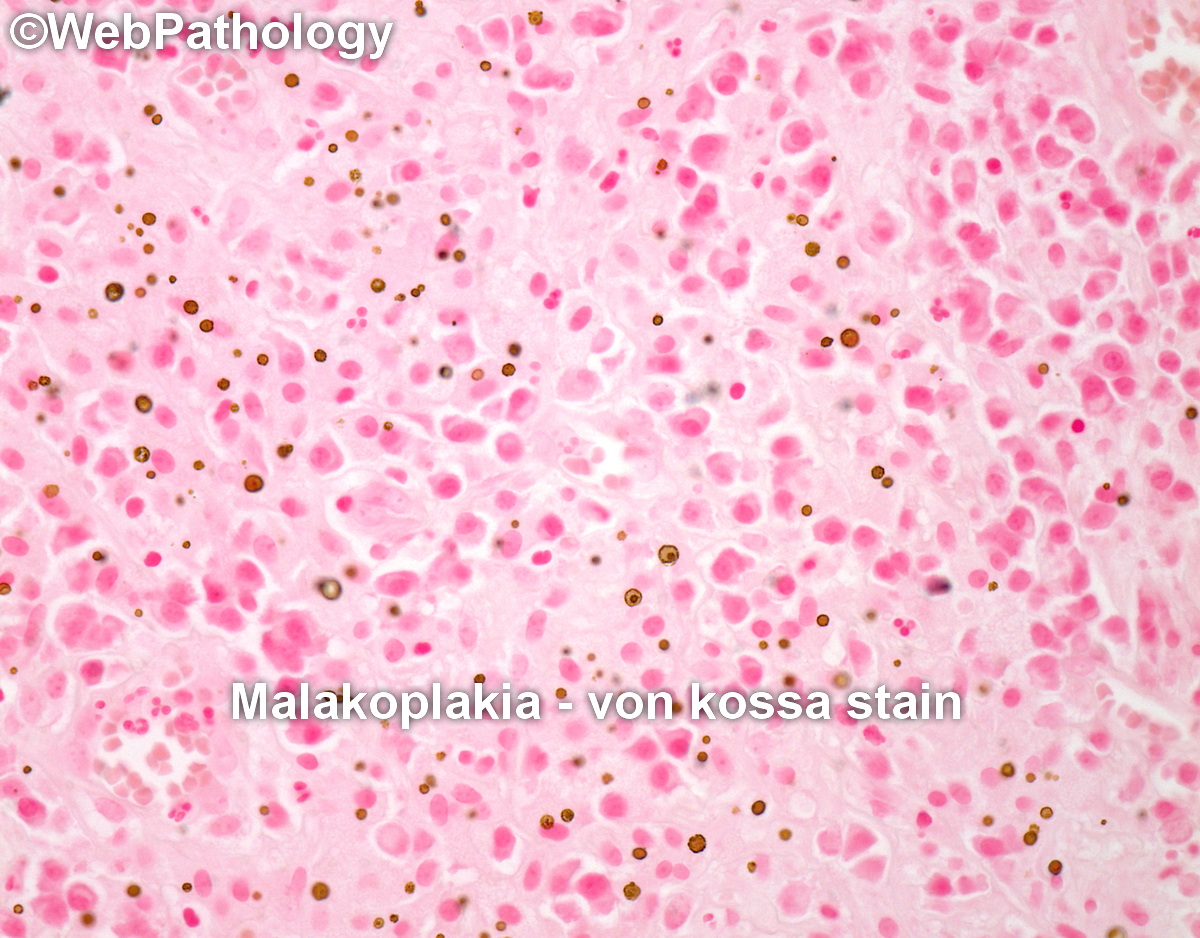
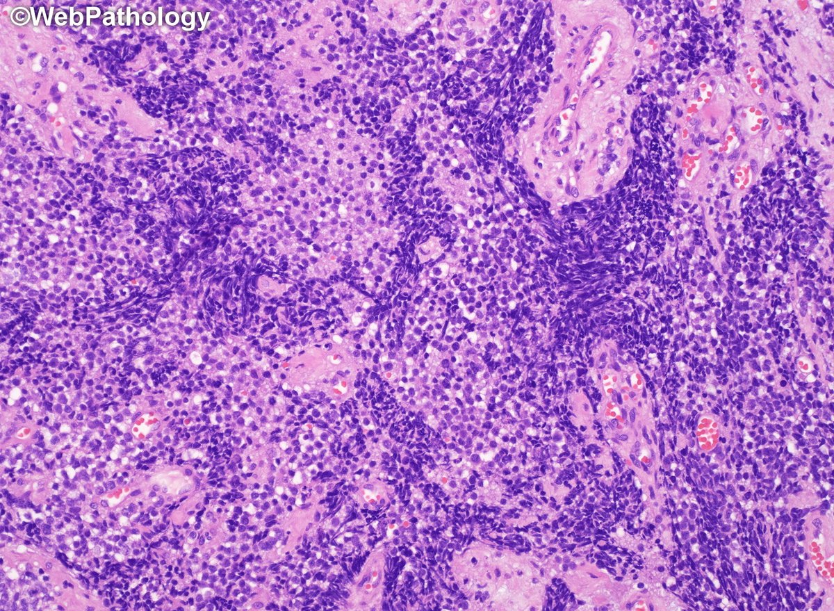
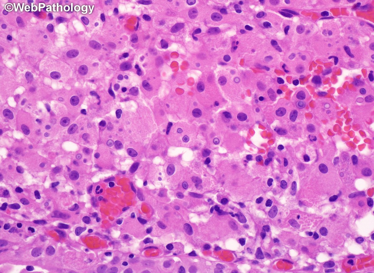
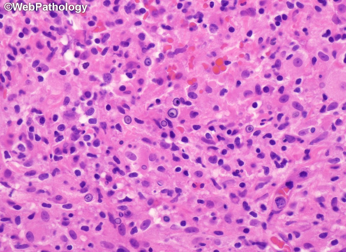
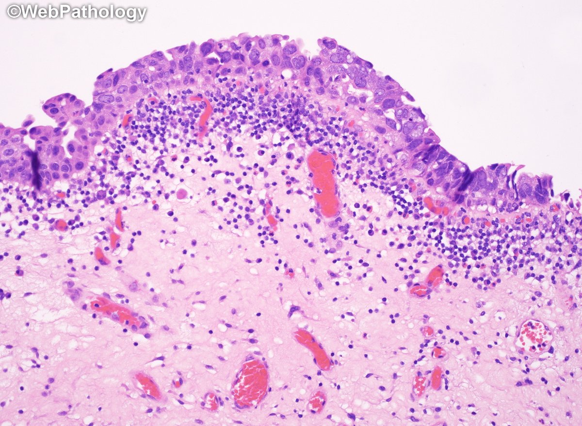
Adult female with history of high-grade uroth. CA. Surveillance cystoscopy showed multiple yellow-tan plaques near trigone. What's your diagnosis? #gupath for #pathresidents Diagnosis & additional images in the comment below.
This bladder tumor had small cell neuroendocrine CA (90%) combined with conventional inv. high-grade Uroth. CA (10%), areas of CIS (flat and micropapillary) and adenoCIS. Here are additional images with immunos. #GUpath