@Andi_Microscopy and I structurally characterized beta cell cilia by #vEM and #UExM. It's out today in @NatureComms! We describe axoneme ultrastructural organization, as well as some novel physical contact between beta cell cilia and neurons. Link below!
https://t.co/WLdKiIT2Lu
After thousands of microscope hours and several years of painstaking optimisation, @glynnca93 and I are very excited to share our latest manuscript. We outline our approach to investigate the molecular organisation within hippocampus tissue using cryo-ET.
Hey #teamtomo!Our 1st❄️🔬work @humantechnopole is finally out!🥳https://t.co/Q75zt93UV4 Serialized On-grid Lift-In Sectioning for Tomography(SOLIST) enables a biopsy at the nanoscale and high-res❄️STA from🧍♀️organoids and🐭liver, brain and heart tissue!Thread by @PeroneGaia below!
How photosynthetic genes are transcribed 🧬🌿 I’m really pleased to share the first work from my group at @JohnInnesCentre published today in @CellCellPress: https://t.co/1GyoYtTIlg
Since people like 🇮🇹 salami slicing😉: I am very happy to share with you the first work of my lab @humantechnopole: Serialized On-grid Lift-In Sectioning for Tomography - SOLIST🔬❄️If you're into cryo-ET of 🧠 organoids, look no further!
Work by Gaia Perone and Jasmien Nguyen!🎵
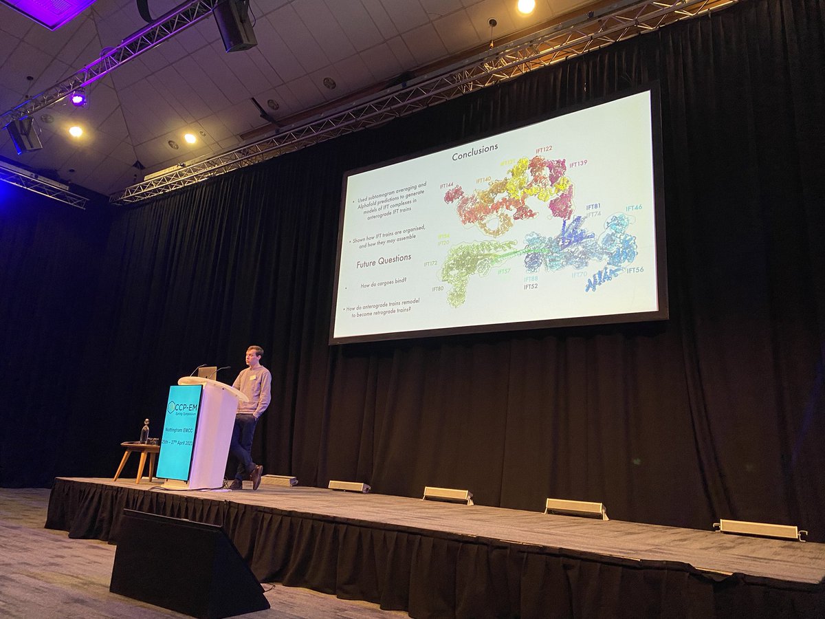
Sam Lacey became one of my favourite scientists this morning - absolutely incredible work on IFT trains with @GaiaPigino@humantechnopole and a beautifully clear presentation
We are looking for labs who got either Thermo’s iFLM or DELMIC’s METEOR installed on their Aquilos. Would love to reach out for honest feedback (feel free to PM or [email protected]). ☕️ or🍷(or both) in reward 😁!!!
🚨Looking for a postdoc 🚨
Want to investigate the link between tubulin posttranslational modifications and #ciliopathies? @ERC_Research
Join us @humantechnopole to discover how the #tubulincode regulates #cilia assembly, transport and mechanics.
👉 https://t.co/4Os9xf7M92
@ERC_Research council-funded #postdocjobs in my group @humantechnopole to unravel the mysteries of #thyroid hormone regulation, using cell biology, light microscopy and #cryoEM ! Please apply here 👉 https://t.co/yNXoGcRCqC. Feel free to get in touch by email for more details!
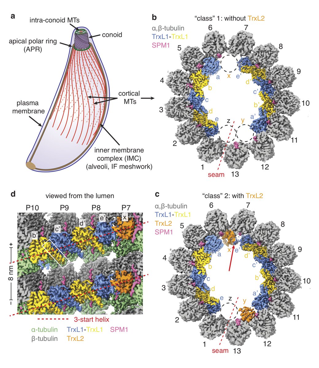
We are very excited to share our latest work, the high-resolution cryo-EM structure of the ex vivo cortical #microtubules from human parasite #Toxoplasma gondii. This work is in collaboration with David Sibley lab and @alanbrownhms lab.
https://t.co/lqnOvMT7Zv
Fascinating microtubule structures solved by sub-tomogram averaging using EMAN (@sludtke and Muyuan Chen), imaging in @chiulab, segmentation with Segger! Identified tubulin proteins in different arrangements along with other components https://t.co/QtJUG9xWgb
One fiducial to suit CLEM all!
We present a bimodal endocytic probe for several 2D- & 3D-CLEM applications with Tokuyasu cryosections, resin-embedded material, and cryo-EM.
A huge thanks to all the coauthors on Twitter @JKlumperman@Carter_Lab@_Helen_Foster_@psirez and beyond!
Cryo-ET in situ is going to revolutionize cell biology! I'm very intrigued by this data that shows virus-like capsids in axons. This is actually consistent with data from @Campbell_RNALab showing that Arc is released from DRGs! https://t.co/B7Ecp9rgnY
Very excited these papers have been released today! One describes the intra-cellular compartments in mammalian axons (https://t.co/6LaBG2OSMQ) and the other focusses on the microtubule cytoskeleton (https://t.co/qNny8xWZgY). Safe to say the second part of my PhD was an adventure!
Two papers from @_Helen_Foster_ . Previously a technician, then PhD student in my lab. Now with @GaiaPigino (thank you for letting her finish these, Gaia!). Also thank you to @CamillaVenturaS for her brilliant contributions.
https://t.co/9o77IM4OCN
https://t.co/Vw2Ol1BHUz