🚨New ESP webinar alert!
#Cancerpatientcare is an evolving field with new treatment strategies been implemented.
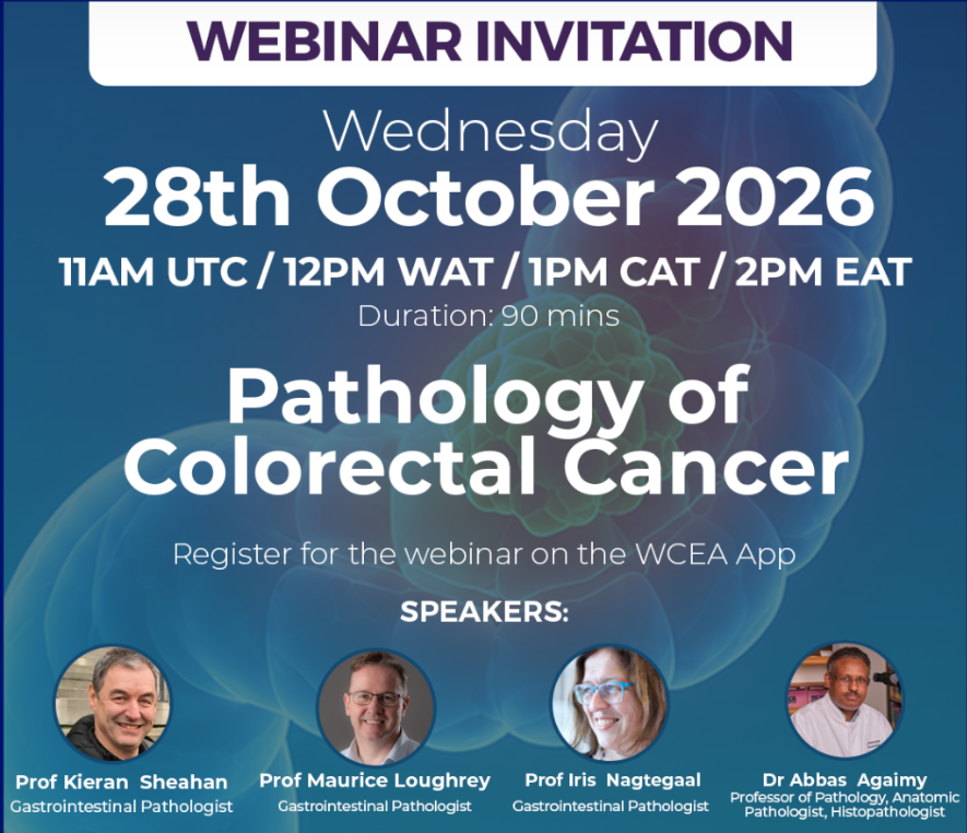
Join us on 18 November at 15:00-16:00 CET to learn from three distinguished speakers about Solid Tumours Biomarker Testing!
Registration: https://t.co/Fl5UVmKNiZ
Out today!
An AI model infers spatial proteomic patterns at the single-cell level from routine H&E slides to predict molecular biomarkers, prognosis and immunotherapy response
https://t.co/WKFZWefYPd
Published in the October 2026 issue of European Urology @EUPlatinum (Impact Factor: 29), this collaborative work was led by Roger Li @UrogerliMD, Can Aydogdu @CanDAydogdu, Laura Bukavina @LauraBukavinaMD A must-read for anyone interested in bladder cancer! Great honor to be part of this work.
@UroDocAsh@AndreaNecchi@apolo_andrea@SpiessPhilippe
This comprehensive review provides an integrated overview of the pathology, molecular characteristics, and clinical management of major bladder cancer subtypes, including squamous, adenocarcinoma/glandular, micropapillary, sarcomatoid, plasmacytoid, nested, and small cell neuroendocrine carcinoma. These tumors are biologically heterogeneous, often clinically aggressive, and frequently understaged on transurethral resection, supporting consideration of early radical cystectomy in selected high-risk patients.
Importantly, treatment response varies substantially by histology. Small cell neuroendocrine carcinoma demonstrates the clearest benefit from platinum/etoposide-based chemotherapy, while other subtypes show variable responses to conventional systemic therapies. Molecular profiling reveals potentially actionable alterations, including ERBB2 in micropapillary carcinoma, CDH1 loss in plasmacytoid carcinoma, FGFR3 alterations in nested carcinoma, and TP53/RB1 alterations in small cell carcinoma. Emerging evidence also supports potential roles for immune checkpoint inhibitors and antibody-drug conjugates.
We emphasizes that variant bladder cancers should not be managed using a uniform approach. Accurate histologic classification, quantification of subtype components, and molecular profiling are increasingly important for personalized treatment and future subtype-specific clinical trials.
Full article available at https://t.co/KLNDkGNVNv
🎙️ Introducing the #ISLBPodcast
We are proud to announce the launch of the ISLB Podcast, the official podcast of the International Society of Liquid Biopsy.
Each episode features a conversation with the authors of a recent, cutting-edge publication on the latest developments in liquid biopsy. We look at the key findings, their clinical relevance, and the technologies and biomarkers shaping the future of precision oncology.
The podcast is an initiative of the ISLB Young Committee, supported by the Executive Committee. It aims to share knowledge across our global community, connect clinicians, scientists and trainees, and amplify the research published in the @jlb_islb
🎧 Listen now on Spotify and on the ISLB Library: https://t.co/rCE9gvn6JO
Follow the podcast and turn on notifications so you don’t miss our upcoming episodes.
@UmbertoMalapel1@ChristianRolfo@ElJanLe@MJOSESERRANO19@drgandara @NatashaLeighl @RobertoBoreaMD@ddemiguelperez@ReduzziCarol@ErulEnes@MariantoniaNac1@PasqualePisapia@DrMirallas
🔬 Beyond the microscope. Closer to the patient.
Join Dr. Héctor Enrique Torres Rivas 🇪🇸 for an exciting #pathCast on Interventional Pathology.
📅 Oct 20 | 12 PM ET
🔴 LIVE & FREE on YouTube and Facebook
🌐 https://t.co/mXBhj3ZgNq
#Pathology#MedEd
Pathologica 🤝 SIAPeC
One scientific community. One shared vision.
Pathologica publishes the SIAPeC Congress proceedings, sharing Italian pathology research worldwide
An English-language congress. A global outlook. Discover https://t.co/hYOhFerxoc
Discover https://t.co/hfoMXg6Gn7
118 YEARS AND STILL LOOKING FORWARD
Founded in 1908 Pathologica is the 4th oldest #pathology journal still in publication
Nobel laureate Golgi contributed to its 1°volume
From microscopy to molecular, digital pathology/AI
A long history, a strong tomorrow!
https://t.co/hYOhFeqZyE
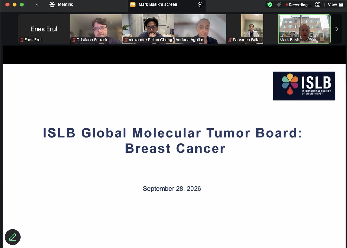
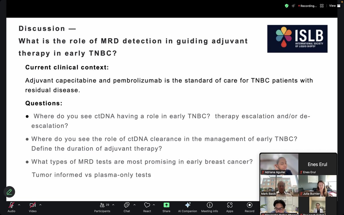
🧬 ISLB Global Molecular Tumor Board: Breast Cancer
Thank you to everyone who joined today’s ISLB Global Molecular Tumor Board – Breast Cancer, part of the ISLB Education Series.
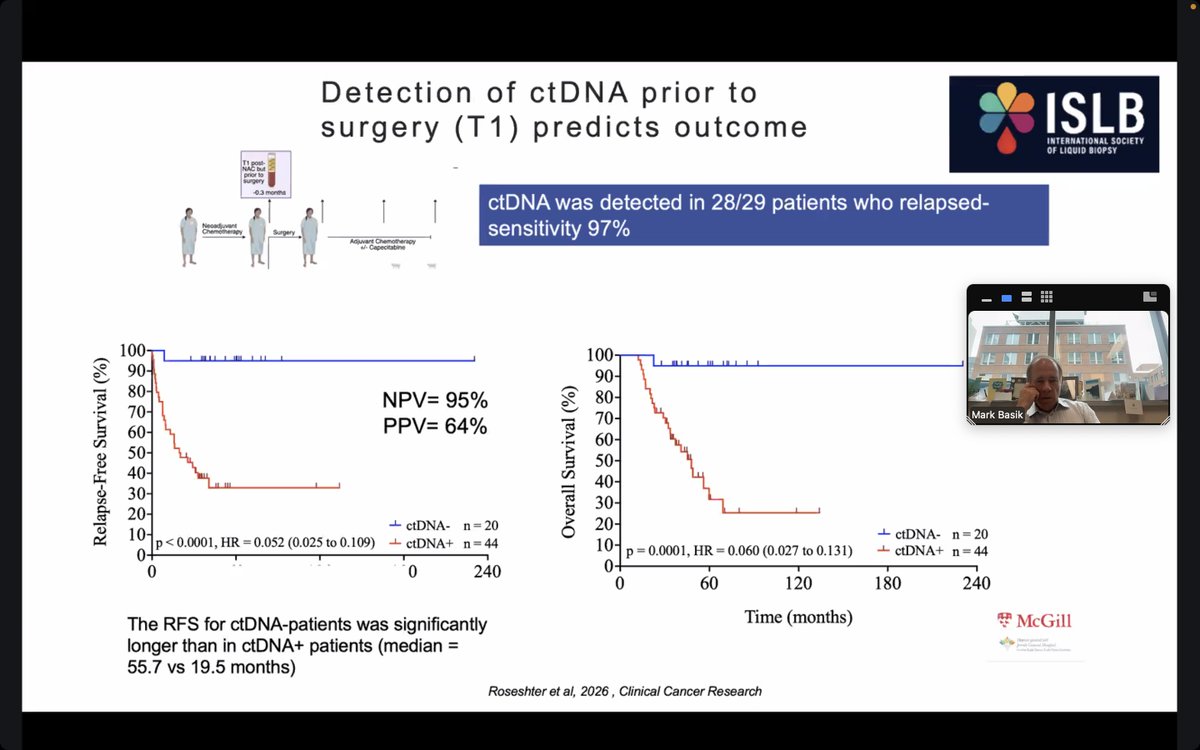
The interactive session explored the evolving role of liquid biopsy and ctDNA in breast cancer, with a particular focus on molecular residual disease (MRD) in early triple-negative breast cancer, ctDNA dynamics around neoadjuvant therapy and surgery, and the potential future role of molecular monitoring in treatment escalation and de-escalation.
A key discussion centered on an increasingly important question:
Can ctDNA move from predicting recurrence to actually guiding treatment decisions in early breast cancer?
Many thanks to our moderators Mark Basik and Adriana Aguilar, presenter Mark Basik, and invited panelists Cristiano Ferrario, @AlexandrePCheng , @jvburnier , and Parvaneh Fallah for an engaging and clinically relevant discussion.
On behalf of the ISLB Education Committee, coordinated by @BrunaPellini and @JordiRemon , thank you to our faculty and participants for making this another highly interactive Global MTB session.
🌍 Advancing molecular diagnostics through global discussion, education, and collaboration.
#ISLB #LiquidBiopsy #BreastCancer #ctDNA #MRD #TNBC #PrecisionOncology #MolecularTumorBoard
@UmbertoMalapel1@ElJanLe@drgandara@MJOSESERRANO19@ChristianRolfo@RobertoBoreaMD@ErulEnes@ReduzziCarol@ddemiguelperez@PasqualePisapia@BenBleibergMD@Al3ssandroRusso@ESaldanhaMD@eleonora_nicolo@DrMirallas@LuisRaezMD@DrYukselUrun
🌍 New @TheLancetOncol Commission: Making Precision Oncology More Equitable
Precision oncology is advancing rapidly but access to cancer genomics remains deeply unequal worldwide.
The new Lancet Oncology Commission reports that:
🧬 7–8 million patients with cancer require molecular testing each year
🌍 Millions still lack access to guideline-indicated biomarker testing
📊 Only 2% of biomarker-based trial–country activity analysed occurred in LMICs
🩸 Liquid biopsy is increasingly integrated into molecular profiling, MRD monitoring and resistance assessment
The message is clear: innovation alone is not enough.
Precision oncology must be implemented with evidence, quality, affordability, workforce capacity, reimbursement and equitable access at its core.
For the liquid biopsy community, this means moving beyond “Can we detect it?” toward:
“Can every eligible patient access testing, interpretation and an effective treatment?”
@DrYukselUrun@UmbertoMalapel1@ElJanLe@drgandara@MJOSESERRANO19@ChristianRolfo@RobertoBoreaMD@ErulEnes@ReduzziCarol@ddemiguelperez@PasqualePisapia@Al3ssandroRusso@ESaldanhaMD@eleonora_nicolo@DrMirallas@LuisRaezMD
#PrecisionOncology #CancerGenomics #LiquidBiopsy #ctDNA #GlobalOncology #HealthEquity #CancerResearch #ISLB
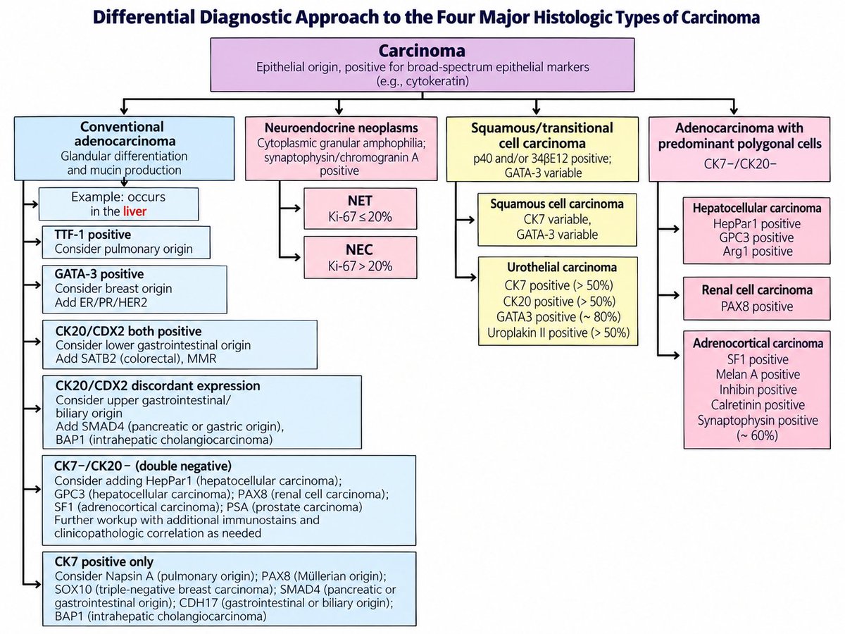
Differential Diagnostic Approach to the Four Major Histologic Types of Carcinoma
The four major histologic types of carcinoma include conventional adenocarcinoma, adenocarcinoma composed predominantly of polygonal cells, squamous/transitional cell carcinoma, and neuroendocrine neoplasms.
Prostate - Gleason pattern 4
90-95% of prostate cancer with metastasis have large cribriform/intraductal carcinoma: Driver of clinical outcomes
Cribriform: see pic 3
Large cribriform: >0.25 mm across the narrow dimension (see pic 4)
Dr. McKinney. Prostatic Adenocarcinoma Grading: 2026 Update #USCAP #pathology #pathX
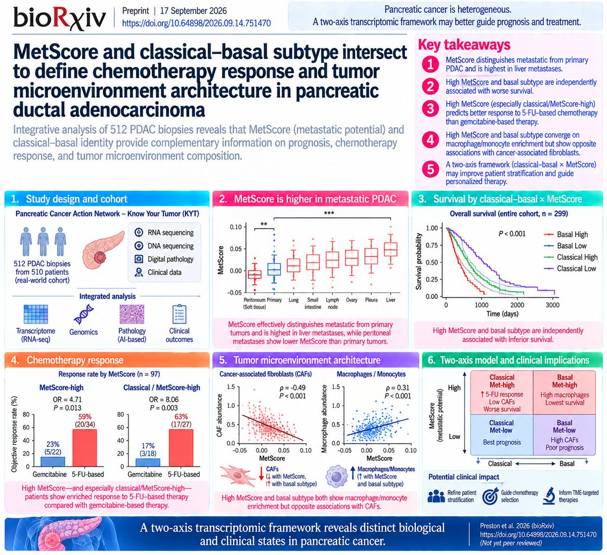
🧬 Pancreatic cancer may need more than a single “classical vs basal” label. A new study suggests PDAC is better understood on TWO independent transcriptomic axes—and their intersection may help explain metastasis, chemotherapy response, and the tumor microenvironment.
A new bioRxiv study analyzed 512 tumor biopsies from 510 patients in the Pancreatic Cancer Action Network Know Your Tumor cohort, integrating RNA-seq, DNA sequencing, clinical outcomes and AI-based digital pathology.
The authors combine the established:
CLASSICAL ↔ BASAL axis
with a second metric:
MetScore LOW ↔ HIGH
MetScore captures a transcriptional cell state associated with metastatic colonization potential and is largely orthogonal to classical–basal differentiation.
This produces four biologically distinct PDAC states:
Classical / Met-low
Classical / Met-high
Basal / Met-low
Basal / Met-high
And that second dimension reveals several important findings.
🔥 1. MetScore tracks systemic metastatic behavior
In the real-world KYT cohort:
Metastatic biopsies had significantly higher MetScores than primary tumors
and this finding replicated independently using another commercial sequencing platform at Emory/Winship.
But the anatomical pattern is particularly interesting.
Liver metastases → highest MetScore
while
peritoneal metastases → lower MetScore than primary tumors.
The plot on page 17 makes this strikingly clear: MetScore progressively rises across several non-peritoneal metastatic sites, with liver metastases showing the strongest signal, whereas peritoneal disease sits below the primary-tumor distribution.
That suggests:
SYSTEMIC dissemination
via hematogenous/lymphatic routes
may require a different tumor-cell state from:
TRANSCOELOMIC dissemination
into the peritoneum.
So “metastatic PDAC” itself may encompass biologically distinct metastatic programs.
📉 2. MetScore and basal subtype independently carry prognostic information
When both variables were included in the same model:
MetScore: HR 1.55 per 1-SD increase
and
Basal vs classical: HR 1.81
for overall mortality.
The Kaplan–Meier curves on page 18 reveal the two-axis structure beautifully:
🟣 Classical / Met-low
→ longest survival
🔴 Basal / Met-high
→ shortest survival
while:
Classical / Met-high
and
Basal / Met-low
occupy intermediate overlapping trajectories.
The associations persisted across primary versus metastatic biopsies and M0 versus M1 disease.
This means MetScore is not simply another way of measuring basal differentiation.
The two axes appear to capture different aspects of PDAC biology.
💊 3. The chemotherapy result is particularly provocative
Among patients with MetScore-high tumors:
5-FU-based therapy
ORR = 59% (20/34)
versus
gemcitabine-based therapy
ORR = 23% (5/22)
with:
OR = 4.71
P = 0.013
and a significant:
treatment × MetScore interaction, P = 0.021.
In contrast, MetScore-low tumors showed no detectable difference between chemotherapy backbones.
Classical/basal subtype alone was less discriminating: classical tumors showed a numerical preference for 5-FU, but the treatment-by-subtype interaction was not significant.
Then the authors combined both axes.
And one group stood out.
CLASSICAL + METSCORE-HIGH
With:
5-FU → ORR 63% (17/27)
versus:
Gemcitabine → ORR 17% (3/18)
OR = 8.06
P = 0.003
The bar graph on page 19, Figure 4C is probably the paper's most immediately clinically interesting figure.
It suggests that adding metastatic-state biology to classical/basal subtype could potentially reveal a chemotherapy-response phenotype invisible to conventional subtyping alone.
🤯 But isolated cancer cells do NOT reproduce the effect
This is perhaps the most biologically interesting result.
The investigators tested five murine classical PDAC cell lines with different MetScores.
Neither:
5-FU sensitivity
nor:
gemcitabine sensitivity
significantly correlated with MetScore.
So why would MetScore predict treatment response in patients but not isolated tumor cells?
That immediately points toward:
THE TUMOR MICROENVIRONMENT
🧱 4. MetScore and basal identity reshape the stroma in opposite directions
This is where the two-axis model becomes especially interesting.
Higher MetScore correlated with:
fibroblasts ↓
ρ = −0.49
whereas basal-state probability correlated with:
fibroblasts ↑
ρ = +0.23.
Both remained independently associated in multivariable analysis.
Consequently:
Classical / Met-high
had the lowest fibroblast abundance
while:
Basal / Met-low
had the highest.
The authors then independently examined H&E slides from:
502 tumors
using CellViT deep-learning cell classification.
Again:
Classical / Met-high → lowest connective/soft-tissue fraction
Basal / Met-low → highest.
The pathology panels and quantitative plots on page 20 provide strong orthogonal support for the RNA-derived observation.
This raises an intriguing hypothesis:
Classical / Met-high
↓
less fibroblast-rich stroma
↓
potentially altered drug delivery / stromal signaling
↓
greater 5-FU response
But that mechanism remains unproven.
🧲 5. Macrophages behave differently from fibroblasts
Unlike fibroblasts, macrophage/monocyte abundance increased with MetScore:
ρ = +0.31, P < 0.001.
Basal-state probability showed a weaker positive relationship.
After modeling both axes simultaneously, MetScore remained independently associated with macrophage/monocyte abundance:
β = +0.31, P < 0.001
whereas the basal-state association was no longer significant.
So high MetScore creates an unusual microenvironmental architecture:
FIBROBLASTS ↓
but
MACROPHAGE/MONOCYTE SIGNAL ↑
This is important because “stroma-rich” and “immune-rich” PDAC cannot necessarily be treated as interchangeable biological states.
🧬 The two-axis model therefore creates four different ecosystems
Conceptually:
🟣 CLASSICAL / MET-LOW
lower metastatic program
lower myeloid signal
more favorable prognosis
🔵 CLASSICAL / MET-HIGH
systemic metastatic program ↑
fibroblasts ↓��↓
myeloid cells ↑
strongest observed 5-FU response enrichment
🟢 BASAL / MET-LOW
basal differentiation
fibroblasts ↑↑
lower MetScore-associated metastatic program
🔴 BASAL / MET-HIGH
basal differentiation
metastatic program ↑↑
myeloid enrichment ↑
poorest observed survival
This is a much richer framework than:
CLASSICAL vs BASAL
alone.
🧬 6. MetScore also intersects with PDAC genomics
Among 499 evaluable tumors:
88.6% carried KRAS mutations.
KRAS-mutant tumors had significantly higher MetScores than KRAS-WT tumors.
In particular:
KRAS G12D
was enriched among MetScore-high tumors:
41% vs 29%.
The authors also examined MTAP deletion, present in approximately:
11.8%
of evaluable tumors.
MTAP-deleted tumors showed higher MetScores, although multivariable analysis suggested this relationship was largely attributable to neighboring CDKN2A co-deletion rather than MTAP loss itself.
The genomic plots on page 21 summarize these relationships nicely.
This potentially links transcriptional state to emerging therapeutic vulnerabilities involving:
KRAS-directed therapy
and
MTAP/PRMT5 synthetic lethality.
But these associations do not establish treatment sensitivity.
🎯 The larger conceptual message
PDAC precision oncology has traditionally focused heavily on:
DNA mutations
and increasingly on:
CLASSICAL ↔ BASAL transcriptional subtype.
This paper argues that neither alone fully captures tumor behavior.
A more informative framework may be:
AXIS 1 — DIFFERENTIATION
Classical ←────────→ Basal
plus
AXIS 2 — METASTATIC POTENTIAL
MetScore-low ←────────→ MetScore-high
Together these axes describe different dimensions:
metastatic route
prognosis
chemotherapy response
fibroblast architecture
myeloid infiltration
and potentially therapeutic vulnerabilities.
That creates a clinically attractive model:
PDAC biopsy
↓
RNA-seq
↓
PurIST subtype + MetScore
↓
2D molecular state
↓
integrate with DNA biomarkers
↓
therapy stratification
Rather than asking:
“Is this PDAC classical or basal?”
the more informative question may eventually become:
“Where does this tumor sit in the classical–basal × metastatic-potential space?”
⚠️ But the chemotherapy result needs particular caution
This is a bioRxiv preprint and has not yet been peer reviewed.
More importantly, the chemotherapy analysis was:
retrospective
nonrandomized
real-world
and involved only
97 response-evaluable first-line patients.
The authors explicitly state that the findings require prospective validation in a randomized comparison of standardized regimens using PFS or OS and powered for treatment–biomarker interaction.
The classical/Met-high 63% vs 17% comparison is particularly interesting, but the reported subgroup P values were nominal and not corrected for multiple comparisons.
So this should not yet be interpreted as evidence to choose FOLFIRINOX over gemcitabine/nab-paclitaxel for an individual patient based on MetScore.
Instead, it provides a strong biomarker hypothesis for prospective testing.
🔥 The next experiment is obvious—and important
A prospective PDAC trial could stratify patients using:
PurIST + MetScore
before treatment and test whether the two-axis state predicts benefit from:
mFOLFIRINOX / NALIRIFOX
versus
gemcitabine + nab-paclitaxel.
But mechanistically, I would go one step further.
The most interesting unresolved question is:
Why does Classical/Met-high PDAC respond better to 5-FU-based therapy in patients but not in isolated cancer cells?
That discrepancy points directly toward the microenvironment.
The obvious candidates are:
CAF abundance/subtype
SPP1⁺ macrophages
drug penetration
ECM architecture
myeloid–tumor interactions
and these should now be dissected with single-cell + spatial transcriptomics + multiplex imaging in pretreatment biopsies.
If validated, the study's most important contribution may not be another PDAC classifier.
It may be the realization that:
CHEMOTHERAPY RESPONSE IS AN EMERGENT PROPERTY OF TUMOR STATE × METASTATIC STATE × MICROENVIRONMENT.
📄 Preston RE, Huang H, Hall MA, et al.
MetScore and classical–basal subtype intersect to define chemotherapy response and tumor microenvironment architecture in pancreatic ductal adenocarcinoma.
bioRxiv. Posted September 17, 2026.
DOI: 10.64898/2026.09.14.751470
#PancreaticCancer #PDAC #MetScore #PrecisionOncology #FOLFIRINOX #5FU #Gemcitabine #TumorMicroenvironment #CancerAssociatedFibroblasts #Macrophages #KRAS #MTAP #Transcriptomics #LiquidBiopsy #CancerResearch
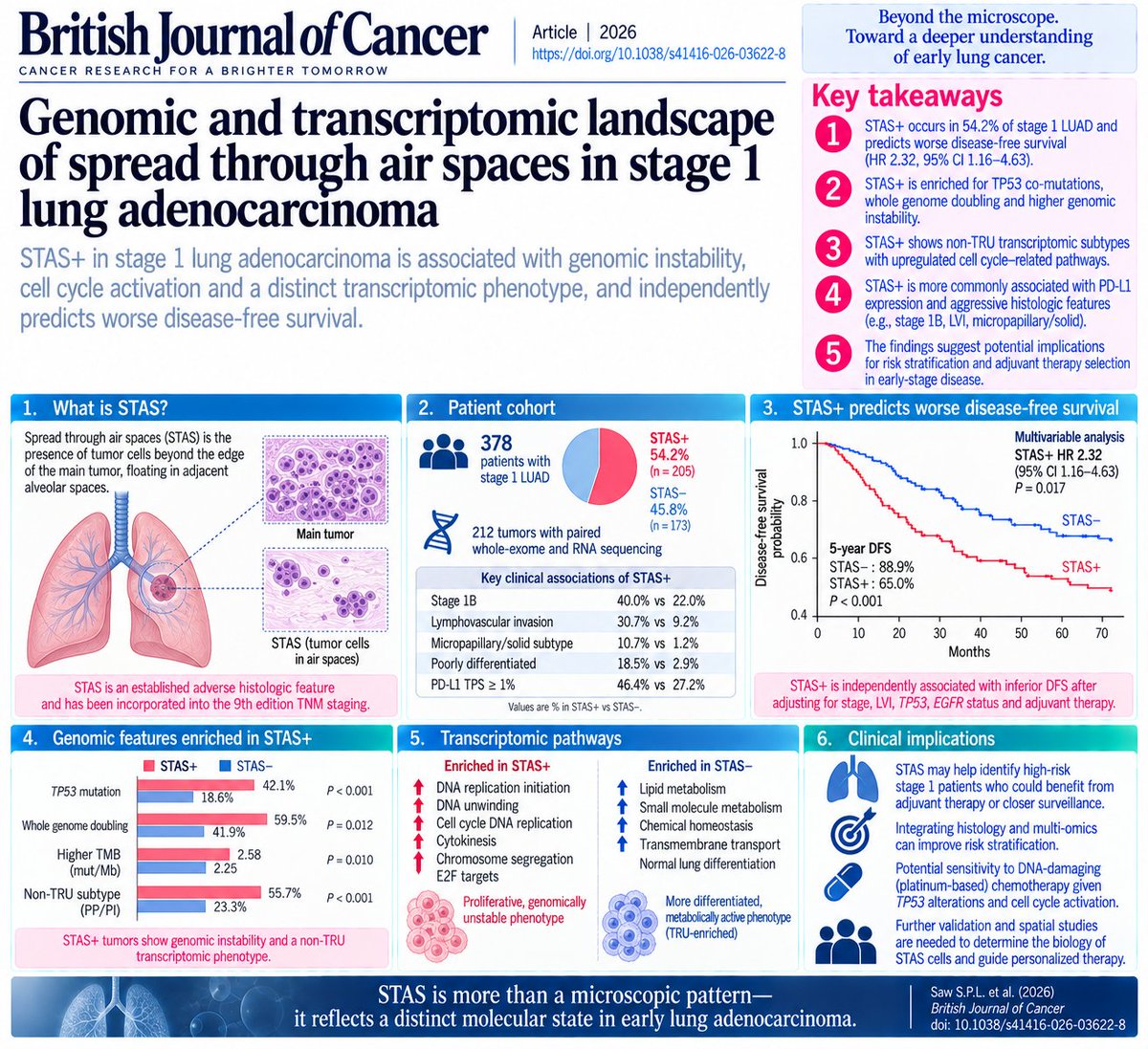
🫁🧬 Spread through air spaces (STAS) in early-stage lung adenocarcinoma may be more than a microscopic pattern of invasion—it appears to mark a distinct, genomically unstable and highly proliferative tumor state.
A new British Journal of Cancer study provides an integrated whole-exome + transcriptomic analysis of STAS specifically in stage I lung adenocarcinoma (LUAD).
The emerging picture is striking:
STAS+ LUAD
→ TP53 co-mutation ↑
→ whole-genome doubling ↑
→ genomic instability ↑
→ non-TRU phenotype ↑
→ DNA replication/cell-cycle programs ↑
→ PD-L1 expression ↑
→ recurrence risk ↑
This suggests STAS may be the histologic manifestation of an underlying aggressive molecular state—not merely tumor cells mechanically floating into adjacent alveoli.
🔬 378 patients with stage I LUAD
The cohort included:
378 surgically resected stage I LUADs
205 (54.2%) STAS+
280 (74.1%) EGFR-mutant
Importantly, STAS prevalence was similar between EGFR-mutant and EGFR-wild-type tumors.
STAS+ tumors were strongly enriched for conventional aggressive pathological features:
Stage IB
40.0% vs 22.0%
Lymphovascular invasion
30.7% vs 9.2%
Poor differentiation
18.5% vs 2.9%
Micropapillary/solid predominance
10.7% vs 1.2%.
They also more frequently expressed PD-L1 TPS ≥1%: 46.4% vs 27.2%.
So STAS already sits within a more aggressive clinicopathologic ecosystem.
But does it independently matter?
📉 STAS strongly predicts recurrence
Yes.
The Kaplan–Meier curves in Figure 1, page 3 show a striking separation.
2-year DFS
STAS+: 85.5%
STAS−: 94.0%
5-year DFS
STAS+: 65.0%
STAS−: 88.9%
P < 0.001 for the 5-year comparison.
And the association survived multivariable adjustment for:
stage
surgery type
TP53 status
lymphovascular invasion
EGFR status
adjuvant therapy.
STAS+ → HR 2.32 for inferior DFS
95% CI 1.16–4.63; P=0.017.
Interestingly, sublobar resection was also independently associated with inferior DFS:
HR 2.64
95% CI 1.12–6.22.
That reinforces the potential importance of recognizing STAS when considering limited resection.
🧬 EGFR mutation itself did NOT explain the phenotype
This is particularly relevant in an Asian LUAD cohort.
Overall DFS was essentially similar between:
EGFR-mutant vs EGFR-WT
P=0.9.
But within EGFR-mutant tumors:
STAS+ → 5-year DFS 62.8%
versus
STAS− → 93.4%
P < 0.001.
The EGFR-WT subgroup showed the same directional trend, although the smaller sample size limited statistical power.
So:
STAS biology appears largely orthogonal to EGFR driver status.
That becomes even clearer when the investigators examine the tumor genome.
🔥 TP53 is strongly enriched in STAS+
Among the 212 tumors with WES + RNA-seq:
TP53 mutations
STAS+: 42.1%
vs
STAS−: 18.6%
P < 0.001.
Additional mutations enriched in STAS+ included:
CUX1
BRCA2
BCL11B
ATM
although each occurred at relatively low frequencies.
The OncoPrint on page 5, Figure 2 is particularly informative: STAS+ tumors visually cluster with TP53 mutation, WGD and non-TRU transcriptional states.
This raises an obvious question:
Is STAS simply a surrogate for TP53-mutant aggressive cancer?
Apparently not entirely.
Cell-cycle pathway activation remained present in STAS+ tumors even after stratification by TP53 status.
💥 Whole-genome doubling provides another clue
STAS+ tumors showed substantially more:
Whole-genome doubling
59.5% vs 41.9%
P=0.012
and slightly higher:
Tumor mutational burden
2.58 vs 2.25 mutations/Mb
P=0.010.
The overall genomic instability index was also higher in STAS+ disease.
That creates an interesting biological model:
TP53 dysfunction
↓
genome-doubling tolerance
↓
chromosomal/genomic instability
↓
proliferative plasticity
↓
STAS phenotype + recurrence potential
The study does not establish this causal sequence, but the molecular associations make it highly testable.
🧬 The transcriptome changes dramatically too
Perhaps the most interesting molecular finding is the shift away from the:
TRU — terminal respiratory unit phenotype
toward the more aggressive:
PP — proximal proliferative
and
PI — proximal inflammatory
states.
PP/PI occurred in:
55.7% of STAS+
versus
23.3% of STAS− tumors.
Conversely, TRU accounted for:
76.7% of STAS−
versus
44.3% of STAS+.
P < 0.001.
So histologically visible STAS is accompanied by a major shift in tumor-cell transcriptional identity.
⚙️ And the dominant program is CELL CYCLE
GSEA provides perhaps the clearest biological signal in the paper.
STAS+ tumors were enriched for:
DNA replication initiation
DNA unwinding
cell-cycle DNA replication
regulation of DNA-dependent DNA replication
cytokinesis
while STAS− tumors were relatively enriched for:
lipid metabolism
small-molecule metabolism
chemical homeostasis
transmembrane transport.
Figure 3 on page 6 captures this beautifully: the pathway plot shows a near mirror-image biology—
STAS+
PROLIFERATION / DNA REPLICATION
versus
STAS−
METABOLISM / HOMEOSTASIS.
Even more importantly, cell-cycle activation persisted regardless of TP53 mutation status.
Thus:
STAS ≠ simply TP53 mutation
It appears to identify a broader proliferative state.
🧠 This changes how we might conceptualize STAS
The conventional picture is largely anatomical:
primary tumor
→ cells detach
→ enter alveolar air spaces
→ establish microscopic clusters beyond tumor edge.
But the molecular data suggest another layer:
genomic instability
cell-cycle activation
non-TRU transcriptional identity
high-grade histology
↓
STAS
↓
greater recurrence potential
In other words:
STAS may be morphology revealing tumor biology.
The pathology slide may already be telling us something that genomics subsequently confirms.
🎯 Could this influence adjuvant treatment?
This is where the study becomes clinically provocative.
Current stage I LUAD treatment decisions rely heavily on:
tumor size/stage
resection type
pathological risk factors
and selected molecular features.
But the authors propose that STAS might identify a subgroup with sufficiently aggressive biology to warrant investigation of:
adjuvant therapy
or
intensified surveillance.
There is an especially interesting therapeutic hypothesis.
STAS+ tumors show:
TP53 alterations
WGD
ATM/BRCA2 enrichment
DNA-replication pathway activation.
Could that phenotype create greater vulnerability to:
DNA-damaging platinum chemotherapy?
The authors specifically raise this possibility, citing previous retrospective and meta-analytic evidence, but emphasize that it requires prospective testing.
This could eventually turn STAS from a purely prognostic pathological descriptor into a treatment-selection biomarker.
But we are not there yet.
🔬 An even more interesting next experiment: spatial omics
Bulk RNA-seq cannot tell us whether the molecular signature comes specifically from:
STAS cells floating in alveoli
or from the primary tumor as a whole.
The authors explicitly identify:
SPATIAL TRANSCRIPTOMICS OF STAS CELLS
as a logical next step.
Imagine profiling:
primary tumor core
vs
invasive edge
vs
STAS cluster
within the same patient.
That could answer whether STAS cells acquire:
EMT/plasticity programs
anoikis resistance
oxidative stress adaptations
cell-cycle activation
immune-evasion programs
or unique metabolic dependencies.
Single-cell/spatial analysis could therefore determine whether STAS represents merely a marker of aggressive tumors—or an actual metastasis-like cellular state within the lung airspace.
⚠️ Important limitations
This is a single-center Singapore cohort, and 74.1% of patients were EGFR-mutant. That is much higher than expected in many Western LUAD populations and limits generalizability.
Only 212/378 tumors underwent WES/RNA-seq, and sequencing was intentionally enriched for EGFR-mutant disease.
The EGFR-WT population was therefore underpowered for detailed molecular subgroup analyses, and external validation was limited by the lack of comparable stage I cohorts with both multi-omics and annotated STAS status.
Most importantly:
the study demonstrates association, not causation.
TP53 mutation, WGD and proliferative signaling may help generate STAS—or STAS may simply occur preferentially within tumors already possessing these aggressive characteristics.
Resolving that distinction will require spatial and functional experiments.
🔥 The broader message
This paper provides a useful bridge between:
PATHOLOGY
and
MOLECULAR ONCOLOGY.
The integrated model is:
Stage I LUAD
↓
STAS+
↓
TP53 co-mutation ↑
+
WGD ↑
+
genomic instability ↑
+
PP/PI transcriptional states ↑
+
DNA replication/cell cycle ↑
+
PD-L1 ↑
↓
aggressive tumor biology
↓
DFS ↓
And remarkably, much of this information begins with something a pathologist can already see under the microscope.
STAS may therefore be more than:
“tumor cells beyond the edge of the main tumor.”
It may be a readily observable histologic readout of an underlying genomically unstable, proliferative LUAD state.
If prospectively validated, the important question may shift from:
“Is STAS present?”
to:
“Which STAS+ stage I patients harbor enough biological risk to justify adjuvant treatment or intensified surveillance?”
📄 Saw SPL, Pang M, Patteri C, et al.
Genomic and transcriptomic landscape of spread through air spaces in stage 1 lung adenocarcinoma.
British Journal of Cancer. 2026.
DOI: 10.1038/s41416-026-03622-8
#LungCancer #NSCLC #LungAdenocarcinoma #LUAD #STAS #EGFR #TP53 #Genomics #Transcriptomics #WholeGenomeDoubling #PrecisionOncology #ThoracicOncology #Pathology #CancerGenomics #BritishJournalOfCancer