Today marks a significant milestone in my life, a day I've eagerly anticipated for so long. I've poured my heart and soul into reaching this point,I've witnessed countless match days pass by, each one instilling in me a sense of hope and resilience. I matched #Match2024
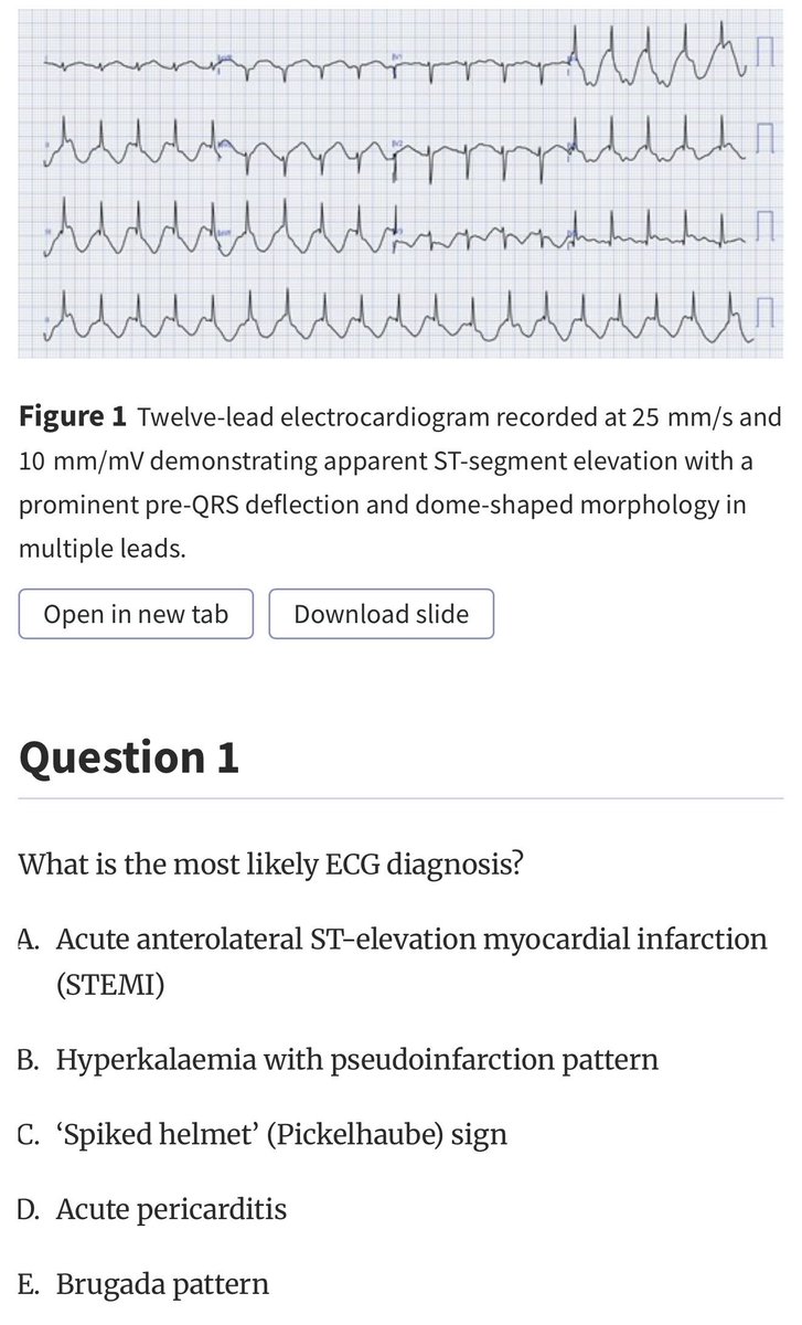
A closer look at V3–V6 shows fusion of:
🛑QRS complex
🛑ST segment
🛑T wave
This creates the characteristic triangular waveform known as the:
Shark fin sign (also called the lambda-wave pattern)
The cardiology community has lost a giant. We honor the extraordinary legacy of Eugene Braunwald, MD, MACC, a visionary leader and pioneer whose outstanding contributions shaped the foundation of cardiovascular medicine as we know it today.
Read more: https://t.co/uaC2n4m5gD
AHFTC training covers an enormous clinical landscape — MCS, transplant, shock, palliative care, etc. — often in a single fellowship year.
I created the Heart Failure Fellow Lecture Series to help close the education gap.
Free. No paywall. No sign-up. 🎓
https://t.co/0oOG9Ekrlx
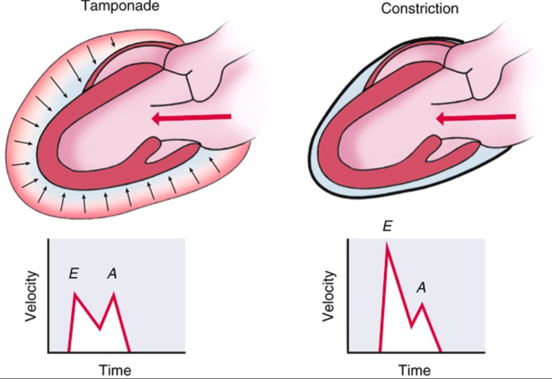
Pericardial Tamponade vs Constrictive Pericarditis ➡️Spot the Difference
Tamponade
⬇️
Accumulation of fluid in the pericardial space
⬇️
↑ intrapericardial pressure
⬇️
impairs diastolic filling throughout the cardiac cycle (early and late diastole).
Doppler shows blunted E and A waves due to restricted filling.
Constrictive Pericarditis
⬇️
Thickened, non-compliant pericardium
⬇️
rapid early diastolic filling that abruptly halts once volume limit is hit.
Doppler shows prominent E wave, reduced A wave, classic for constriction.
Recognizing the echocardiographic filling patterns is essential for diagnosis.
Ref: Catherine M. Otto, Textbook of Clinical Echocardiography
Summary of the different types of ventricular complexes.
1️⃣ Interpolated PVCs
✅ A PVC occurs between two normal beats without disrupting the sinus rhythm.
✅ No compensatory pause.
✅ Often seen in bradycardic patients.
2️⃣ PVCs in Bigeminy
✅ Every normal beat (sinus beat) is followed by a PVC → Regular pattern.
✅ The PVC is premature and typically has a wide QRS complex.
3️⃣ PVCs in Trigeminy
✅ Two normal beats followed by a PVC, repeating in a cycle.
✅ A predictable ventricular ectopy pattern.
4️⃣ Multifocal PVCs
✅ PVCs originate from multiple foci, showing different QRS morphologies.
✅ Indicates more ventricular instability than unifocal PVCs.
5️⃣ Paired or Back-to-Back PVCs
✅ Two consecutive PVCs occurring without an intervening sinus beat.
6️⃣ Nonsustained Monomorphic Ventricular Tachycardia (NSVT)
✅ Three or more consecutive PVCs at a rate >100 bpm, but lasting <30 sec.
✅ Monomorphic: All QRS complexes have the same shape (single ectopic focus).
7️⃣ Nonsustained Polymorphic Ventricular Tachycardia (NSVT)
✅ Three or more PVCs lasting <30 sec, but QRS morphology varies (multiple ectopic foci).
✅ Often associated with long QT syndrome, ischemia, or electrolyte abnormalities.
8️⃣ Ventricular Fibrillation (VF) – A Medical Emergency!
✅ Chaotic, disorganized electrical activity with no identifiable QRS complexes.
✅ The heart is quivering instead of pumping, leading to no cardiac output.
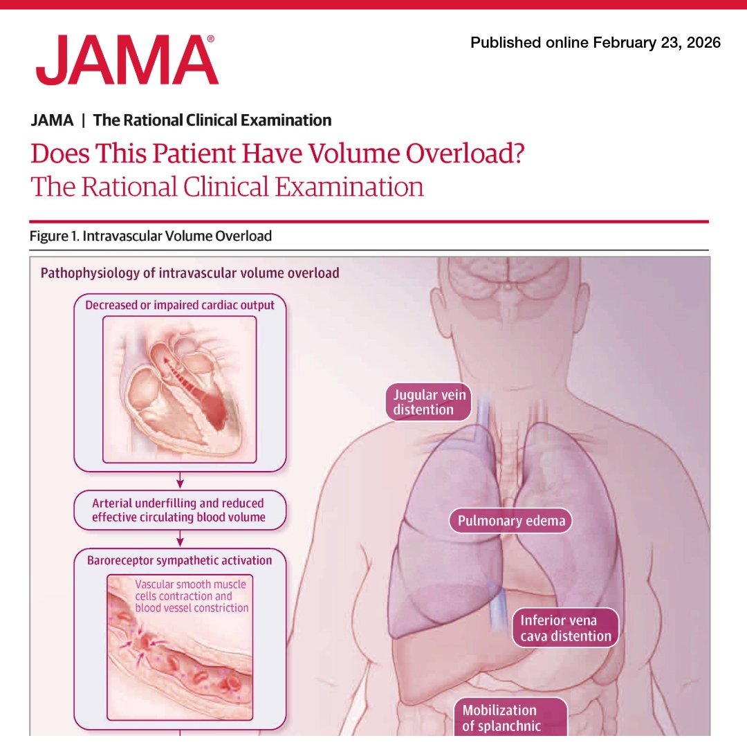
How can volume overload be reliably excluded at the bedside?
📊 This Rational Clinical Examination evaluates the diagnostic accuracy of clinical examination, radiographic imaging, and laboratory testing in assessing volume overload in nonintubated patients.
https://t.co/NycBzCLjqS
Hello, Moon. It’s great to be back.
Here’s a taste of what the Artemis II astronauts photographed during their flight around the Moon. Check out more photos from the mission: https://t.co/rzM1P0QbOl
Coronary artery territories
The main epicardial coronary arteries supply distinct regions of the myocardium, which can be mapped and assessed during an ultrasound examination. For standardization, the left ventricle (LV) is divided along its long axis into four quadrants: anterior, inferior, septal, and lateral.
At the basal and midventricular levels, the septal and lateral walls are further subdivided into anterior and inferior segments. Each wall is then divided in short-axis views into basal, mid, and apical thirds. Beyond the LV cavity, the distal apex forms a cap segment. Altogether, this results in a total of 17 myocardial segments.
Most of the heart’s blood supply comes from the left main coronary artery, which divides into the left anterior descending (LAD) and left circumflex (LCx) arteries. The LAD supplies most of the anterior ventricular wall, while its septal branches supply the anterior two-thirds of the interventricular septum. Its diagonal branches supply the anterolateral wall. In some cases, a large LAD may wrap around the apex and supply the distal portion of the inferior wall.
The LCx travels in the atrioventricular groove, and its obtuse marginal branches supply the inferolateral wall. The right coronary artery (RCA) supplies the inferior third of the septum and the inferior wall, and it also provides blood to the right ventricle.
Adapted from Bulwer BE, Rivero JM, eds. Echocardiography Pocket Guide: The Transthoracic Examination
Spontaneous Coronary Artery Dissection (SCAD) Types
Three classic angiographic patterns:
Type 1: Contrast dye staining (classic double lumen).
● Type 2: Long diffuse smooth narrowing (most common)
》Subtypes:
2A: Normal segments proximal & distal
2B: Extends to distal tip of vessel.
● Type 3: Focal stenosis mimicking atherosclerosis.
What are Risk Factors for SCAD?!
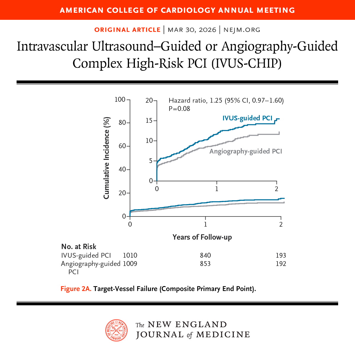
Presented at #ACC26:
In the IVUS-CHIP trial involving patients undergoing complex high-risk PCI, the risk of target-vessel failure (a composite outcome) was not lower with intravascular ultrasound guidance than with angiography guidance. Full trial results: https://t.co/oqHofLJUi6
Editorial: IVUS — A Zigzag Path to Success https://t.co/hb7PwRPlR3
@ACCinTouch
New Orleans is buzzing and so is #ACC26! 🎺
Good morning, New Orleans! Kick off Day 1⃣ with 📹 What’s Brewing at #ACC26 today ☕️ and get the inside scoop on today’s highlights, sessions, and experiences you won’t want to miss.
Hi Everyone -
🥸Here are all the 27 late breaking clinical trials presented at @ACCinTouch (ACC.26) with session number, day, time, and objective.
😱See you in NOLA:
👇👇👇
Among patients with supraventricular tachycardia, use of a handheld Valsalva assist device resulted in greater rates of sinus rhythm restoration compared with the standard Valsalva maneuver (63% vs 29% within 1 minute after up to two attempts). https://t.co/nzVVdHNO7a
Among older patients with #AtrialFibrillation, abelacimab reduced major or clinically relevant bleeding vs rivaroxaban, with a greater absolute risk reduction in those aged ≥75 years. https://t.co/0OpaHcW4QP