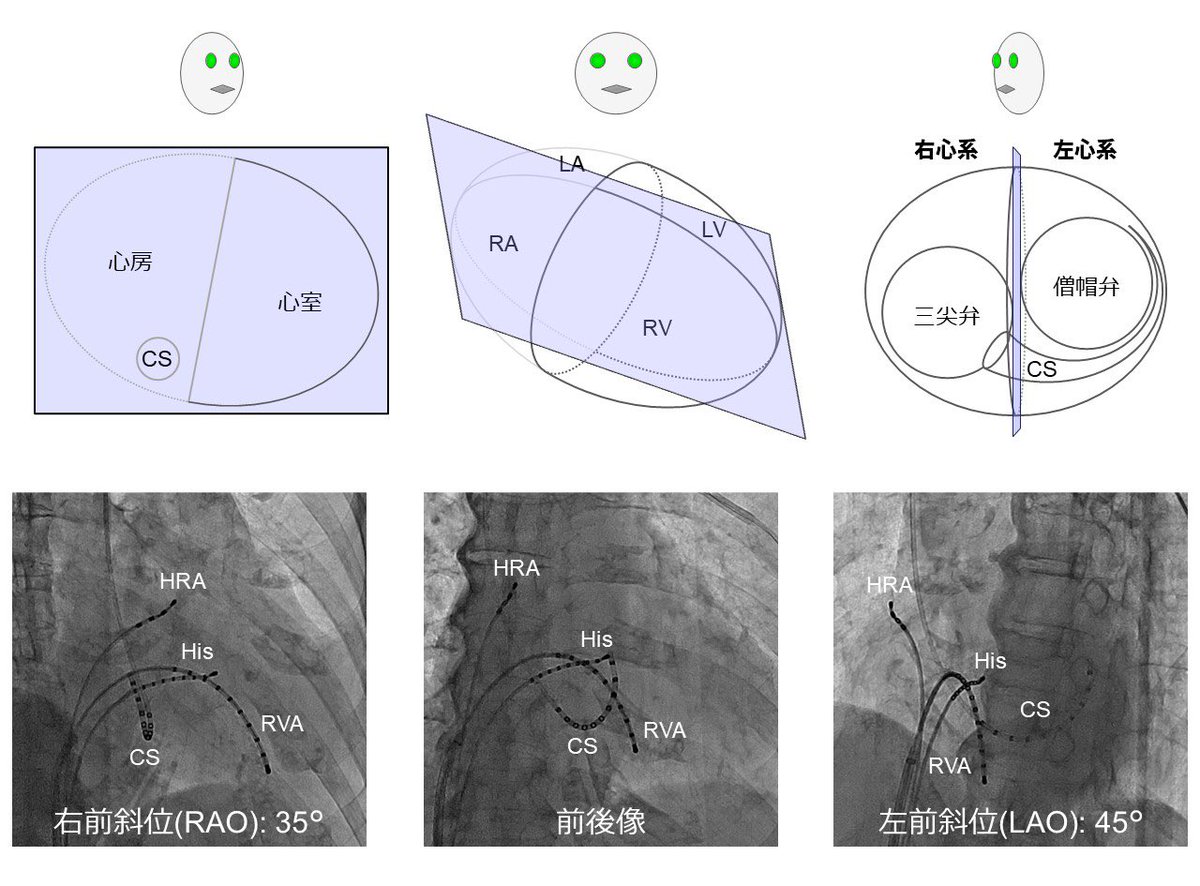
Rao, P.S.; Rao, N.S. Diagnosis of Dextrocardia with a Pictorial Rendition of Terminology and Diagnosis. Children 2022, 9, 1977.
CC-BY 4.0
https://t.co/DVHTsuUBWr
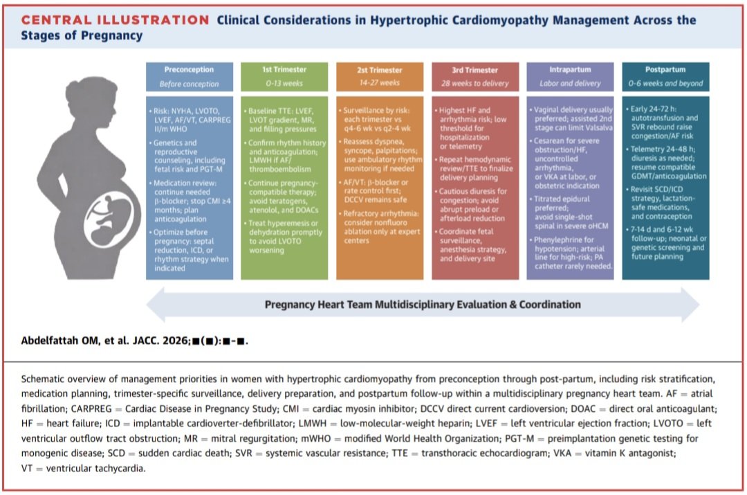
Miocardiopatía Hipertrófica & Embarazo. 🫀💥🤰
🔷️La mayoría de las mujeres con MCH pueden tener embarazos exitosos, siempre que exista una adecuada estratificación de riesgo y seguimiento por un equipo cardio-obstétrico especializado.
🔷️El momento más importante no es el embarazo, sino la consulta preconcepcional. Antes de intentar la gestación debe evaluarse la presencia de obstrucción del tracto de salida del VI, función sistólica, carga arrítmica, historia de IC y riesgo de muerte súbita.
🔷️La combinación de las escalas CARPREG II y mWHO permite identificar a las pacientes que requerirán vigilancia estrecha o incluso aconsejar diferir el embarazo si presentan FEVI <50%, síntomas NYHA III-IV o gradientes obstructivos severos.
🔷️Tto: los Ɓ-bloqueadores continúan siendo la piedra angular, con preferencia por metoprolol (o propranolol), mientras que debe evitarse atenolol por su asociación con restricción del crecimiento fetal. En MCH obstructiva sintomática debe considerarse miectomía septal o ablación septal antes de la concepción, ya que los inhibidores de miosina (mavacamten y aficamten) están contraindicados durante el embarazo por su potencial efecto teratogénico (se recomienda suspender mavacamten al menos 4 meses antes de buscar embarazo).
📚🆓️⤵️ State-Of-The-Art Review2026 @JACCJournals 💯
https://t.co/QjiDicw1WT
https://t.co/8pQRUjvPoL
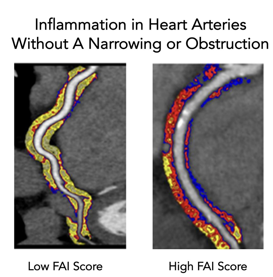
The first AI to quantify coronary artery inflammation was approved by the FDA yesterday @CaristoHeart
This is an important step forward for preventing heart disease that I've written about
https://t.co/foSaaTYmNw
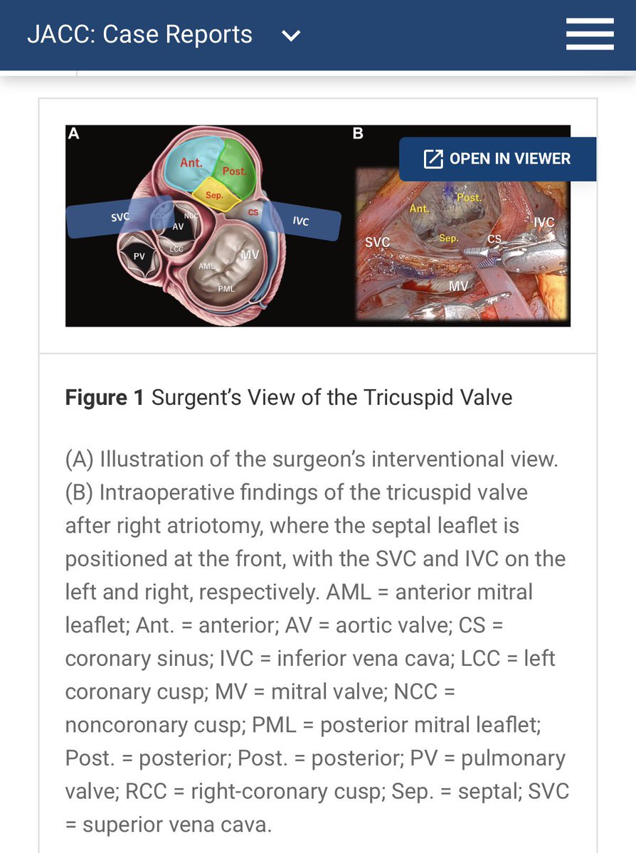
Nice educational tool about the TEE(R) anatomy and spatial relationships of TV leaflets. Though I have to admit, I spent a minute wondering who the “surgent” was… until I realized it must be the upgraded version of the surgeon’s view 😂 #echofirst#TEER
https://t.co/nnrsZVvMWp
Acute Coronary Syndrome: How clinicians rapidly decide who has a myocardial infarction
When a patient presents with chest pain, the diagnosis is never made from a single test. Instead, clinicians combine four key pieces of information to estimate the likelihood of myocardial infarction (MI) and decide whether the patient can be discharged, requires observation, or needs immediate treatment.
The process begins with the clinical assessment.
The patient's symptoms, physical examination, and vital signs provide the first estimate of risk.
Low-risk features include mild or atypical chest discomfort with stable vital signs.
High-risk features include:
- Typical crushing chest pain
- Pain radiating to the arm, jaw, or back
- Diaphoresis, nausea, or dyspnea
- Hypotension or shock
- Cardiac arrest or hemodynamic instability
The next step is the 12-lead ECG.
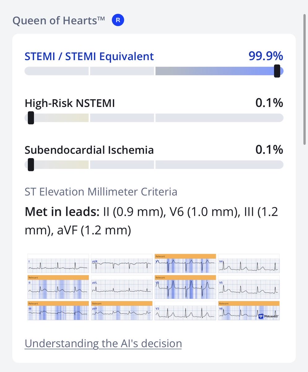
The ECG provides immediate information about ongoing myocardial ischemia.
Typical findings include:
🟥 Normal ECG: lowers the probability of MI but does not exclude it.
🟥 Mild ST-segment depression: may indicate early ischemia.
🟥 Significant ST-segment depression: increases suspicion for NSTEMI or extensive subendocardial ischemia.
🟥 ST-segment elevation: strongly suggests acute coronary artery occlusion (STEMI) and usually requires immediate reperfusion therapy.
The third component is the cardiac troponin level at presentation (0 hour).
Cardiac troponin is the most sensitive biomarker of myocardial injury.
Importantly, troponin should be interpreted as a quantitative marker.
In general:
- The higher the initial troponin level, the greater the likelihood of myocardial infarction.
- A normal initial troponin does not exclude very early MI.
For this reason, serial testing is essential.
The fourth component is the change in troponin over time, usually after 1–3 hours.
An acute rise or fall in troponin supports ongoing myocardial injury.
The greater the absolute change, the higher the probability of an acute MI.
These four elements are integrated into a triage strategy.
Rule-out MI Patients with:
- Low-risk symptoms
- Normal or non-ischemic ECG • Low troponin
- No significant change on repeat testing
are unlikely to have an acute myocardial infarction. Alternative diagnoses should be considered, including pneumonia, pneumothorax, musculoskeletal chest pain, or gastrointestinal disorders.
Observe Patients with intermediate-risk findings or equivocal troponin results require additional evaluation.
Possible diagnoses include:
- Unstable angina
- Early NSTEMI
- Myocarditis
- Takotsubo syndrome
- Heart failure
- Other cardiac disorders
Further ECGs, repeat troponins, imaging, or coronary evaluation may be needed.
Rule-in MI Patients with:
- Ischemic symptoms
- Dynamic ischemic ECG changes
- Elevated troponin with a significant rise or fall
have a high likelihood of acute myocardial infarction.
Those with persistent ST-segment elevation are treated as STEMI, requiring urgent reperfusion.
Those without ST elevation but with elevated troponin are diagnosed with NSTEMI and managed according to their risk profile.
Patients presenting with cardiac arrest, shock, or severe hemodynamic instability require immediate ECG and bedside echocardiography to identify life-threatening cardiac causes. If the presentation suggests aortic dissection or pulmonary embolism, dedicated diagnostic pathways should be followed rather than assuming acute coronary syndrome.
The key message is simple: Acute MI is diagnosed by integrating clinical presentation, ECG findings, and serial cardiac troponin measurements, not by relying on any single test in isolation.
Reference: Braunwald's Heart Disease: A Textbook of Cardiovascular Medicine (Diagnostic approach to acute coronary syndrome; adapted from the 2020 ESC Guidelines for the management of acute coronary syndromes).
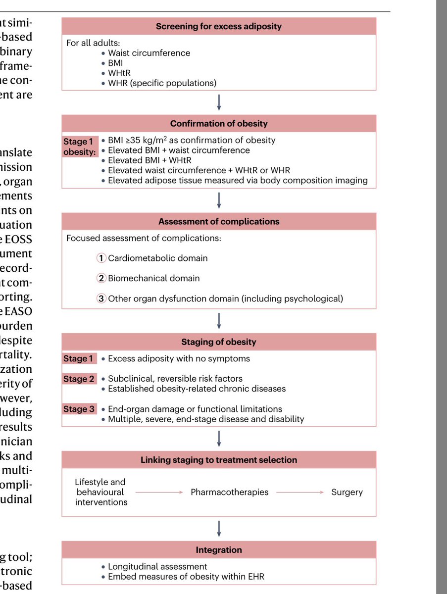
Este paper es probablemente la mejor síntesis hasta ahora en obesidad. No es una nueva guía. Compara las más recientes EASO (2024), (2025), ACC (2025) y AACE (2025) en un paper.
Obesidad deja de ser definida por IMC y se define ahora por exceso de adiposidad que genera daño.
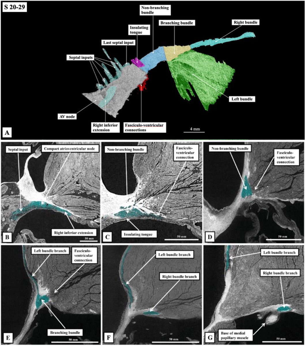
Unravelling the enigmatic morphology of the atrioventricular conduction axis using Hierarchical Phase-Contrast Tomography (HiP-CT) #OpenAccess@Chloe_Li_@aaalmehandi@UCL_ICS
https://t.co/YvNpPRyFnL
@ElNutriDice Com o Vinho tinha essa falacia de beneficios que durou por muito tempo . Isso aí não passa de uma mentira contada com uma roupagem de medicina baseada em evidencias.
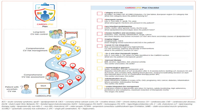
📚 New #ECRJournal Article: "CARDIOLIPID Plan: A Decision Pathway for Dyslipidaemia Management"
📚 Just Published!
🔓Access it here 👉 https://t.co/zyBrwGGjKp
#MedEd#CardioEd#VascularDisease