🫀Does albumin save lives in septic shock?
For years, albumin has been one of the most debated therapies in septic shock. Although current guidelines suggest crystalloids as first-line resuscitation fluids, the question has never been completely settled for patients with established septic shock.
This new systematic review and dual frequentist-Bayesian meta-analysis revisited the evidence by including only randomized trials focused on septic shock. Seven randomized trials involving 3,273 patients were analyzed to determine whether albumin-based resuscitation influences mortality.
The frequentist analysis demonstrated a 10% relative reduction in all-cause mortality with albumin compared with crystalloid resuscitation (RR 0.90, 95% CI 0.83-0.99; p = 0.02), with essentially no statistical heterogeneity (I² = 0%).
The Bayesian analysis provides an even more clinically intuitive interpretation. Rather than simply asking whether the result reached statistical significance, it estimated the probability that albumin truly reduces mortality. Using the primary model, there was a 94.7% probability that albumin reduces mortality (RR <1.0), although the probability of achieving a clinically important reduction greater than 5% was lower at 73.9%.
However, this is not the final answer.
The certainty of evidence was rated low using the GRADE framework because much of the evidence came from septic shock subgroups within larger sepsis trials rather than studies designed exclusively for septic shock. The estimated benefit corresponds to approximately 36 fewer deaths per 1,000 treated patients, but the confidence interval remains wide enough that the true effect could range from minimal to clinically important.
Interestingly, subgroup analyses found no convincing evidence that the effect depended on albumin concentration (4-5% versus 20-25%), dosing strategy, or baseline serum albumin. Likewise, sensitivity analyses consistently showed the same direction of benefit, supporting the robustness of the findings.
Clinical implications
This study does not support replacing balanced crystalloids with albumin as the universal first-line fluid in septic shock.
Instead, it strengthens the hypothesis that albumin may provide a meaningful survival advantage in selected patients, particularly after substantial crystalloid administration or in patients with persistent septic shock. At the same time, the available evidence is still insufficient to justify a strong guideline recommendation, and further adequately powered randomized trials remain necessary.
Until then, albumin should probably be viewed as a targeted adjunct rather than a routine replacement for crystalloid resuscitation.
Reference 📚
Mendes, H. (2026). Mortality effect of albumin fluid resuscitation in adults with septic shock: A systematic review and dual frequentist-Bayesian meta-analysis of randomised trials. Critical Care. Advance online publication. https://t.co/zDV8gUn4Yl
Critical Physiology Series #13
Monitoring Left Ventricular Function in Critical Care: From Pressure to Power
How can we know whether the left ventricle is functioning adequately in a critically ill patient?
Blood pressure is usually the first variable we observe. However, blood pressure tells us mainly about pressure generation, not how much blood the ventricle is actually ejecting.
A patient with severe vasoconstriction may maintain an acceptable arterial pressure despite a very low cardiac output. Another patient may have a low blood pressure because of vasodilation while the left ventricle ejects a normal or even increased volume.
Pressure alone cannot define LV function.
Cardiac index adds information about flow. It represents cardiac output adjusted for body surface area. A low cardiac index suggests that the heart is delivering insufficient blood for the patient’s size, but it does not explain why.
The problem may be impaired contractility, inadequate preload, excessive afterload, tachycardia, bradycardia, or mechanical obstruction.
Cardiac power index combines pressure and flow.
It can be approximated as mean arterial pressure multiplied by cardiac index and divided by 451. This produces a value expressed in watts per square metre.
This is physiologically attractive because the ventricle must generate both flow and pressure. A patient may have an acceptable cardiac index but produce it at a very low pressure. Another may maintain pressure through vasoconstriction while flow remains critically reduced. Cardiac power integrates both dimensions.
Left ventricular ejection fraction describes the proportion of end diastolic volume ejected during each contraction.
However, LVEF is highly dependent on loading conditions.
A low LVEF may reflect impaired contractility, but it can also be influenced by increased afterload. A normal or hyperdynamic LVEF does not guarantee a normal stroke volume. A small, underfilled ventricle can eject a large percentage of a very small volume.
This is why a hyperdynamic ventricle in septic shock should not automatically be interpreted as excellent cardiac function.
Left ventricular stroke work examines how much work the ventricle performs during each beat. True stroke work corresponds to the area enclosed by the pressure volume loop. At the bedside, it can be estimated using stroke volume and the pressure against which the ventricle ejects.
Indexed LV stroke work therefore combines pressure generation with the volume ejected during one contraction.
Each variable answers a different question. The most useful assessment comes from integrating them.
References 📚
Fincke, 2004, https://t.co/6vBJb89L9D
Jentzer, 2020, https://t.co/bg8bAiYvVt
Huygh, 2016, https://t.co/3bWmK0Lpcp
🫁 Mechanical power is changing the way we understand ventilator induced lung injury.
For decades, ventilator induced lung injury (VILI) has been explained through four mechanisms: volutrauma, barotrauma, atelectrauma, and biotrauma. These concepts remain fundamental, but they describe only the consequences of an even deeper process.
This excellent review proposes a modern framework in which mechanical power represents the common pathway linking ventilator settings to biological injury. Rather than focusing on tidal volume or airway pressure alone, mechanical power integrates tidal volume, respiratory rate, airway pressures, driving pressure, and flow into the total energy transferred to the lung every minute. Excessive energy delivery activates mechanosensitive pathways that transform physical stress into inflammation and cellular dysfunction.
Mechanical stretch is sensed by mechanoreceptors such as Piezo channels and integrins, initiating intracellular signalling through NF κB, MAPK, and other inflammatory pathways. Alveolar epithelial cells and macrophages release IL 1β, IL 6, and TNF α, while neutrophil recruitment amplifies tissue injury. Mechanical ventilation therefore becomes not only a respiratory support technique but also a biological stimulus capable of modifying immune behaviour.
A central element of this new concept is mitochondrial dysfunction. Excessive mechanical energy generates reactive oxygen species, damages mitochondrial DNA, activates the NLRP3 inflammasome, reduces ATP production, and shifts cellular metabolism toward glycolysis. The result is a vicious cycle in which oxidative stress and inflammation continuously reinforce each other.
Mechanical injury also disrupts the alveolar capillary barrier through apoptosis, pyroptosis, and ferroptosis. These different forms of programmed cell death interact with one another, accelerating epithelial disruption, pulmonary oedema, and worsening gas exchange.
From a clinical perspective, this review reinforces that mechanical power should be considered alongside driving pressure and tidal volume when individualising ventilation. Lung protection is no longer simply about limiting pressure or volume. It is about minimising unnecessary energy transfer to vulnerable lungs. The authors highlight lung protective ventilation using tidal volumes below 6 mL/kg predicted body weight, driving pressure below 15 cmH₂O, and mechanical power ideally below approximately 12 J/min as the physiological targets of this integrated strategy.
Reference 📚
Luo, Y., Wu, Z., Wu, J., You, Z., Chen, Y., Wei, X., Liang, Y., Yang, J., Luo, M., Chen, J., & Dai, H. (2026). Revisiting ventilator induced lung injury: From mechanical power to immunometabolic interaction. Frontiers in Immunology, 17, 1754983. https://t.co/ttqhnUXnXx
Que pacientes en “choque” séptico se mueren más?
Los que persisten en choque aún a pesar de los vasopresores (+ fluidos o lo que ya les hayan puesto)
Quienes persistan con datos de hipoperfusión (llenado capilar retardado >3s, lactato que no baja) son aquellos con peor pronóstico (por mucho)
El choque séptico refractario, pues.
Nuevo estudio subanalisis del A2 del Prof. @edu_kattan 👏👌🙌🏼
https://t.co/EEr6zLAzFY
🧵🥣⚠️ 𝗡𝘂𝘁𝗿𝗶𝗰𝗶𝗼́𝗻 𝗲𝗻 𝗨𝗖𝗜: 𝗲𝗻 𝗹𝗮 𝗳𝗮𝘀𝗲 𝗮𝗴𝘂𝗱𝗮, “𝗺𝗮́𝘀 𝗰𝗮𝗹𝗼𝗿𝗶́𝗮𝘀 𝘆 𝗺𝗮́𝘀 𝗽𝗿𝗼𝘁𝗲𝗶́𝗻𝗮” 𝗻𝗼 𝘀𝗶𝗴𝗻𝗶𝗳𝗶𝗰𝗮 𝗺𝗲𝗷𝗼𝗿 𝗿𝗲𝗰𝘂𝗽𝗲𝗿𝗮𝗰𝗶𝗼́𝗻 🚨
@NEJM
👇🏼👇🏼👇🏼👇🏼
📑🔗🔑🔓
https://t.co/89ht2mBRSA
⬇️⬇️⬇️⬇️
🧵👇
Review de 𝙉𝙀𝙅𝙈 𝟮𝟬𝟮𝟲 que cambia el enfoque clásico.
Durante los primeros días de enfermedad crítica predominan:
🔥 catabolismo intenso
🧬 inflamación
💪 pérdida muscular acelerada
🦠 disfunción intestinal
📉 resistencia anabólica
👉 La prioridad inicial no es “cubrir el 100%”, sino evitar sobrealimentación mientras se preserva la función intestinal.
🫁 𝙇𝙖 𝙫𝙞́𝙖 𝙚𝙣𝙩𝙚𝙧𝙖𝙡 𝙨𝙞𝙜𝙪𝙚 𝙨𝙞𝙚𝙣𝙙𝙤 𝙥𝙧𝙚𝙛𝙚𝙧𝙞𝙙𝙖… 𝙥𝙚𝙧𝙤 𝙡𝙖 𝙣𝙪𝙩𝙧𝙞𝙘𝙞𝙤́𝙣 𝙥𝙖𝙧𝙚𝙣𝙩𝙚𝙧𝙖𝙡 𝙩𝙚𝙢𝙥𝙧𝙖𝙣𝙖 𝙮𝙖 𝙣𝙤 𝙙𝙚𝙗𝙚 𝙫𝙚𝙧𝙨𝙚 𝙘𝙤𝙢𝙤 𝙪𝙣 𝙛𝙧𝙖𝙘𝙖𝙨𝙤
Cuando el tubo digestivo funciona, la nutrición enteral precoz —idealmente en 𝟮𝟰–𝟯𝟲 𝙝𝙤𝙧𝙖𝙨— ayuda a preservar:
✅ barrera intestinal
✅ arquitectura vellosa
✅ microbiota
✅ inmunidad mucosa
Pero grandes ensayos mostraron que la 𝙣𝙪𝙩𝙧𝙞𝙘𝙞𝙤́𝙣 𝙥𝙖𝙧𝙚𝙣𝙩𝙚𝙧𝙖𝙡 𝙩𝙚𝙢𝙥𝙧𝙖𝙣𝙖 𝙖 𝙘𝙤𝙧𝙩𝙤 𝙥𝙡𝙖𝙯𝙤 tiene mortalidad similar y no aumenta infecciones cuando la vía enteral está contraindicada o no es viable.
👉 Enteral primero, pero parenteral temprana es una alternativa segura en el paciente adecuado.
⚡ 𝙀𝙣 𝙡𝙖 𝙛𝙖𝙨𝙚 𝙖𝙜𝙪𝙙𝙖, 𝙚𝙡 𝙤𝙗𝙟𝙚𝙩𝙞𝙫𝙤 𝙙𝙚𝙗𝙚𝙧𝙞́𝙖 𝙨𝙚𝙧 𝙧𝙚𝙨𝙩𝙧𝙞𝙘𝙩𝙞𝙫𝙤, 𝙣𝙤 𝙘𝙤𝙢𝙥𝙡𝙚𝙩𝙤
Estrategia por fases:
🔹 𝙁𝙖𝙨𝙚 𝙖𝙜𝙪𝙙𝙖 𝙩𝙚𝙢𝙥𝙧𝙖𝙣𝙖: ~25% del objetivo energético
🔹 𝙁𝙖𝙨𝙚 𝙖𝙜𝙪𝙙𝙖 𝙩𝙖𝙧𝙙𝙞́𝙖: progresar gradualmente a 50–80%
🔹 𝙁𝙖𝙨𝙚 𝙙𝙚 𝙧𝙚𝙘𝙪𝙥𝙚𝙧𝙖𝙘𝙞𝙤́𝙣: alcanzar 80–100%
Los ensayos EDEN, PERMIT, TARGET y NUTRIREA-3 no demostraron beneficio del aporte completo precoz y sí encontraron más:
🤢 vómito y diarrea
💉 necesidad de insulina
⚠️ complicaciones gastrointestinales
🫀 riesgo de isquemia intestinal en shock
🔥 En UCI, alimentar agresivamente demasiado pronto puede añadir estrés metabólico al estrés de la enfermedad.
💪 𝙇𝙖 𝙥𝙧𝙤𝙩𝙚𝙞́𝙣𝙖 𝙚𝙣 𝙙𝙤𝙨𝙞𝙨 𝙖𝙡𝙩𝙖𝙨 𝙩𝙖𝙢𝙥𝙤𝙘𝙤 𝙢𝙚𝙟𝙤𝙧𝙤́ 𝙡𝙤𝙨 𝙙𝙚𝙨𝙚𝙣𝙡𝙖𝙘𝙚𝙨… 𝙮 𝙥𝙪𝙚𝙙𝙚 𝙥𝙚𝙧𝙟𝙪𝙙𝙞𝙘𝙖𝙧 𝙚𝙣 𝙡𝙚𝙨𝙞𝙤́𝙣 𝙧𝙚𝙣𝙖𝙡 𝙖𝙜𝙪𝙙𝙖
Los ensayos 𝙀𝙁𝙁𝙊𝙍𝙏 𝙋𝙧𝙤𝙩𝙚𝙞𝙣, 𝙋𝙍𝙀𝘾𝙄𝙎𝙀 𝙮 𝙏𝘼𝙍𝙂𝙀𝙏 𝙋𝙧𝙤𝙩𝙚𝙞𝙣 compararon dosis altas frente a estándar.
Resultado:
🚫 sin reducción de mortalidad
🚫 sin mayor número de días vivos fuera del hospital
🚫 sin mejor recuperación funcional consistente
⚠️ señales de daño en pacientes con 𝘼𝙆𝙄, mayor gravedad o nueva TRR
👉 La estrategia más razonable es avanzar gradualmente hacia aproximadamente 𝟭.𝟯 𝙜/𝙠𝙜/𝙙𝙞́𝙖, no iniciar de rutina con >2 g/kg/día durante la fase aguda.
🎯 𝙏𝙖𝙠𝙚-𝙝𝙤𝙢𝙚: 𝙡𝙖 𝙣𝙪𝙩𝙧𝙞𝙘𝙞𝙤́𝙣 𝙘𝙧𝙞́𝙩𝙞𝙘𝙖 𝙙𝙚𝙗𝙚 𝙨𝙚𝙧 𝙥𝙧𝙚𝙘𝙤𝙯, 𝙥𝙧𝙤𝙜𝙧𝙚𝙨𝙞𝙫𝙖 𝙮 𝙜𝙪𝙞𝙖𝙙𝙖 𝙥𝙤𝙧 𝙡𝙖 𝙛𝙖𝙨𝙚 𝙢𝙚𝙩𝙖𝙗𝙤́𝙡𝙞𝙘𝙖
Puntos prácticos:
✅ iniciar enteral precoz si es segura
✅ usar dosis tróficas de 𝟭𝟬–𝟮𝟬 𝙢𝙇/𝙝 al inicio
✅ evitar nutrición enteral completa con vasopresores altos
✅ mantener glucosa <𝟭𝟴𝟬 𝙢𝙜/𝙙𝙇
✅ prevenir realimentación con tiamina y vigilancia de P, Mg y K
🚫 evitar monitoreo rutinario del residuo gástrico
🔥 𝙈𝙚𝙣𝙨𝙖𝙟𝙚 𝙛𝙞𝙣𝙖𝙡: durante la fase aguda, la nutrición no debe perseguir déficits acumulados a cualquier costo. El objetivo es 𝙥𝙧𝙤𝙩𝙚𝙜𝙚𝙧 𝙞𝙣𝙩𝙚𝙨𝙩𝙞𝙣𝙤, 𝙚𝙫𝙞𝙩𝙖𝙧 𝙨𝙤𝙗𝙧𝙚𝙖𝙡𝙞𝙢𝙚𝙣𝙩𝙖𝙘𝙞𝙤́𝙣 𝙮 𝙖𝙫𝙖𝙣𝙯𝙖𝙧 𝙚𝙣𝙚𝙧𝙜𝙞́𝙖 𝙮 𝙥𝙧𝙤𝙩𝙚𝙞́𝙣𝙖 𝙖 𝙢𝙚𝙙𝙞𝙙𝙖 𝙦𝙪𝙚 𝙙𝙞𝙨𝙢𝙞𝙣𝙪𝙮𝙚𝙣 𝙞𝙣𝙛𝙡𝙖𝙢𝙖𝙘𝙞𝙤́𝙣 𝙮 𝙘𝙖𝙩𝙖𝙗𝙤𝙡𝙞𝙨𝙢𝙤.
𝙉𝙀𝙅𝙈 2026 | 𝘿𝙊𝙄: 𝟭𝟬.𝟭𝟬𝟱𝟲/𝙉𝙀𝙅𝙈𝙧𝙖𝟮𝟱𝟬𝟲𝟭𝟭𝟭
📚📖 Más en 𝕏 @MarlonVFZR y en el blog 👉 [https://t.co/kN4nAf4QOR]
‼️Si te sirve: ❤️ Me gusta | 🔁 Repost | ➕ Follow para más👇🏼👇🏼👇🏼👇🏼
📚📖#ClubCrit👨🏻⚕️👨🏻🏫🧠🫶
#NutriciónCrítica #NutriciónEnteral #Proteína #RefeedingSyndrome
#FOAMed #FOAMcc #CriticalCare #CriticalCare #CuidadoCritico #MedTwitter #CritCare #icu #intensivecare #diagnosis #management #MedicinaBasadaEnEvidencia #MedEd #MedX #IntensiveCare #MedIntensiva #MedXCommunity #MedED #ICUmanagement #MustRead #LecturaRecomendada
🫀Macrocirculation is only the beginning.
For decades, haemodynamic resuscitation has focused on restoring blood pressure, cardiac output, and oxygen delivery. These variables remain essential, but they do not guarantee that oxygen actually reaches the cells that need it.
Tissue oxygenation is a sequential physiological process with three interconnected levels. First, the macrocirculation transports oxygenated blood through the heart and large vessels. Second, the microcirculation distributes that oxygen according to local metabolic demand through arterioles, capillaries, and venules. Finally, the mitochondria use oxygen as the final electron acceptor in oxidative phosphorylation to generate ATP. Organ function depends on the integrity of every step in this cascade, not simply on blood pressure or cardiac output.
This explains why a patient with a mean arterial pressure of 70 mmHg and a normal cardiac output may still develop tissue hypoxia and multiple organ dysfunction. Oxygen delivery can appear adequate while microvascular flow is severely impaired, a phenomenon known as loss of haemodynamic coherence. This is particularly relevant in sepsis, major trauma, burns, and acute pancreatitis, where optimization of macrocirculatory variables alone frequently fails to restore tissue perfusion.
The determinants of oxygen delivery are also worth remembering. Oxygen delivery (DO₂) is the product of cardiac output and arterial oxygen content (CaO₂). Arterial oxygen content depends predominantly on haemoglobin concentration and arterial oxygen saturation, while dissolved oxygen contributes only minimally under physiological conditions. Increasing FiO₂ without correcting anaemia or low cardiac output often has far less impact than expected.
Even when oxygen reaches the tissues, the final determinant of survival is mitochondrial function. If mitochondria cannot utilise oxygen efficiently because of inflammation or metabolic dysfunction, ATP production falls dramatically and cells switch toward anaerobic metabolism with lactate generation. Cellular oxygen utilisation is therefore as important as oxygen delivery itself.
The clinical implication is profound. Modern haemodynamic management should move beyond treating numbers. Mean arterial pressure, cardiac output, haemoglobin, tissue perfusion, lactate, venous oxygen saturation, and, increasingly, microcirculatory assessment should all be interpreted together. The goal is not simply restoring circulation but ensuring that oxygen successfully completes its journey from the lungs to the mitochondria.
This shift from macrocirculation to cellular oxygenation represents one of the most important physiological concepts in perioperative and critical care medicine.
Reference 📚
Meier, J. British Journal of Anaesthesia, 136(6), 1761–1775. https://t.co/PRM3vEd1W3
Serum lactate is used as a surrogate measure of tissue hypoperfusion
#Nephpearls#AKIinICU
🪦 In the setting of shock, it is marker of increased in-hospital mortality
🔮 Elevated lactate is a predictor of mortality even in the absence of hypotension, making this value useful in patients on the cusp of developing shock or already on vasopressors
🧪 If elevated (typically >2 mmol/L), this should be repeated every 2 to 4 hours until normalized
👉 https://t.co/9bDeDkMOhE
👉 https://t.co/QxUgpV0dag
👉 https://t.co/zX07dF2UW1
📣 JAMA Clinical Guidelines Synopsis: The 2026 #Dyslipidemia guideline by @ACCinTouch and @American_Heart recommends earlier risk assessment, lifetime risk estimation, and measurement of new #biomarkers, including apolipoprotein B and lipoprotein(a) [Lp(a)], for individualized #atherosclerotic #cardiovascular disease (#ASCVD) prevention.
https://t.co/aLCpves3mt
🦠🫀Septic shock is far more than vasoplegia.
For years, septic shock has been viewed primarily as a disease of profound vasodilation. While loss of vascular tone remains a hallmark, it is only one component of a much more complex syndrome.
This new review reminds us that septic shock is a multisystem failure involving the microcirculation, cellular metabolism, endocrine regulation, myocardial function, and autonomic nervous system. Treating blood pressure alone cannot fully restore tissue perfusion or cellular function.
One of the earliest abnormalities occurs within the microcirculation. Functional capillary density decreases, blood flow becomes highly heterogeneous, and endothelial injury with glycocalyx degradation increases vascular permeability and interstitial oedema. Interestingly, despite these profound microvascular abnormalities, extensive cellular necrosis is often absent. Instead, cells may reduce oxygen consumption through a protective "hibernation-like" metabolic response that preserves viability while limiting ATP demand.
The relationship between the macrocirculation and microcirculation also changes throughout sepsis. Early fluid resuscitation can recruit the microvasculature and improve perfusion, whereas excessive fluid administration later in the disease may worsen tissue oedema and further impair capillary blood flow. This helps explain why identical haemodynamic interventions can produce very different physiological responses depending on the timing of treatment. Bedside assessment of capillary refill time (CRT) remains a valuable dynamic marker of tissue perfusion, and personalised CRT-guided resuscitation has shown clinical benefit.
Beyond the circulation, septic shock induces profound bioenergetic dysfunction. Despite apparently adequate oxygen delivery, mitochondrial dysfunction limits ATP generation and shifts cells toward metabolic reprogramming. Endocrine disturbances, including relative vasopressin deficiency, impaired angiotensin II signalling, corticosteroid abnormalities, and the sick euthyroid syndrome, further contribute to haemodynamic instability.
Sepsis-induced cardiomyopathy adds another layer of complexity. Inflammatory mediators, oxidative stress, calcium dysregulation, mitochondrial dysfunction, and impaired β adrenergic signalling reduce myocardial contractility while decreasing responsiveness to both inotropes and vasopressors. Treatment therefore extends beyond increasing blood pressure and requires prompt source control, careful fluid management, vasopressors, and selective use of inotropes when hypoperfusion persists.
Reference 📚
Hunsicker, O., Schaller, S. J., & Singer, M. (2026). Pathophysiology of distributive shock in sepsis: Beyond vasoplegia. Intensive Care Medicine. Advance online publication. https://t.co/xClmEvjiLS
🚨 NEW MUST READ in New England Journal of Medicine: Critical Care Nutrition Review🚨
How should we feed critically ill patients? The answer continues to evolve.
💬 What do you think is the biggest misconception about ICU nutrition? Share your thoughts before reading below.
⬇️
This outstanding new review by Dr. Jayshil Patel and Dr. Stephen McClave summarizes the latest evidence on nutrition therapy in critically ill adults.
Key takeaways:
✅ Early enteral nutrition remains the preferred approach when feasible, helping support gut integrity and the microbiome.
✅ When enteral feeding isn’t possible, parenteral nutrition is a safe alternative.
✅ More calories are not necessarily better early in critical illness—restrictive energy delivery may reduce gastrointestinal and metabolic complications.
✅ Higher protein doses have not consistently improved outcomes and may be harmful in some patients, particularly those with acute kidney injury.
✅ Preventing refeeding syndrome, optimizing glycemic control, and delivering nutrition safely are just as important as deciding what to feed.
✅ The future is precision nutrition—tailoring nutrition to the patient’s phase of illness, biology, and recovery goals to better preserve muscle and improve long-term outcomes.
Critical care nutrition is no longer “one size fits all.” Personalized, phase-specific therapy is becoming the next frontier.
📖 Reference:
Patel JJ, McClave SA. Nutrition Therapy in Critically Ill Adults. New England Journal of Medicine. 2026. doi:10.1056/NEJMra2506111
https://t.co/5iA9dSxUpo
@ICUnutrition@AuSPENfeed
📚 A New Era in Critical Care Nutrition
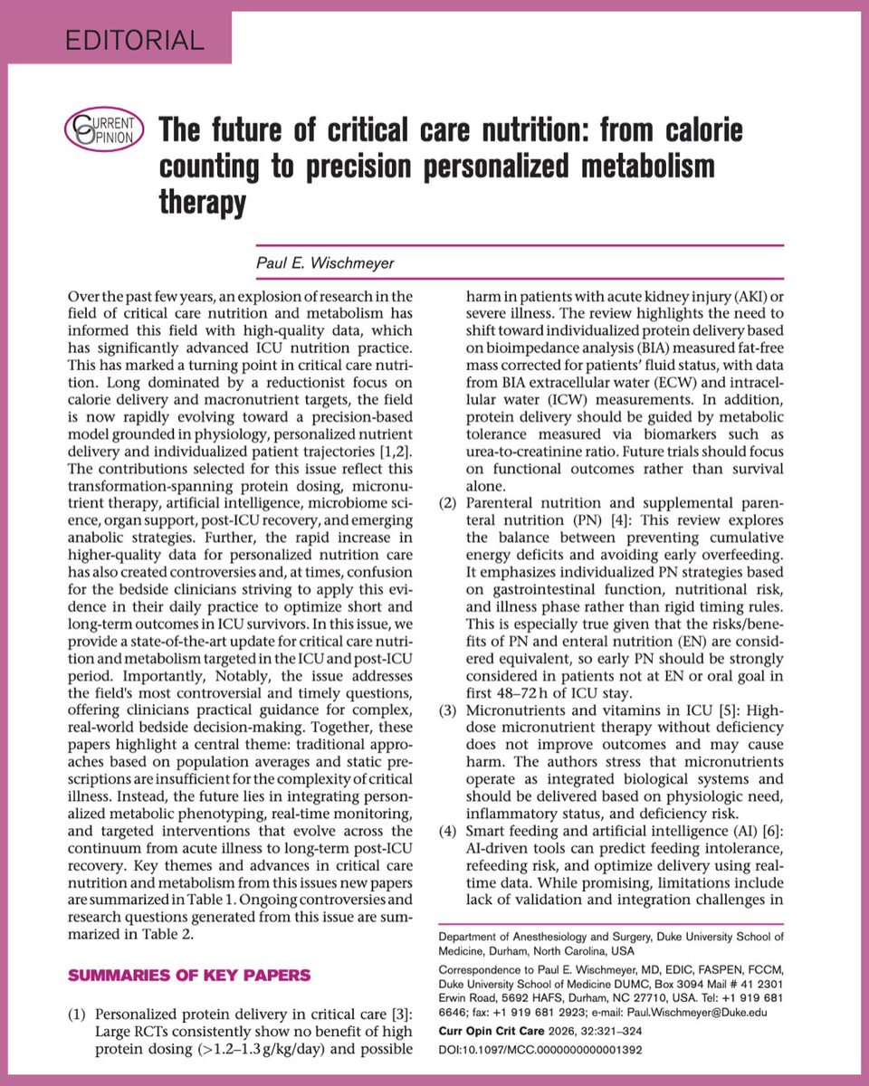
I’m honored to serve as Guest Editor for the new Nutrition & Metabolism issue of Current Opinion in Critical Care. This special issue brings together leading international experts to review the latest science transforming nutrition and metabolism in critically ill patients.
For decades, ICU nutrition focused on calories and protein targets. Today, the field is rapidly evolving toward precision, personalized metabolism therapy—using physiology, technology, and individualized interventions to improve both survival and long-term recovery.
The articles and topics in the issue are highlighted below and include:
🥩 Personalized Protein Delivery — Nienke Overwater, Marjolein Paulus, Linda Schepers & Arthur van Zanten @ArthurvanZanten
💉 Parenteral & Supplemental Parenteral Nutrition — Tam Lac, Lee-anne Chapple & Emma Ridley
@icunutrition
💊 Micronutrient Trials in Critical Illness — Renee Blaauw & Mette Berger
🤖 Smart Feeding: Artificial Intelligence & Integrated Nutrition Platforms — Pierre Singer & Oren Raphaeli
🦠 The Leaky Gut & Microbiome in Critical Illness — Natalie Daniels, Emily Wilson & Mara Serbanescu
🥣 Practical Nutrition (coming in the next issue) — Stephen McClave & Robert Martindale
🫀 Nutrition During ECMO & CRRT — Christian Stoppe, Stefano Marelli & Elisabeth De Waele @ElisabethWaele
🏃 Individualizing Post-ICU Nutrition (coming in the next issue) — Olivier Leeman, Zoë Rosseel, Claude Pichard, and colleagues
💪 Testosterone & Anabolic Therapy to Recover Strength, Function & Quality of Life After Critical Illness — Paul E. Wischmeyer
The central message is clear: critical care nutrition is no longer just about delivering calories—it’s about delivering the right metabolic therapy to the right patient at the right time.
A sincere thank you to all of these outstanding authors for their exceptional contributions to this special issue.
📖 Read the complete Nutrition & Metabolism issue here:
https://t.co/OBEupVIIXm
💬 Which topic do you think will have the greatest impact on improving outcomes in critical illness over the next decade? @Duke_Anesthesia@ICUnutrition
#ICUNutrition #Sccm #criticalcare #icurehab #nutritionmatters
@NutrioSci
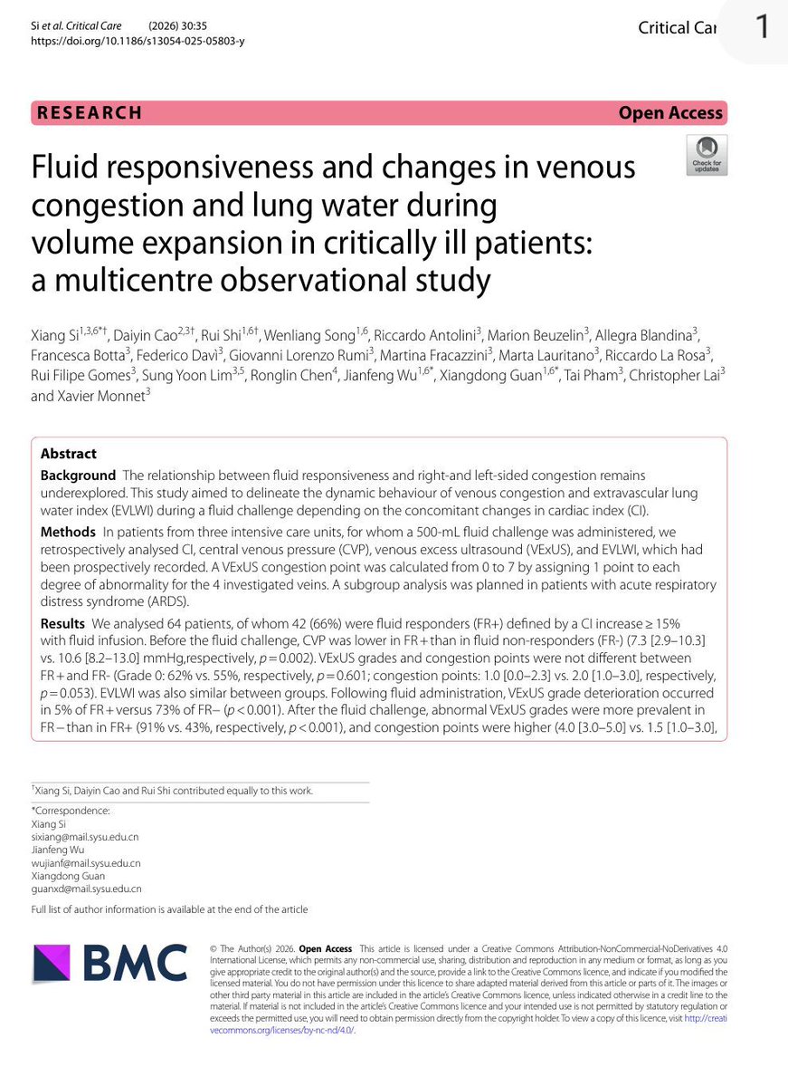
💧 Fluid Responsiveness ≠ Fluid Tolerance
For years, critical care clinicians have focused on a fundamental question:
Will this patient increase cardiac output if I give fluids?
A new multicentre study from France and China suggests we may need to ask a second question:
What price will the patient pay for that fluid?
In this observational study of 64 critically ill patients receiving a standardized 500 mL fluid challenge, investigators evaluated not only cardiac index (CI), but also venous congestion using VExUS and pulmonary congestion using extravascular lung water (EVLWI).
The findings are striking.
Before fluid administration, venous congestion could coexist with fluid responsiveness. In fact, many patients remained preload responsive despite already demonstrating evidence of systemic venous congestion.
However, what happened after fluid administration was even more important.
Among fluid responders:
✅ Cardiac index increased significantly
✅ Venous congestion remained largely stable
✅ Only 5% experienced worsening VExUS grade
✅ Lung water remained relatively unchanged
Among fluid non-responders:
❌ Cardiac output barely changed
❌ Venous congestion worsened dramatically
❌ VExUS deterioration occurred in 73%
❌ EVLWI increased significantly
❌ Congestion became evident across hepatic, portal, and renal venous territories
Perhaps the most important physiological message is this:
When the heart cannot convert preload into flow, the fluid has to go somewhere.
And where it goes is congestion.
The study also found a strong correlation between CVP and VExUS, suggesting that while advanced ultrasound provides valuable organ-level information, a carefully interpreted CVP may still remain a useful bedside marker of right-sided congestion.
The ARDS subgroup provides another important lesson.
Even fluid-responsive ARDS patients accumulated more extravascular lung water after fluid administration than non-ARDS patients, highlighting the role of pulmonary permeability in determining fluid tolerance.
This study reinforces a concept that is increasingly central to modern hemodynamic management:
The goal is not simply to identify fluid responsiveness.
The goal is to identify patients who are both:
✔ Fluid responsive
✔ Fluid tolerant
A fluid challenge that increases congestion without increasing flow is not resuscitation.
It is fluid accumulation.
The future of hemodynamic management may lie at the intersection of:
• Fluid responsiveness
• Venous congestion assessment
• Pulmonary permeability
• Organ-specific fluid tolerance
Because the best fluid is not the one that can be given.
It is the one that provides benefit without causing harm.
Reference , 📚
Si X, Critical Care. 2026;30:35.
Central Venous Pressure Revisited: Physiology, Pitfalls, Misconceptions, and Modern Clinical Interpretation in Critical Care
CCR Journal Watch
https://t.co/Sp06oA6IDG
Today's Paper of the Day is:
Heart-Lungs interactions: the basics and clinical implications
https://t.co/JKgcYjlUQ5
Join us to read 1 paper per day and stay up-to-date as we cover the spectrum of critical care across 2026
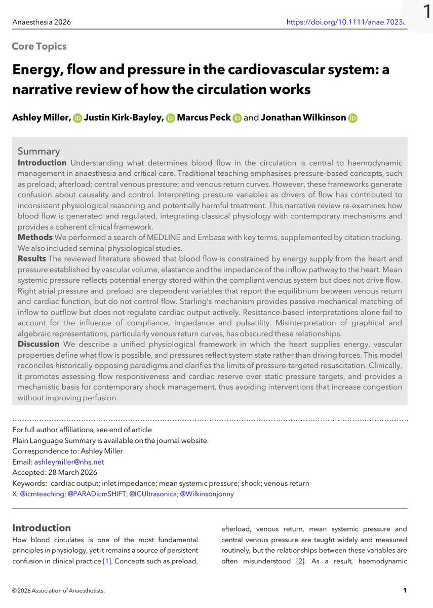
🫀🤓Pressure does not move blood. Energy does.
This outstanding review challenges one of the most deeply rooted concepts in haemodynamic management: the idea that pressure variables are the primary drivers of circulation. Instead, the authors propose a physiology framework where the heart supplies energy, the vasculature defines constraints, and pressures merely reflect system state.
Several concepts deserve special attention for critical care clinicians:
• Mean systemic pressure does not “drive” flow
• Right atrial pressure is a dependent variable, not a therapeutic target
• Venous return depends on inflow acceptance and inlet impedance
• Raising pressure without improving flow may worsen congestion
• Shock should be interpreted as either impaired venous delivery or impaired cardiac acceptance
Clinically, this framework helps explain why:
• CVP-guided fluid loading often fails
• Vasopressors may normalize MAP without restoring perfusion
• Congestion can coexist with preserved arterial pressure
• Flow responsiveness matters more than static pressure targets
One of the strongest messages of the paper is simple but powerful:
“Pressure is not perfusion.”
For intensivists, anesthesiologists, and cardiogenic shock teams, this review is worth reading in full. It reconnects bedside haemodynamics with first-principles physiology.
Miller A, Anaesthesia. 2026. https://t.co/ejjvREUe7c
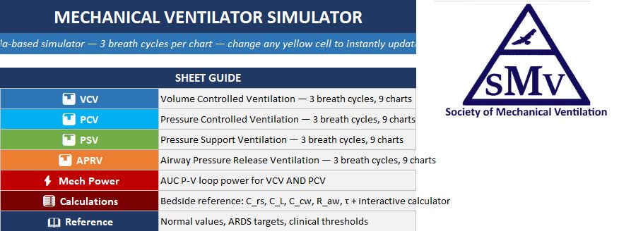
We are excited to share a mechanical ventilator simulator built in Excel! It models VC, PC, PS, APRV, and SIMV ventilation with real-time waveforms for airway pressure, flow, volume, esophageal pressure, transpulmonary pressure, and alveolar pressure.
https://t.co/Dw4u64UF8E


![MarlonVFZR's tweet photo. 🧵🥣⚠️ 𝗡𝘂𝘁𝗿𝗶𝗰𝗶𝗼́𝗻 𝗲𝗻 𝗨𝗖𝗜: 𝗲𝗻 𝗹𝗮 𝗳𝗮𝘀𝗲 𝗮𝗴𝘂𝗱𝗮, “𝗺𝗮́𝘀 𝗰𝗮𝗹𝗼𝗿𝗶́𝗮𝘀 𝘆 𝗺𝗮́𝘀 𝗽𝗿𝗼𝘁𝗲𝗶́𝗻𝗮” 𝗻𝗼 𝘀𝗶𝗴𝗻𝗶𝗳𝗶𝗰𝗮 𝗺𝗲𝗷𝗼𝗿 𝗿𝗲𝗰𝘂𝗽𝗲𝗿𝗮𝗰𝗶𝗼́𝗻 🚨
@NEJM
👇🏼👇🏼👇🏼👇🏼
📑🔗🔑🔓
https://t.co/89ht2mBRSA
⬇️⬇️⬇️⬇️
🧵👇
Review de 𝙉𝙀𝙅𝙈 𝟮𝟬𝟮𝟲 que cambia el enfoque clásico.
Durante los primeros días de enfermedad crítica predominan:
🔥 catabolismo intenso
🧬 inflamación
💪 pérdida muscular acelerada
🦠 disfunción intestinal
📉 resistencia anabólica
👉 La prioridad inicial no es “cubrir el 100%”, sino evitar sobrealimentación mientras se preserva la función intestinal.
🫁 𝙇𝙖 𝙫𝙞́𝙖 𝙚𝙣𝙩𝙚𝙧𝙖𝙡 𝙨𝙞𝙜𝙪𝙚 𝙨𝙞𝙚𝙣𝙙𝙤 𝙥𝙧𝙚𝙛𝙚𝙧𝙞𝙙𝙖… 𝙥𝙚𝙧𝙤 𝙡𝙖 𝙣𝙪𝙩𝙧𝙞𝙘𝙞𝙤́𝙣 𝙥𝙖𝙧𝙚𝙣𝙩𝙚𝙧𝙖𝙡 𝙩𝙚𝙢𝙥𝙧𝙖𝙣𝙖 𝙮𝙖 𝙣𝙤 𝙙𝙚𝙗𝙚 𝙫𝙚𝙧𝙨𝙚 𝙘𝙤𝙢𝙤 𝙪𝙣 𝙛𝙧𝙖𝙘𝙖𝙨𝙤
Cuando el tubo digestivo funciona, la nutrición enteral precoz —idealmente en 𝟮𝟰–𝟯𝟲 𝙝𝙤𝙧𝙖𝙨— ayuda a preservar:
✅ barrera intestinal
✅ arquitectura vellosa
✅ microbiota
✅ inmunidad mucosa
Pero grandes ensayos mostraron que la 𝙣𝙪𝙩𝙧𝙞𝙘𝙞𝙤́𝙣 𝙥𝙖𝙧𝙚𝙣𝙩𝙚𝙧𝙖𝙡 𝙩𝙚𝙢𝙥𝙧𝙖𝙣𝙖 𝙖 𝙘𝙤𝙧𝙩𝙤 𝙥𝙡𝙖𝙯𝙤 tiene mortalidad similar y no aumenta infecciones cuando la vía enteral está contraindicada o no es viable.
👉 Enteral primero, pero parenteral temprana es una alternativa segura en el paciente adecuado.
⚡ 𝙀𝙣 𝙡𝙖 𝙛𝙖𝙨𝙚 𝙖𝙜𝙪𝙙𝙖, 𝙚𝙡 𝙤𝙗𝙟𝙚𝙩𝙞𝙫𝙤 𝙙𝙚𝙗𝙚𝙧𝙞́𝙖 𝙨𝙚𝙧 𝙧𝙚𝙨𝙩𝙧𝙞𝙘𝙩𝙞𝙫𝙤, 𝙣𝙤 𝙘𝙤𝙢𝙥𝙡𝙚𝙩𝙤
Estrategia por fases:
🔹 𝙁𝙖𝙨𝙚 𝙖𝙜𝙪𝙙𝙖 𝙩𝙚𝙢𝙥𝙧𝙖𝙣𝙖: ~25% del objetivo energético
🔹 𝙁𝙖𝙨𝙚 𝙖𝙜𝙪𝙙𝙖 𝙩𝙖𝙧𝙙𝙞́𝙖: progresar gradualmente a 50–80%
🔹 𝙁𝙖𝙨𝙚 𝙙𝙚 𝙧𝙚𝙘𝙪𝙥𝙚𝙧𝙖𝙘𝙞𝙤́𝙣: alcanzar 80–100%
Los ensayos EDEN, PERMIT, TARGET y NUTRIREA-3 no demostraron beneficio del aporte completo precoz y sí encontraron más:
🤢 vómito y diarrea
💉 necesidad de insulina
⚠️ complicaciones gastrointestinales
🫀 riesgo de isquemia intestinal en shock
🔥 En UCI, alimentar agresivamente demasiado pronto puede añadir estrés metabólico al estrés de la enfermedad.
💪 𝙇𝙖 𝙥𝙧𝙤𝙩𝙚𝙞́𝙣𝙖 𝙚𝙣 𝙙𝙤𝙨𝙞𝙨 𝙖𝙡𝙩𝙖𝙨 𝙩𝙖𝙢𝙥𝙤𝙘𝙤 𝙢𝙚𝙟𝙤𝙧𝙤́ 𝙡𝙤𝙨 𝙙𝙚𝙨𝙚𝙣𝙡𝙖𝙘𝙚𝙨… 𝙮 𝙥𝙪𝙚𝙙𝙚 𝙥𝙚𝙧𝙟𝙪𝙙𝙞𝙘𝙖𝙧 𝙚𝙣 𝙡𝙚𝙨𝙞𝙤́𝙣 𝙧𝙚𝙣𝙖𝙡 𝙖𝙜𝙪𝙙𝙖
Los ensayos 𝙀𝙁𝙁𝙊𝙍𝙏 𝙋𝙧𝙤𝙩𝙚𝙞𝙣, 𝙋𝙍𝙀𝘾𝙄𝙎𝙀 𝙮 𝙏𝘼𝙍𝙂𝙀𝙏 𝙋𝙧𝙤𝙩𝙚𝙞𝙣 compararon dosis altas frente a estándar.
Resultado:
🚫 sin reducción de mortalidad
🚫 sin mayor número de días vivos fuera del hospital
🚫 sin mejor recuperación funcional consistente
⚠️ señales de daño en pacientes con 𝘼𝙆𝙄, mayor gravedad o nueva TRR
👉 La estrategia más razonable es avanzar gradualmente hacia aproximadamente 𝟭.𝟯 𝙜/𝙠𝙜/𝙙𝙞́𝙖, no iniciar de rutina con >2 g/kg/día durante la fase aguda.
🎯 𝙏𝙖𝙠𝙚-𝙝𝙤𝙢𝙚: 𝙡𝙖 𝙣𝙪𝙩𝙧𝙞𝙘𝙞𝙤́𝙣 𝙘𝙧𝙞́𝙩𝙞𝙘𝙖 𝙙𝙚𝙗𝙚 𝙨𝙚𝙧 𝙥𝙧𝙚𝙘𝙤𝙯, 𝙥𝙧𝙤𝙜𝙧𝙚𝙨𝙞𝙫𝙖 𝙮 𝙜𝙪𝙞𝙖𝙙𝙖 𝙥𝙤𝙧 𝙡𝙖 𝙛𝙖𝙨𝙚 𝙢𝙚𝙩𝙖𝙗𝙤́𝙡𝙞𝙘𝙖
Puntos prácticos:
✅ iniciar enteral precoz si es segura
✅ usar dosis tróficas de 𝟭𝟬–𝟮𝟬 𝙢𝙇/𝙝 al inicio
✅ evitar nutrición enteral completa con vasopresores altos
✅ mantener glucosa <𝟭𝟴𝟬 𝙢𝙜/𝙙𝙇
✅ prevenir realimentación con tiamina y vigilancia de P, Mg y K
🚫 evitar monitoreo rutinario del residuo gástrico
🔥 𝙈𝙚𝙣𝙨𝙖𝙟𝙚 𝙛𝙞𝙣𝙖𝙡: durante la fase aguda, la nutrición no debe perseguir déficits acumulados a cualquier costo. El objetivo es 𝙥𝙧𝙤𝙩𝙚𝙜𝙚𝙧 𝙞𝙣𝙩𝙚𝙨𝙩𝙞𝙣𝙤, 𝙚𝙫𝙞𝙩𝙖𝙧 𝙨𝙤𝙗𝙧𝙚𝙖𝙡𝙞𝙢𝙚𝙣𝙩𝙖𝙘𝙞𝙤́𝙣 𝙮 𝙖𝙫𝙖𝙣𝙯𝙖𝙧 𝙚𝙣𝙚𝙧𝙜𝙞́𝙖 𝙮 𝙥𝙧𝙤𝙩𝙚𝙞́𝙣𝙖 𝙖 𝙢𝙚𝙙𝙞𝙙𝙖 𝙦𝙪𝙚 𝙙𝙞𝙨𝙢𝙞𝙣𝙪𝙮𝙚𝙣 𝙞𝙣𝙛𝙡𝙖𝙢𝙖𝙘𝙞𝙤́𝙣 𝙮 𝙘𝙖𝙩𝙖𝙗𝙤𝙡𝙞𝙨𝙢𝙤.
𝙉𝙀𝙅𝙈 2026 | 𝘿𝙊𝙄: 𝟭𝟬.𝟭𝟬𝟱𝟲/𝙉𝙀𝙅𝙈𝙧𝙖𝟮𝟱𝟬𝟲𝟭𝟭𝟭
📚📖 Más en 𝕏 @MarlonVFZR y en el blog 👉 [https://t.co/kN4nAf4QOR]
‼️Si te sirve: ❤️ Me gusta | 🔁 Repost | ➕ Follow para más👇🏼👇🏼👇🏼👇🏼
📚📖#ClubCrit👨🏻⚕️👨🏻🏫🧠🫶
#NutriciónCrítica #NutriciónEnteral #Proteína #RefeedingSyndrome
#FOAMed #FOAMcc #CriticalCare #CriticalCare #CuidadoCritico #MedTwitter #CritCare #icu #intensivecare #diagnosis #management #MedicinaBasadaEnEvidencia #MedEd #MedX #IntensiveCare #MedIntensiva #MedXCommunity #MedED #ICUmanagement #MustRead #LecturaRecomendada](https://pbs.twimg.com/media/HNrEG2wXgAACDtH.jpg)
![MarlonVFZR's tweet photo. 🧵🥣⚠️ 𝗡𝘂𝘁𝗿𝗶𝗰𝗶𝗼́𝗻 𝗲𝗻 𝗨𝗖𝗜: 𝗲𝗻 𝗹𝗮 𝗳𝗮𝘀𝗲 𝗮𝗴𝘂𝗱𝗮, “𝗺𝗮́𝘀 𝗰𝗮𝗹𝗼𝗿𝗶́𝗮𝘀 𝘆 𝗺𝗮́𝘀 𝗽𝗿𝗼𝘁𝗲𝗶́𝗻𝗮” 𝗻𝗼 𝘀𝗶𝗴𝗻𝗶𝗳𝗶𝗰𝗮 𝗺𝗲𝗷𝗼𝗿 𝗿𝗲𝗰𝘂𝗽𝗲𝗿𝗮𝗰𝗶𝗼́𝗻 🚨
@NEJM
👇🏼👇🏼👇🏼👇🏼
📑🔗🔑🔓
https://t.co/89ht2mBRSA
⬇️⬇️⬇️⬇️
🧵👇
Review de 𝙉𝙀𝙅𝙈 𝟮𝟬𝟮𝟲 que cambia el enfoque clásico.
Durante los primeros días de enfermedad crítica predominan:
🔥 catabolismo intenso
🧬 inflamación
💪 pérdida muscular acelerada
🦠 disfunción intestinal
📉 resistencia anabólica
👉 La prioridad inicial no es “cubrir el 100%”, sino evitar sobrealimentación mientras se preserva la función intestinal.
🫁 𝙇𝙖 𝙫𝙞́𝙖 𝙚𝙣𝙩𝙚𝙧𝙖𝙡 𝙨𝙞𝙜𝙪𝙚 𝙨𝙞𝙚𝙣𝙙𝙤 𝙥𝙧𝙚𝙛𝙚𝙧𝙞𝙙𝙖… 𝙥𝙚𝙧𝙤 𝙡𝙖 𝙣𝙪𝙩𝙧𝙞𝙘𝙞𝙤́𝙣 𝙥𝙖𝙧𝙚𝙣𝙩𝙚𝙧𝙖𝙡 𝙩𝙚𝙢𝙥𝙧𝙖𝙣𝙖 𝙮𝙖 𝙣𝙤 𝙙𝙚𝙗𝙚 𝙫𝙚𝙧𝙨𝙚 𝙘𝙤𝙢𝙤 𝙪𝙣 𝙛𝙧𝙖𝙘𝙖𝙨𝙤
Cuando el tubo digestivo funciona, la nutrición enteral precoz —idealmente en 𝟮𝟰–𝟯𝟲 𝙝𝙤𝙧𝙖𝙨— ayuda a preservar:
✅ barrera intestinal
✅ arquitectura vellosa
✅ microbiota
✅ inmunidad mucosa
Pero grandes ensayos mostraron que la 𝙣𝙪𝙩𝙧𝙞𝙘𝙞𝙤́𝙣 𝙥𝙖𝙧𝙚𝙣𝙩𝙚𝙧𝙖𝙡 𝙩𝙚𝙢𝙥𝙧𝙖𝙣𝙖 𝙖 𝙘𝙤𝙧𝙩𝙤 𝙥𝙡𝙖𝙯𝙤 tiene mortalidad similar y no aumenta infecciones cuando la vía enteral está contraindicada o no es viable.
👉 Enteral primero, pero parenteral temprana es una alternativa segura en el paciente adecuado.
⚡ 𝙀𝙣 𝙡𝙖 𝙛𝙖𝙨𝙚 𝙖𝙜𝙪𝙙𝙖, 𝙚𝙡 𝙤𝙗𝙟𝙚𝙩𝙞𝙫𝙤 𝙙𝙚𝙗𝙚𝙧𝙞́𝙖 𝙨𝙚𝙧 𝙧𝙚𝙨𝙩𝙧𝙞𝙘𝙩𝙞𝙫𝙤, 𝙣𝙤 𝙘𝙤𝙢𝙥𝙡𝙚𝙩𝙤
Estrategia por fases:
🔹 𝙁𝙖𝙨𝙚 𝙖𝙜𝙪𝙙𝙖 𝙩𝙚𝙢𝙥𝙧𝙖𝙣𝙖: ~25% del objetivo energético
🔹 𝙁𝙖𝙨𝙚 𝙖𝙜𝙪𝙙𝙖 𝙩𝙖𝙧𝙙𝙞́𝙖: progresar gradualmente a 50–80%
🔹 𝙁𝙖𝙨𝙚 𝙙𝙚 𝙧𝙚𝙘𝙪𝙥𝙚𝙧𝙖𝙘𝙞𝙤́𝙣: alcanzar 80–100%
Los ensayos EDEN, PERMIT, TARGET y NUTRIREA-3 no demostraron beneficio del aporte completo precoz y sí encontraron más:
🤢 vómito y diarrea
💉 necesidad de insulina
⚠️ complicaciones gastrointestinales
🫀 riesgo de isquemia intestinal en shock
🔥 En UCI, alimentar agresivamente demasiado pronto puede añadir estrés metabólico al estrés de la enfermedad.
💪 𝙇𝙖 𝙥𝙧𝙤𝙩𝙚𝙞́𝙣𝙖 𝙚𝙣 𝙙𝙤𝙨𝙞𝙨 𝙖𝙡𝙩𝙖𝙨 𝙩𝙖𝙢𝙥𝙤𝙘𝙤 𝙢𝙚𝙟𝙤𝙧𝙤́ 𝙡𝙤𝙨 𝙙𝙚𝙨𝙚𝙣𝙡𝙖𝙘𝙚𝙨… 𝙮 𝙥𝙪𝙚𝙙𝙚 𝙥𝙚𝙧𝙟𝙪𝙙𝙞𝙘𝙖𝙧 𝙚𝙣 𝙡𝙚𝙨𝙞𝙤́𝙣 𝙧𝙚𝙣𝙖𝙡 𝙖𝙜𝙪𝙙𝙖
Los ensayos 𝙀𝙁𝙁𝙊𝙍𝙏 𝙋𝙧𝙤𝙩𝙚𝙞𝙣, 𝙋𝙍𝙀𝘾𝙄𝙎𝙀 𝙮 𝙏𝘼𝙍𝙂𝙀𝙏 𝙋𝙧𝙤𝙩𝙚𝙞𝙣 compararon dosis altas frente a estándar.
Resultado:
🚫 sin reducción de mortalidad
🚫 sin mayor número de días vivos fuera del hospital
🚫 sin mejor recuperación funcional consistente
⚠️ señales de daño en pacientes con 𝘼𝙆𝙄, mayor gravedad o nueva TRR
👉 La estrategia más razonable es avanzar gradualmente hacia aproximadamente 𝟭.𝟯 𝙜/𝙠𝙜/𝙙𝙞́𝙖, no iniciar de rutina con >2 g/kg/día durante la fase aguda.
🎯 𝙏𝙖𝙠𝙚-𝙝𝙤𝙢𝙚: 𝙡𝙖 𝙣𝙪𝙩𝙧𝙞𝙘𝙞𝙤́𝙣 𝙘𝙧𝙞́𝙩𝙞𝙘𝙖 𝙙𝙚𝙗𝙚 𝙨𝙚𝙧 𝙥𝙧𝙚𝙘𝙤𝙯, 𝙥𝙧𝙤𝙜𝙧𝙚𝙨𝙞𝙫𝙖 𝙮 𝙜𝙪𝙞𝙖𝙙𝙖 𝙥𝙤𝙧 𝙡𝙖 𝙛𝙖𝙨𝙚 𝙢𝙚𝙩𝙖𝙗𝙤́𝙡𝙞𝙘𝙖
Puntos prácticos:
✅ iniciar enteral precoz si es segura
✅ usar dosis tróficas de 𝟭𝟬–𝟮𝟬 𝙢𝙇/𝙝 al inicio
✅ evitar nutrición enteral completa con vasopresores altos
✅ mantener glucosa <𝟭𝟴𝟬 𝙢𝙜/𝙙𝙇
✅ prevenir realimentación con tiamina y vigilancia de P, Mg y K
🚫 evitar monitoreo rutinario del residuo gástrico
🔥 𝙈𝙚𝙣𝙨𝙖𝙟𝙚 𝙛𝙞𝙣𝙖𝙡: durante la fase aguda, la nutrición no debe perseguir déficits acumulados a cualquier costo. El objetivo es 𝙥𝙧𝙤𝙩𝙚𝙜𝙚𝙧 𝙞𝙣𝙩𝙚𝙨𝙩𝙞𝙣𝙤, 𝙚𝙫𝙞𝙩𝙖𝙧 𝙨𝙤𝙗𝙧𝙚𝙖𝙡𝙞𝙢𝙚𝙣𝙩𝙖𝙘𝙞𝙤́𝙣 𝙮 𝙖𝙫𝙖𝙣𝙯𝙖𝙧 𝙚𝙣𝙚𝙧𝙜𝙞́𝙖 𝙮 𝙥𝙧𝙤𝙩𝙚𝙞́𝙣𝙖 𝙖 𝙢𝙚𝙙𝙞𝙙𝙖 𝙦𝙪𝙚 𝙙𝙞𝙨𝙢𝙞𝙣𝙪𝙮𝙚𝙣 𝙞𝙣𝙛𝙡𝙖𝙢𝙖𝙘𝙞𝙤́𝙣 𝙮 𝙘𝙖𝙩𝙖𝙗𝙤𝙡𝙞𝙨𝙢𝙤.
𝙉𝙀𝙅𝙈 2026 | 𝘿𝙊𝙄: 𝟭𝟬.𝟭𝟬𝟱𝟲/𝙉𝙀𝙅𝙈𝙧𝙖𝟮𝟱𝟬𝟲𝟭𝟭𝟭
📚📖 Más en 𝕏 @MarlonVFZR y en el blog 👉 [https://t.co/kN4nAf4QOR]
‼️Si te sirve: ❤️ Me gusta | 🔁 Repost | ➕ Follow para más👇🏼👇🏼👇🏼👇🏼
📚📖#ClubCrit👨🏻⚕️👨🏻🏫🧠🫶
#NutriciónCrítica #NutriciónEnteral #Proteína #RefeedingSyndrome
#FOAMed #FOAMcc #CriticalCare #CriticalCare #CuidadoCritico #MedTwitter #CritCare #icu #intensivecare #diagnosis #management #MedicinaBasadaEnEvidencia #MedEd #MedX #IntensiveCare #MedIntensiva #MedXCommunity #MedED #ICUmanagement #MustRead #LecturaRecomendada](https://pbs.twimg.com/media/HNrEGzxXsAAHe1H.jpg)





![MarlonVFZR's tweet photo. 🧵🥣⚠️ 𝗡𝘂𝘁𝗿𝗶𝗰𝗶𝗼́𝗻 𝗲𝗻 𝗨𝗖𝗜: 𝗲𝗻 𝗹𝗮 𝗳𝗮𝘀𝗲 𝗮𝗴𝘂𝗱𝗮, “𝗺𝗮́𝘀 𝗰𝗮𝗹𝗼𝗿𝗶́𝗮𝘀 𝘆 𝗺𝗮́𝘀 𝗽𝗿𝗼𝘁𝗲𝗶́𝗻𝗮” 𝗻𝗼 𝘀𝗶𝗴𝗻𝗶𝗳𝗶𝗰𝗮 𝗺𝗲𝗷𝗼𝗿 𝗿𝗲𝗰𝘂𝗽𝗲𝗿𝗮𝗰𝗶𝗼́𝗻 🚨
@NEJM
👇🏼👇🏼👇🏼👇🏼
📑🔗🔑🔓
https://t.co/89ht2mBRSA
⬇️⬇️⬇️⬇️
🧵👇
Review de 𝙉𝙀𝙅𝙈 𝟮𝟬𝟮𝟲 que cambia el enfoque clásico.
Durante los primeros días de enfermedad crítica predominan:
🔥 catabolismo intenso
🧬 inflamación
💪 pérdida muscular acelerada
🦠 disfunción intestinal
📉 resistencia anabólica
👉 La prioridad inicial no es “cubrir el 100%”, sino evitar sobrealimentación mientras se preserva la función intestinal.
🫁 𝙇𝙖 𝙫𝙞́𝙖 𝙚𝙣𝙩𝙚𝙧𝙖𝙡 𝙨𝙞𝙜𝙪𝙚 𝙨𝙞𝙚𝙣𝙙𝙤 𝙥𝙧𝙚𝙛𝙚𝙧𝙞𝙙𝙖… 𝙥𝙚𝙧𝙤 𝙡𝙖 𝙣𝙪𝙩𝙧𝙞𝙘𝙞𝙤́𝙣 𝙥𝙖𝙧𝙚𝙣𝙩𝙚𝙧𝙖𝙡 𝙩𝙚𝙢𝙥𝙧𝙖𝙣𝙖 𝙮𝙖 𝙣𝙤 𝙙𝙚𝙗𝙚 𝙫𝙚𝙧𝙨𝙚 𝙘𝙤𝙢𝙤 𝙪𝙣 𝙛𝙧𝙖𝙘𝙖𝙨𝙤
Cuando el tubo digestivo funciona, la nutrición enteral precoz —idealmente en 𝟮𝟰–𝟯𝟲 𝙝𝙤𝙧𝙖𝙨— ayuda a preservar:
✅ barrera intestinal
✅ arquitectura vellosa
✅ microbiota
✅ inmunidad mucosa
Pero grandes ensayos mostraron que la 𝙣𝙪𝙩𝙧𝙞𝙘𝙞𝙤́𝙣 𝙥𝙖𝙧𝙚𝙣𝙩𝙚𝙧𝙖𝙡 𝙩𝙚𝙢𝙥𝙧𝙖𝙣𝙖 𝙖 𝙘𝙤𝙧𝙩𝙤 𝙥𝙡𝙖𝙯𝙤 tiene mortalidad similar y no aumenta infecciones cuando la vía enteral está contraindicada o no es viable.
👉 Enteral primero, pero parenteral temprana es una alternativa segura en el paciente adecuado.
⚡ 𝙀𝙣 𝙡𝙖 𝙛𝙖𝙨𝙚 𝙖𝙜𝙪𝙙𝙖, 𝙚𝙡 𝙤𝙗𝙟𝙚𝙩𝙞𝙫𝙤 𝙙𝙚𝙗𝙚𝙧𝙞́𝙖 𝙨𝙚𝙧 𝙧𝙚𝙨𝙩𝙧𝙞𝙘𝙩𝙞𝙫𝙤, 𝙣𝙤 𝙘𝙤𝙢𝙥𝙡𝙚𝙩𝙤
Estrategia por fases:
🔹 𝙁𝙖𝙨𝙚 𝙖𝙜𝙪𝙙𝙖 𝙩𝙚𝙢𝙥𝙧𝙖𝙣𝙖: ~25% del objetivo energético
🔹 𝙁𝙖𝙨𝙚 𝙖𝙜𝙪𝙙𝙖 𝙩𝙖𝙧𝙙𝙞́𝙖: progresar gradualmente a 50–80%
🔹 𝙁𝙖𝙨𝙚 𝙙𝙚 𝙧𝙚𝙘𝙪𝙥𝙚𝙧𝙖𝙘𝙞𝙤́𝙣: alcanzar 80–100%
Los ensayos EDEN, PERMIT, TARGET y NUTRIREA-3 no demostraron beneficio del aporte completo precoz y sí encontraron más:
🤢 vómito y diarrea
💉 necesidad de insulina
⚠️ complicaciones gastrointestinales
🫀 riesgo de isquemia intestinal en shock
🔥 En UCI, alimentar agresivamente demasiado pronto puede añadir estrés metabólico al estrés de la enfermedad.
💪 𝙇𝙖 𝙥𝙧𝙤𝙩𝙚𝙞́𝙣𝙖 𝙚𝙣 𝙙𝙤𝙨𝙞𝙨 𝙖𝙡𝙩𝙖𝙨 𝙩𝙖𝙢𝙥𝙤𝙘𝙤 𝙢𝙚𝙟𝙤𝙧𝙤́ 𝙡𝙤𝙨 𝙙𝙚𝙨𝙚𝙣𝙡𝙖𝙘𝙚𝙨… 𝙮 𝙥𝙪𝙚𝙙𝙚 𝙥𝙚𝙧𝙟𝙪𝙙𝙞𝙘𝙖𝙧 𝙚𝙣 𝙡𝙚𝙨𝙞𝙤́𝙣 𝙧𝙚𝙣𝙖𝙡 𝙖𝙜𝙪𝙙𝙖
Los ensayos 𝙀𝙁𝙁𝙊𝙍𝙏 𝙋𝙧𝙤𝙩𝙚𝙞𝙣, 𝙋𝙍𝙀𝘾𝙄𝙎𝙀 𝙮 𝙏𝘼𝙍𝙂𝙀𝙏 𝙋𝙧𝙤𝙩𝙚𝙞𝙣 compararon dosis altas frente a estándar.
Resultado:
🚫 sin reducción de mortalidad
🚫 sin mayor número de días vivos fuera del hospital
🚫 sin mejor recuperación funcional consistente
⚠️ señales de daño en pacientes con 𝘼𝙆𝙄, mayor gravedad o nueva TRR
👉 La estrategia más razonable es avanzar gradualmente hacia aproximadamente 𝟭.𝟯 𝙜/𝙠𝙜/𝙙𝙞́𝙖, no iniciar de rutina con >2 g/kg/día durante la fase aguda.
🎯 𝙏𝙖𝙠𝙚-𝙝𝙤𝙢𝙚: 𝙡𝙖 𝙣𝙪𝙩𝙧𝙞𝙘𝙞𝙤́𝙣 𝙘𝙧𝙞́𝙩𝙞𝙘𝙖 𝙙𝙚𝙗𝙚 𝙨𝙚𝙧 𝙥𝙧𝙚𝙘𝙤𝙯, 𝙥𝙧𝙤𝙜𝙧𝙚𝙨𝙞𝙫𝙖 𝙮 𝙜𝙪𝙞𝙖𝙙𝙖 𝙥𝙤𝙧 𝙡𝙖 𝙛𝙖𝙨𝙚 𝙢𝙚𝙩𝙖𝙗𝙤́𝙡𝙞𝙘𝙖
Puntos prácticos:
✅ iniciar enteral precoz si es segura
✅ usar dosis tróficas de 𝟭𝟬–𝟮𝟬 𝙢𝙇/𝙝 al inicio
✅ evitar nutrición enteral completa con vasopresores altos
✅ mantener glucosa <𝟭𝟴𝟬 𝙢𝙜/𝙙𝙇
✅ prevenir realimentación con tiamina y vigilancia de P, Mg y K
🚫 evitar monitoreo rutinario del residuo gástrico
🔥 𝙈𝙚𝙣𝙨𝙖𝙟𝙚 𝙛𝙞𝙣𝙖𝙡: durante la fase aguda, la nutrición no debe perseguir déficits acumulados a cualquier costo. El objetivo es 𝙥𝙧𝙤𝙩𝙚𝙜𝙚𝙧 𝙞𝙣𝙩𝙚𝙨𝙩𝙞𝙣𝙤, 𝙚𝙫𝙞𝙩𝙖𝙧 𝙨𝙤𝙗𝙧𝙚𝙖𝙡𝙞𝙢𝙚𝙣𝙩𝙖𝙘𝙞𝙤́𝙣 𝙮 𝙖𝙫𝙖𝙣𝙯𝙖𝙧 𝙚𝙣𝙚𝙧𝙜𝙞́𝙖 𝙮 𝙥𝙧𝙤𝙩𝙚𝙞́𝙣𝙖 𝙖 𝙢𝙚𝙙𝙞𝙙𝙖 𝙦𝙪𝙚 𝙙𝙞𝙨𝙢𝙞𝙣𝙪𝙮𝙚𝙣 𝙞𝙣𝙛𝙡𝙖𝙢𝙖𝙘𝙞𝙤́𝙣 𝙮 𝙘𝙖𝙩𝙖𝙗𝙤𝙡𝙞𝙨𝙢𝙤.
𝙉𝙀𝙅𝙈 2026 | 𝘿𝙊𝙄: 𝟭𝟬.𝟭𝟬𝟱𝟲/𝙉𝙀𝙅𝙈𝙧𝙖𝟮𝟱𝟬𝟲𝟭𝟭𝟭
📚📖 Más en 𝕏 @MarlonVFZR y en el blog 👉 [https://t.co/kN4nAf4QOR]
‼️Si te sirve: ❤️ Me gusta | 🔁 Repost | ➕ Follow para más👇🏼👇🏼👇🏼👇🏼
📚📖#ClubCrit👨🏻⚕️👨🏻🏫🧠🫶
#NutriciónCrítica #NutriciónEnteral #Proteína #RefeedingSyndrome
#FOAMed #FOAMcc #CriticalCare #CriticalCare #CuidadoCritico #MedTwitter #CritCare #icu #intensivecare #diagnosis #management #MedicinaBasadaEnEvidencia #MedEd #MedX #IntensiveCare #MedIntensiva #MedXCommunity #MedED #ICUmanagement #MustRead #LecturaRecomendada](https://pbs.twimg.com/media/HNrEG3IWQAAJjJx.jpg)


![JAMA_current's tweet photo. 📣 JAMA Clinical Guidelines Synopsis: The 2026 #Dyslipidemia guideline by @ACCinTouch and @American_Heart recommends earlier risk assessment, lifetime risk estimation, and measurement of new #biomarkers, including apolipoprotein B and lipoprotein(a) [Lp(a)], for individualized #atherosclerotic #cardiovascular disease (#ASCVD) prevention.
https://t.co/aLCpves3mt](https://pbs.twimg.com/media/HNrsX8mWUAADaJ6.jpg)