#Sketchpose is out in @MELBAJournal !
Go read it if you are interested in training cell segmentation models using few partial annotations😀
https://t.co/MchVtWrKgM
Thrilled to share MyoFuse, an AI-based workflow for automated skeletal muscle cell fusion quantification! 🥳
This is a collaboration with @Benjamin_Lair_ and @CedricMoro_I2MC (@I2mcT), improving the Fusion Index (FI) measurement.
Preprint: https://t.co/XHtCbpoT4U
After a long journey, Segment Anything for Microscopy is now published in Nature Methods! We significantly improve SAM for interactive and automatic segmentation in light and electron microscopy and build a user-friendly tool. https://t.co/HL19SGMdNt
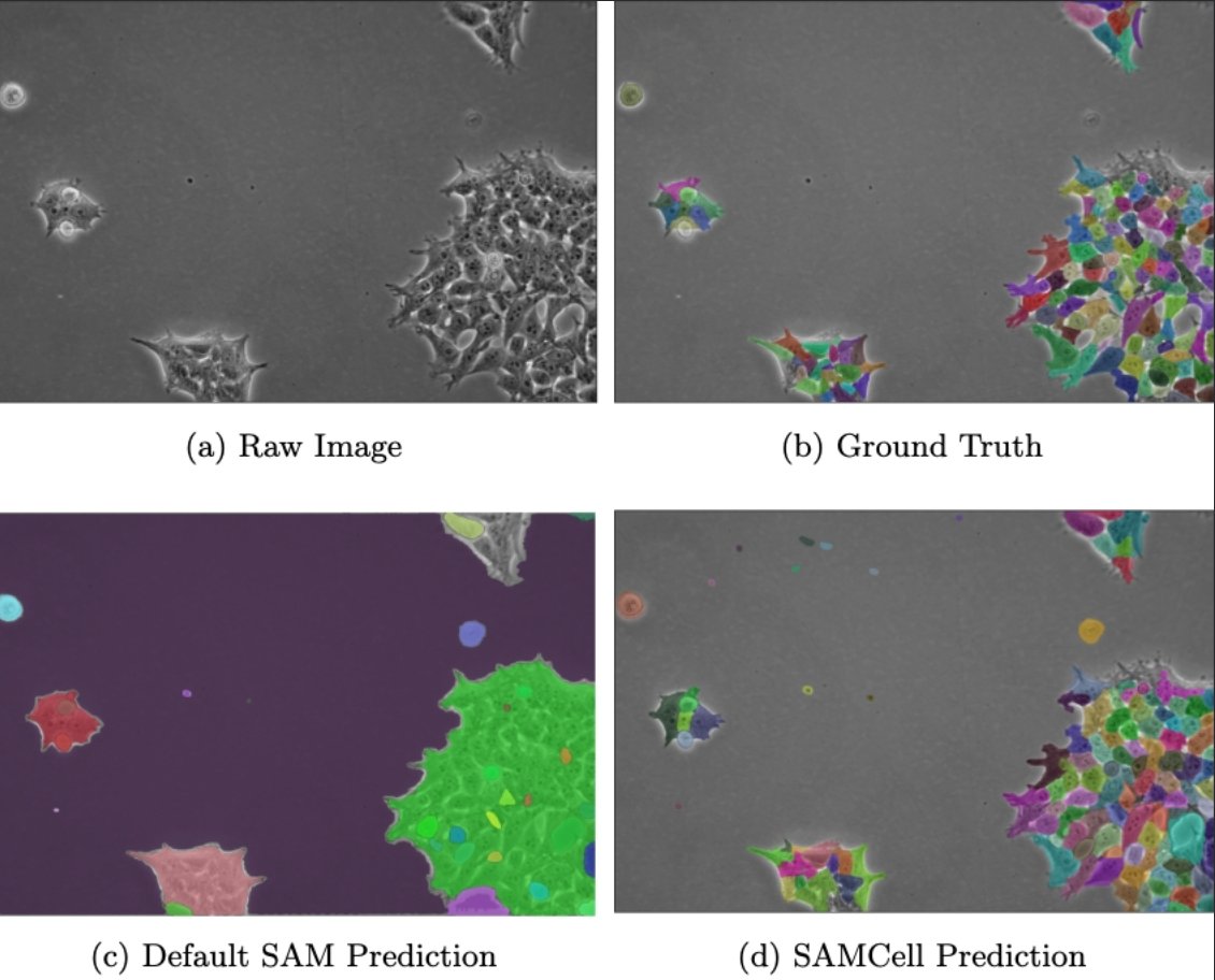
SAMCell: Generalized Label-Free Biological Cell Segmentation with Segment Anything
Alexandra Dunnum VandeLoo, Nathan J. Malta, Emilio Aponte, Caitlin van Zyl, Danfei Xu and Craig Forest
https://t.co/AYYaos9iqp
Researchers at @Imactiv3D and @maths_toulouse aim to streamline the classification of #histopathology segmentation results by minimizing the amount of manual annotation. 🖊️
Learn about the plugin they've created to address this issue in @SciReports: https://t.co/kVazRNr1wg
Last year, we found that diffusion models are nearly bridging the gap between human and machine (diversity vs. recognizability) for drawing https://t.co/ubRUCqxKe4.
This year, we’re exploring which inductive bias best captures human-like creativity in VAE latents 🎨
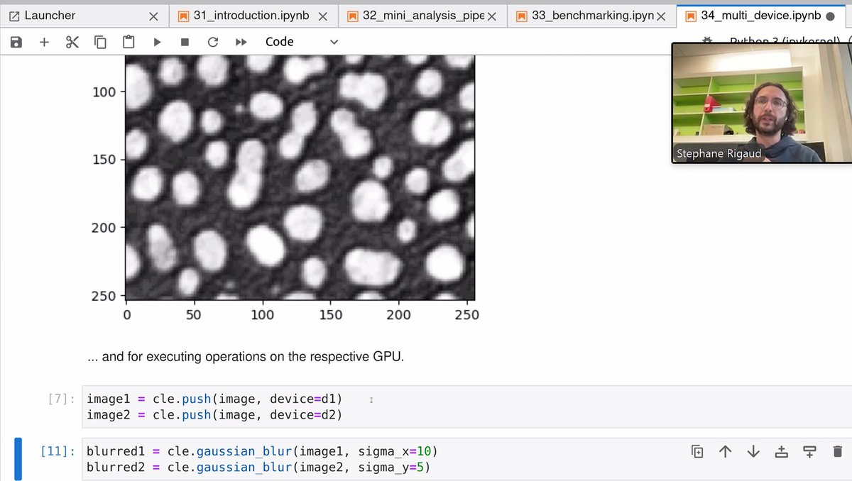
I just uploaded my slides for the webinar about #BioImageAnalysis Code Generation at #I2K2024 - #openaccess. See you in an hour! 🔬🖥️🤖🔓
https://t.co/R9TI8J7b9B
🚀 Thrilled to announce #inTRACKtive: a web-based tool for exploring massive cell-tracking datasets, no software installation required! Just open your browser and dive into terabytes of developmental biology data. We used it to build the virtual https://t.co/ZpTn6MvKtF of tracked embryonic development datasets 🐭🪰🪱🪲🐠, but you can also use it for your own data! 🔬
Preprint:
https://t.co/AGUek2Adom
Repository:
https://t.co/FWT0xixJmv
This project was spear-headed by @TeunHuijben, together with engineers Andrew Sweet and @aganders3 from @cziscience
@czbbiohub #CZBiohubSF #devbio #tracking #web #visualization
🧵1/n
Our Napari plugin Svetlana (https://t.co/Fe75ZCEc2h) for classifying ROIs in segmentation masks has improved. Try it!
1) The unbalance between classes is now managed better.
2) An interactive loss plotting is available in the GUI.
3) An automatic check-for-updates.
I implemented an oldie-but-goodie for Jupyter Notebooks: Interactive plotting and clustering with segmented images side-by-side; new in #stackview 0.7.11: "clusterplot", inspired by the napari-clusters-plotter 🔬💻🤩
https://t.co/l8tFmW2L9u
If you're not already following @Christophe21664, you definitely should! 😉
I'm super proud of this work, and there's more on the way. In case you're curious, Chris created these images without diffusion—just pure gradient ascent on a classifier. A true magician 🐇
So happy to see how far @napari_imaging has gone! The repo was created with @jnuneziglesias on the Caltrain on the way to the @ComputerHistory Museum. Fast forward 6 (!) years and napari is an essential tool for bioimaging and the Python Science ecosystem. Thanks to a thriving and tireless community! Congrats to all contributors, past and present! #SoProud