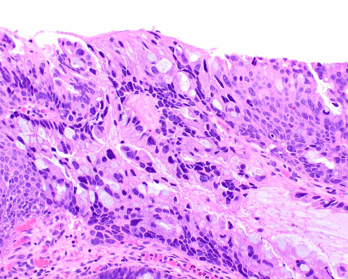
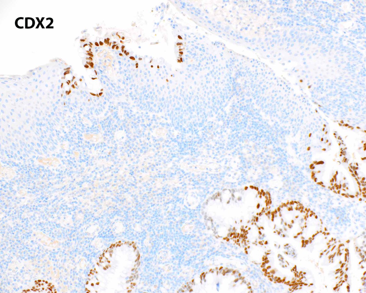
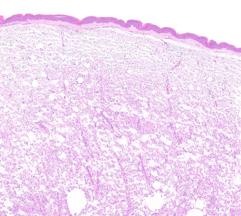
Case Record of the Massachusetts General Hospital (@MassGeneralNews): A 42-year-old pregnant woman presented to the labor and delivery triage unit of the hospital at 20 weeks’ gestation because of recurrent heavy vaginal bleeding. ���� https://t.co/rALCDjoMYt
Macroscopic examination showed a post-trachelectomy gravid hysterectomy specimen (weighing 487 g) with an in situ placenta (measuring 8.5 cm by 7.4 cm) with greater than 90% adherence to the posterior endometrial surface (Panel A). On sectioning, the placenta invaded the myometrium predominantly in the posterior wall (to a depth of up to 0.3 cm in a 2.6-cm wall) and extended to the lower uterine segment (Panel B). On microscopic examination, there were areas with no decidua between the chorionic villi of the placenta and the myometrium, a finding consistent with placental invasion (Panels C and D). Grading of placenta accreta spectrum disorder is based on the extent of invasion and destruction of local tissues. Given the presence of placental invasion with preservation of more than 25% of the total myometrial wall thickness, the criteria were met for placenta accreta spectrum disorder of grade 1–2.
Learn more about the disorder and read more about the case in “A 42-Year-Old Pregnant Woman with Vaginal Hemorrhage,” a Case Record of the Massachusetts General Hospital, by M. Prabhu et al.: https://t.co/rALCDjoMYt
Carcinoid tumors arising in the ovary can be primary, of germ cell origin, but it is important to exclude metastatic low-grade neuroendocrine tumor
This distinction may be aided by molecular testing for loss of heterozygosity (LOH)
Teratomas (and mucinous ovarian neoplasms derived from teratomas) usually show extensive loss of heterozygosity reflecting their germ cell derivation
LOH is likely to be used in the future to help distinguish between primary and metastatic carcinoid tumor of the ovary in those cases where there is uncertainty.
Dr. Gilks. Gynecologic Pathology Masterclass
#USCAP #pathology #pathX
Atypical necroinflammatory interstitial nephritis can be seen in SLE patients. This lesion is similar to lesion seen lymph nodes of KFD patients https://t.co/cxK1eqhAvN
Don’t miss the next Richard Byers Pathology Teaching Series webinar with Diagnexia.
Topic: Haematopathology
Speaker: Richard Byers
Date: October 28 2026
Time: 8am EST | 12pm GMT | 2pm Lusaka | 3pm Nairobi
Access: https://t.co/oP770F0bV4
Earn 1 CPD point. See you there!
#PathologyEducation #Haematopathology
Submandibular gland mass excision in 64 years old male
#ENTPath#PathX
Diagnosis?
Different Patterns in all 4 images…
Comedo, Cribriform, Cystic, Solid.
#PathResidents#Pathologists
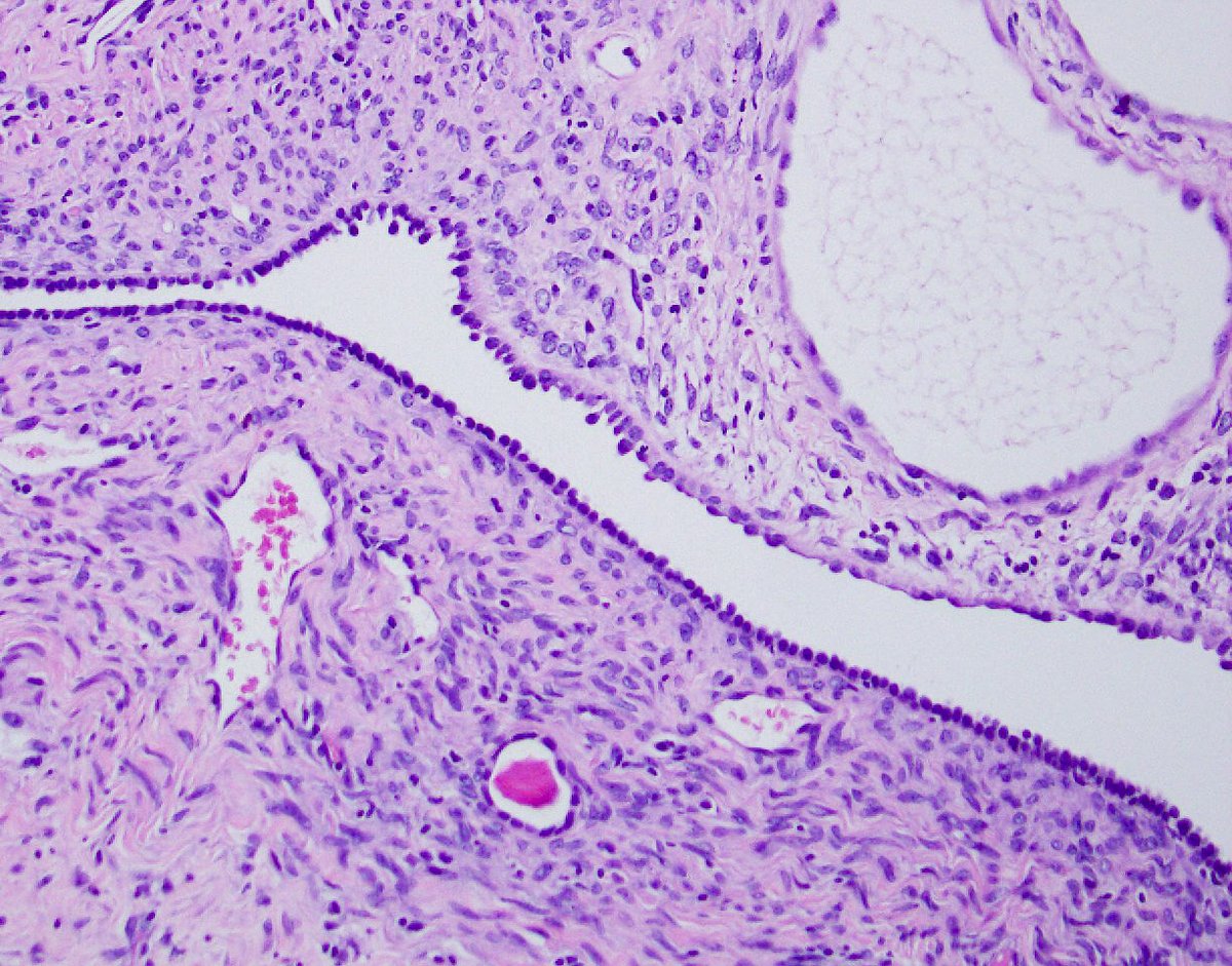
Mixed epithelial and stromal tumor (MEST)/ Adult cystic nephroma.
Adult cystic nephroma is usually more cystic; MEST has more prominent ovarian-type stroma.
Always inspect the stroma: synovial sarcoma may entrap and dilate native tubules, mimicking MEST. 😜
#DxMindMap #KidneyPath #GUPath #PathTwitter #Mimicker @MGBpathology
🔎Happening 10:00am today at #ASCyto26! @IvanaKholova leads Companion Session 4: Cytomorphology – The Clue for Unexpected Diagnosis
See Dr. Kholová's recent work on Navigating the International System for Reporting Serous Fluid Cytopathology:
https://t.co/RGdczd7nJI
#CytoPath
#publicationalert#gupath
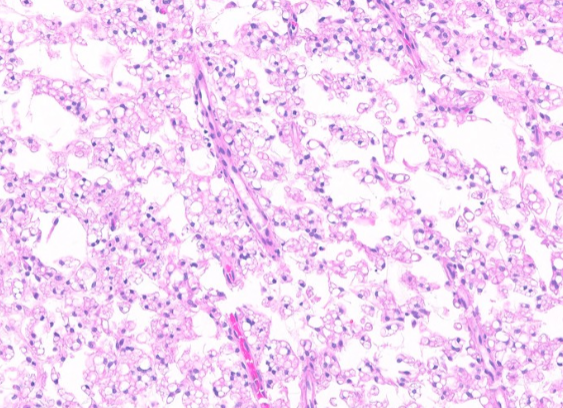
In this multi-institutional study of 51 EVTs, CD56 was positive in 98% of cases, supporting its utility as an adjunct diagnostic marker.
Thank you to the @patholojenMD and @slusagar for including me in this collaborative effort!
Tip of the Week - New-in-Practice Pathologists
63-year-old woman with a 2-year history of painful, burning plaques on the buttocks, refractory to multiple prior treatments
ASDP Interactive Case Study (Dr. Yu et al.). Posted by Dr. Smith, see link
https://t.co/oZUZOlR4LU
#NIPCAP #pathology #dermpath
🧬 Are we underestimating rare prostate cancer histologies simply because we rarely look for them?
https://t.co/wZS6mtThct
Rare prostate cancer histologies: detection, characteristics and treatment
Rare #ProstateCancer histologies comprise less than 5% of cases but represent a clinically important and under-recognized group.
➡️These subtypes often show aggressive behaviour, atypical metastatic patterns and poorer outcomes, while remaining underrepresented in guidelines and trials.
➡️Accurate diagnosis increasingly depends on multiparametric MRI, immunohistochemistry and genomic profiling.
➡️Molecular alterations such as DNA🧬 repair defects, TP53/RB1 loss and mismatch repair deficiency may offer therapeutic opportunities, highlighting the need for subtype-specific evidence and prospective studies.
@bavilima , @UrogerliMD, @katcollmd, @neerajaiims, @PGrivasMDPhD, @Albert0Briganti, @AndreaNecchi, @LiangChengMD, @SpiessPhilippe@Oncoalert 🚨 #LPCCC27
@Silke_Gillessen@AOmlin@ProfKHerrmann@weoncologists
Virchows Archiv reports on eosinophilic vacuolated tumors (EVTs) in kidneys, highlighting the diagnostic role of CD56 immunohistochemistry. The study analyzed 51 EVTs and found CD56 expressed in 98% of cases, aiding differentiation from other tumors.
https://t.co/EtqHPim0S7
🍞 Sign out with me 😉: Daily cases series
A middle-aged woman with a cystic mass in the renal pelvis.
The clinical history may give away the diagnosis.
What would you consider?
#DxMindMap#KidneyPath#GUPath#PathTwitter@MGBpathology
The December 2026 issue of Histopathology@Histo_Journal is out! Honored to have had the opportunity to collaborate and contribute three articles to this issue. Congratulations to all the authors on their excellent work!
🔹 Perivascular epithelioid cell tumours of the genitourinary tract: clinicopathological features and molecular landscape
https://t.co/SjBmnrz2KM https://t.co/ZDMPf8vZ3t
🔹 Histological assessment of treatment effect in lymph node-positive breast cancer after neoadjuvant chemotherapy
https://t.co/DJNpzZTIrV https://t.co/Q2ADx3RAMV
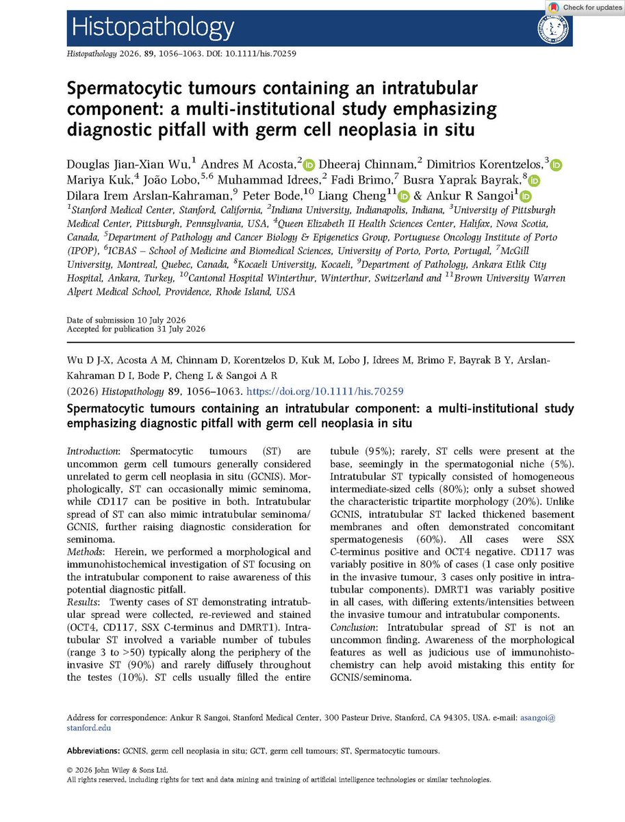
🔹 Spermatocytic tumours containing an intratubular component: a multi-institutional study emphasizing diagnostic pitfall with germ cell neoplasia in situ
https://t.co/DJNpzZTIrV
https://t.co/HQudnvKKut
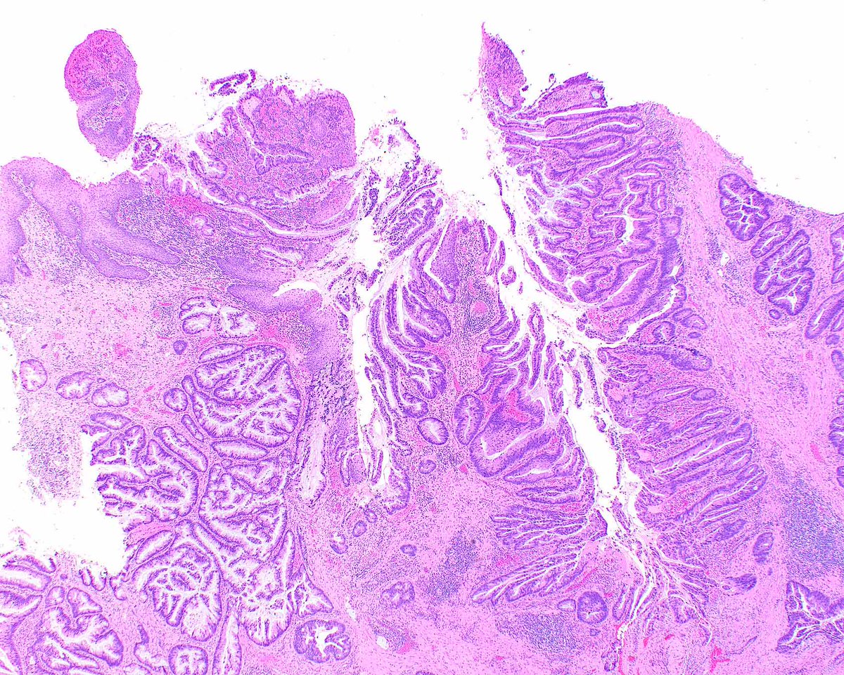
Primary perianal adenocarcinoma with pagetoid intra-epithelial extension - these look a bit like HPV-associated lesions, lack HPV transcripts, and have TP53, ERBB2, and MYC abnormalities, without typical rectal adenocarcinoma mutations (APC, KRAS, BRAF). https://t.co/ajb2kPFkBI
Histological and molecular characterization of lateral extension in papillary urothelial carcinoma: implications for classification and management #gupath https://t.co/FLc5jtzyAb