Pain biomarkers can't prove or disprove someone's pain, but that doesn't make them futile. My comment in Nat Neurosci argues for a pluralistic, bio-psycho-social approach: biomarkers as complementary channels, not replacements for patient voice. https://t.co/W6BfghvVN3
This week, please join us for a talk by @DiegoPizzagalli titled, "Translational and Cross-Species Approaches to Anhedonia: Implications for Treatment Development and Stratification." Wed, June 3, 1:30pm EST at the usual zoom link. DM your email if not on our list for the link.
How does your brain filter out noise and focus enough to learn efficiently?
Our newest preprint uses intracranial recordings + computational modeling to explore how selective attention shapes learning and state representations in the human brain 🧠
https://t.co/5UdiULuAHR
1/🧵
New paper in Imaging Neuroscience by Andrew N. Van, Nico U.F. Dosenbach, et al:
Frame-wise multi-echo distortion correction for superior functional MRI
https://t.co/1VNpk0HTFj
Is resting-state fMRI a good proxy for anatomy? Site-seeded rsFC only weakly correlated with es-fMRI connectivity (median r=0.20). Two sites in the same canonical resting-state network can have distinct anatomical connections. rsFC tracks co-fluctuated activity, not wiring. 12/n
New preprint: "Monosynaptic connections link functionally similar regions in human cortex." We use electrical stimulation + fMRI in epilepsy patients to map whole-brain monosynaptic connectivity at 42 cortical sites. https://t.co/rSSSVsMWCQ 1/n
Lesion network mapping (LNM) has been powerful in linking symptoms and brain functional circuits, but ongoing debates highlight that it is still hard to isolate symptom-specific effects. We came up with a new method, robust LNM (rLNM) — a unified framework combining null models and selective specificity to reveal reliable, symptom-specific networks from background structure. https://t.co/6WHpBRNuQn
@bttyeo@foxmdphd@ndosenbach@club_scan
I'm proud we are releasing LAION-fMRI, a densely sampled 7T fMRI dataset of natural images, with very broad stimulus sampling for testing countless hypotheses & deeply exploring brain representations. It is now available at
https://t.co/hOnILHonf9
What does LAION-fMRI offer? 🧵
Finally, today is the day: Josefine Zerbe will present and release our new multi-echo 7T fMRI dataset LAION-fMRI during #VSS2026, with >30 fMRI session per subject and unprecedented stimulus diversity. Come to Talk Room 1 (Scene perception) today at 5:15. Details after the talk!
🧠 How can personalized brain imaging help advance more targeted treatments for #BipolarDisorder?
Excited to share more about our work with @BD2Discoveries. Our team at @WeillCornell (@immanuel_elbau, Lindsay Victoria, @amykooz), in collaboration with @StanfordMed (Cammie Rolle, @DrCoreyKeller), is using individualized brain imaging to better understand brain circuits involved in mood regulation and to inform more precise stimulation-based interventions.
Grateful to be part of this broader effort to move toward more personalized approaches to bipolar disorder care.
A move toward precision psychiatry: Charles Lynch, PhD and his BD² Discovery team at @WeillCornell, in collaboration with @StanfordMed, are using personalized brain imaging to tailor targeted interventions for #bipolardisorder.
New in Brain Stimulation: Real-time E-field neuronavigation on realistic head models for conventional & multi-locus TMS!
Ana M. Soto et al. present a robust framework accounting for cortical folding.
Full text: https://t.co/6d4ctgufNk
#TMS#Neuroscience#BrainStimulation
📊BD² data release: the largest multi‑modal psychiatric dataset. Built across 6 sites with shared standards—aligned protocols, defined data practices & rigorous QA—so it’s ready to power new questions and collaborations. 🔗 https://t.co/oiKLJhDVKV
I’m excited to share this work from an amazing team @StanfordBrain: @mystrypercy who co-led the study, Zhiyao Gao, Weidong Cai, and Vinod Menon.
https://t.co/k9yMTldiBr [2/12]
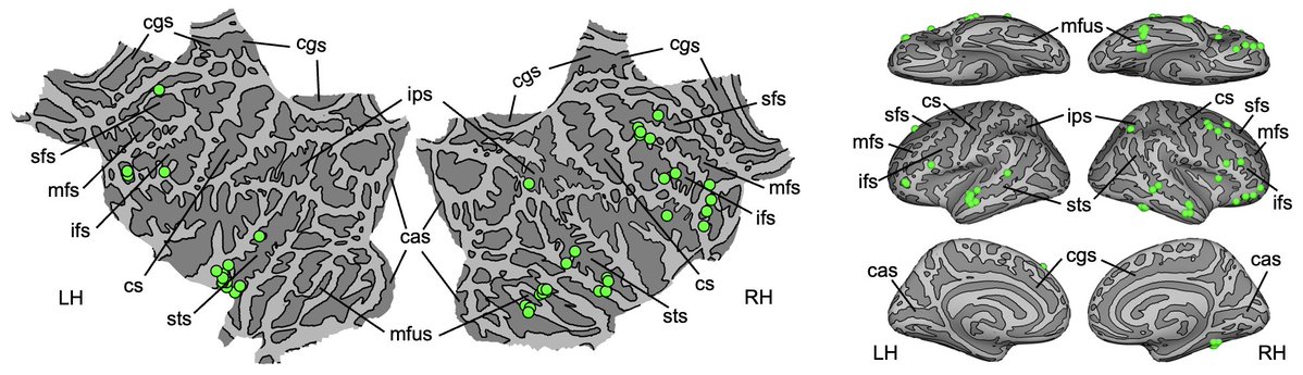
Association cortex connected to the striatum (esp. PFC) is more chained (catenated) than parietal regions belonging to the DMN (e.g. angular gyrus) and context-association network (CAN), with FC to the hippocampus. Why? what's the processing difference?
https://t.co/V7PQ5DtPK1
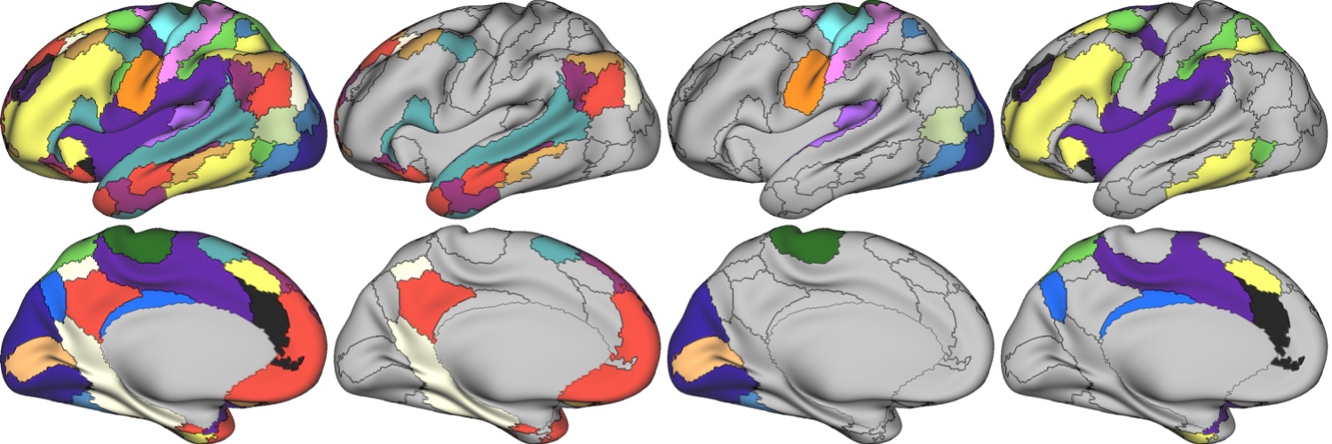
Function & cytoarchitecture don't overlap ... they're orthogonal. Prefrontal cortex is tiled with chains of functional patches mostly known from face processing. Multi-modal parcellations are wrong ... & other insights hidden by group-averaging fMRI data: https://t.co/WEo2Cf9N26