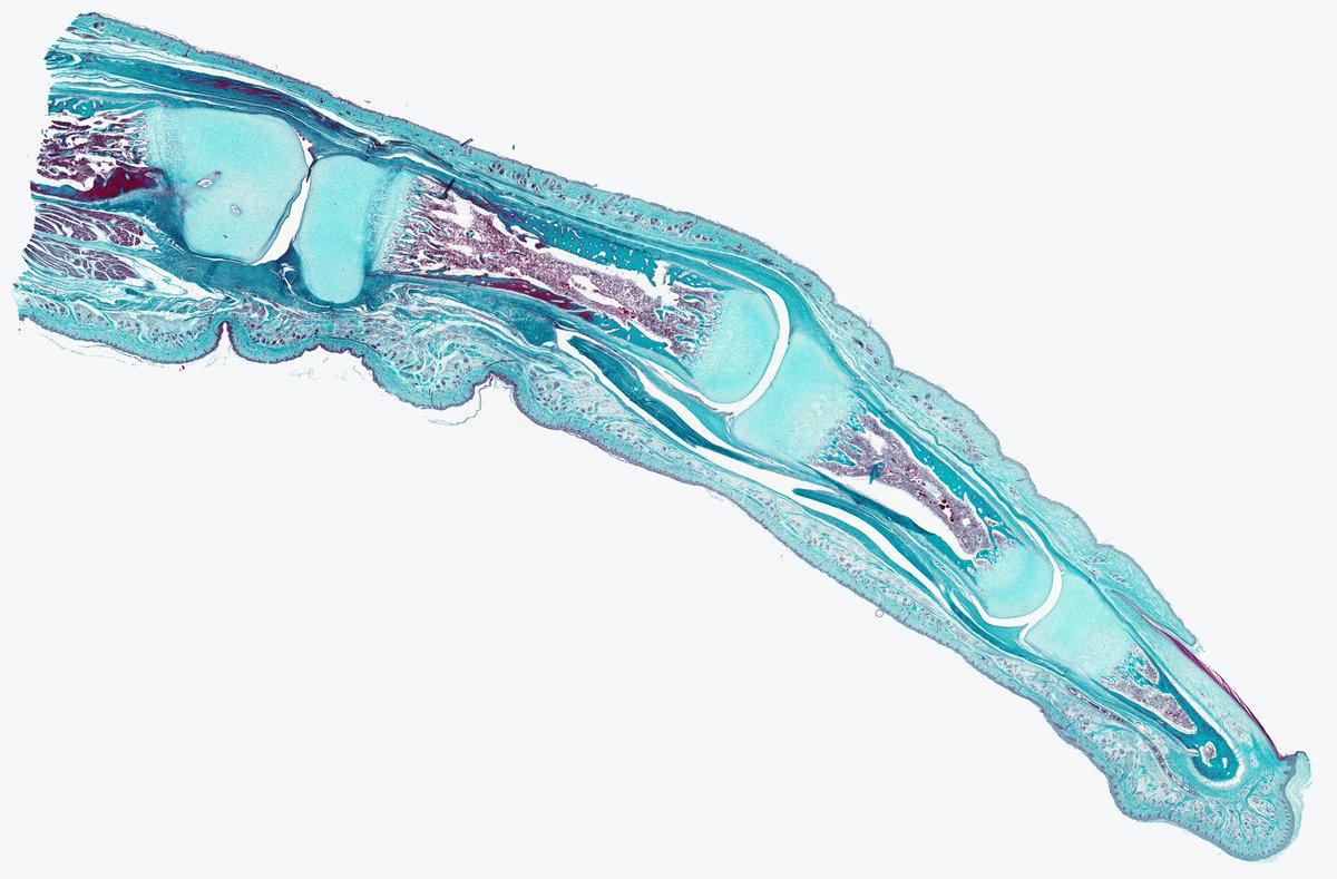
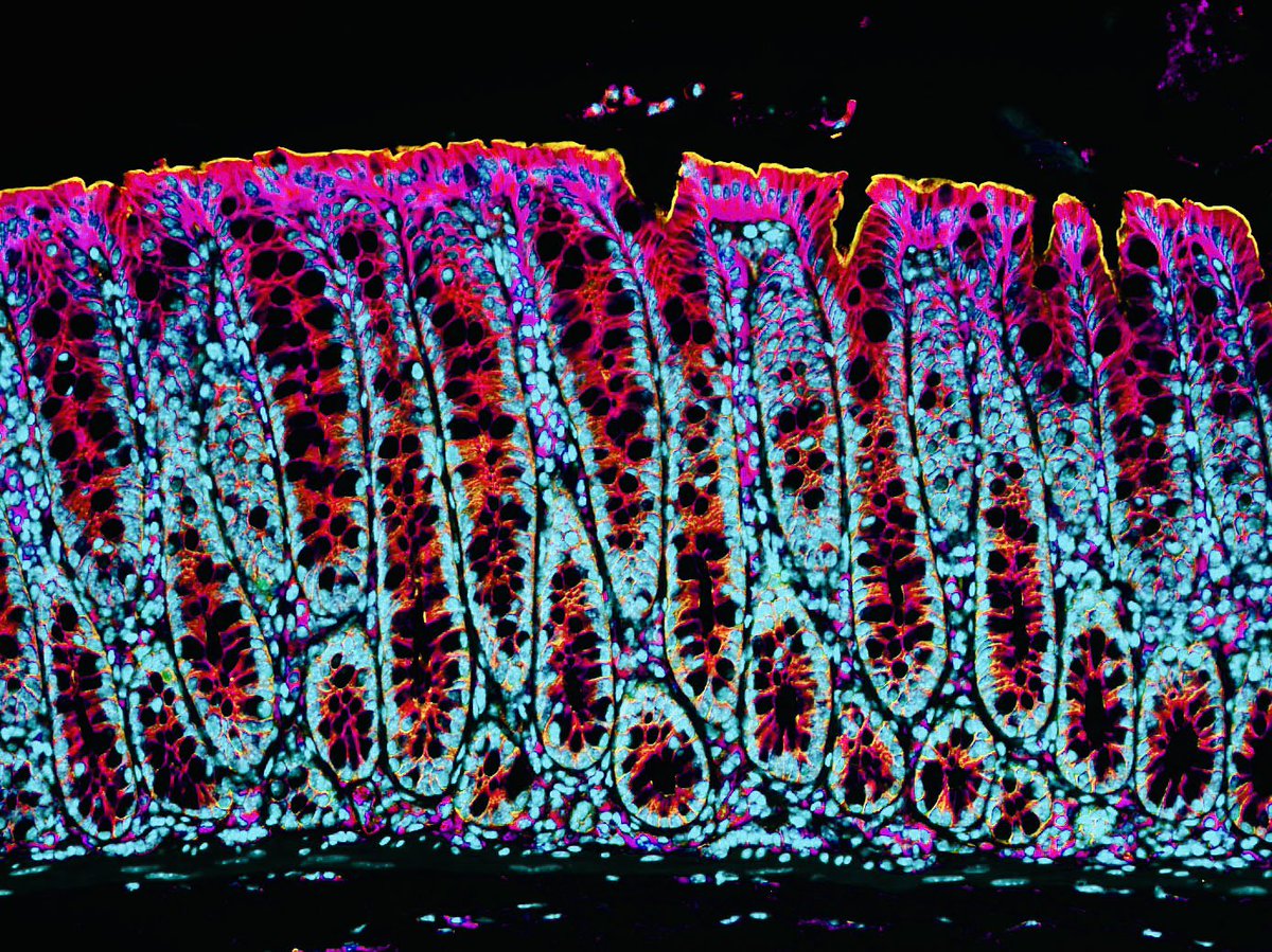
I am using the 1 month break between the end of my #PhD and start of my #PostDoc to put together some resource for the #Drosophila community especially egg #development enthusiasts. Here is a sneak peek. 10 hrs imaging of stage 11-14 #Drosophila egg
For #FluorescenceFriday, we love this #lightsheet image from our @zeiss_micro Z.1! Installed 2 wks before shutdown, now it's popular. Beautiful E9.5 mouse embryo with vascular perfusion from Linh Trinh in @MagnusonMark Lab. Expert imaging with CISR's Nick Mignemi. Scale=200um.
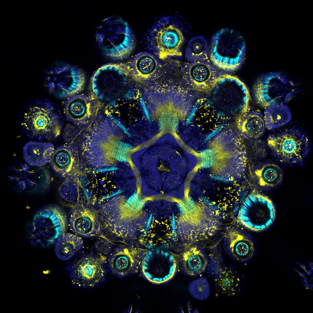
Hundreds of beautiful #microscope images were submitted to this year's Global Image of the Year Award competition. Here's a glimpse at a few of the images that received honorable mentions by our panel of jurors.
Click here to see all winning images - https://t.co/cEsBgLiipm
Happy Monday! Does anyone else see balloons in this picture? These are actually individually labeled axons in an embryonic chick ciliary ganglion. #MondayFunday
https://t.co/pPkUFAnzud
Cred: Dr. Ryo Egawa
Here, we present you a solution to image whole mouse spinal cord from sub-micron to sub hundred nanometer spatial resolution using tiling light sheet microscopy and tissue clearing techniques.
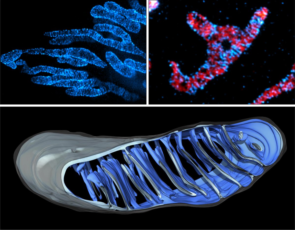
Bonus content! I don't think this movie made the final paper, but I still think it's one of my favorite examples of a metaphase mitochondrion pushed around by an actin comet tail.
cyan hot = mito
orange glow = actin (lifeact)
Loop ~40sec.
More here:
https://t.co/3yhC91anbx
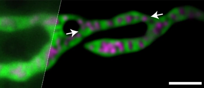
We are super excited to present our newest #publication in the @embojournal : "MICOS assembly controls mitochondrial inner membrane remodeling and crista junction redistribution to mediate cristae formation" https://t.co/mvpGTtfa8g
#mitochondria#superresolution#EM
Out today from Dong Li's lab, a deep learning approach that uses frequency content information in the Fourier domain to improve SIM reconstruction under low-SNR conditions. And an accompanying News and Views from @davephoffman! https://t.co/Mld5qqt5qY https://t.co/yv2J9lsUyT