"ترمب استنتج أن إيران تشكّل تهديدًا وشيكًا".. مديرة الاستخبارات الأميركية تقول إن ترمب هو المسؤول عن تحديد من يمثّل خطرًا محتملًا على البلاد وليست الاستخبارات وهو ما
ما دفع إلى شنّ حملة عسكرية للقضاء على التهديد النووي الإيراني
رئيس أكبر دولة ادعت يوما الديمقراطية وحرية الصحافة ،أسكت صحفي ل #cnn بالقوة الذي افحمه بأسئلة واقعية ومحرجة
_هكذا هو المعتوه #ترامب الدكتاتور يريد أسئلة على مقاسه وتخدمه
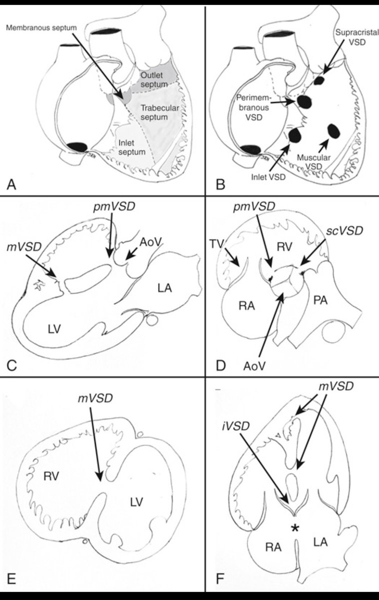
Ventricular Septal Defects (VSDs) Explained! 🫀
The ventricular septum is divided into four key regions: inlet, trabecular, outlet, and membranous. A VSD is an abnormal opening in this septum, allowing blood to shunt between the LV & RV.
🔍 Types of VSDs & Their Locations:
📌 Perimembranous (pmVSD): Below the aortic valve, near the membranous septum
📌 Muscular (mVSD): Located in the trabecular septum; can be multiple (Swiss-cheese VSD)
📌 Inlet (iVSD): Near the AV valves, often linked to endocardial cushion defects
📌 Supracristal (scVSD): Just below the pulmonic valve, may lead to aortic regurgitation
How Do We Diagnose VSDs?
Echocardiography plays a key role! Different echo views help identify VSDs:
🔹 Parasternal long-axis → Best for pmVSD & mVSD
🔹 Parasternal short-axis → Locates pmVSD (10 o’clock) & scVSD (1 o’clock)
🔹 Apical 4-chamber → Ideal for detecting inlet & apical muscular VSDs
Why does this matter?
VSD size, location, and associated defects determine hemodynamic impact & management! Some close spontaneously, while others require surgical or percutaneous closure.
Understanding VSDs = Better diagnosis & treatment!
#Cardiology #VSD #ECHOFacts #HeartDefects #MedTwitter
STEMI re-look. US doppler showed radial occlusion.
Angiogram followed by radial thrombectomy via radial sheath.
Thrombus protuding after aspiration then captured.
We present the oldest living (age, 93 years) asymptomatic patient with marked Apical Hypertrophic Cardiomyopathy (ApHCM).
Check out the @CASEfromASE article, "Never Too Old: A Nonagenarian With Apical Hypertrophic Cardiomyopathy!" https://t.co/qjvbiFZcEY #echofirst