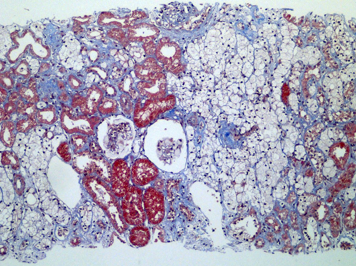
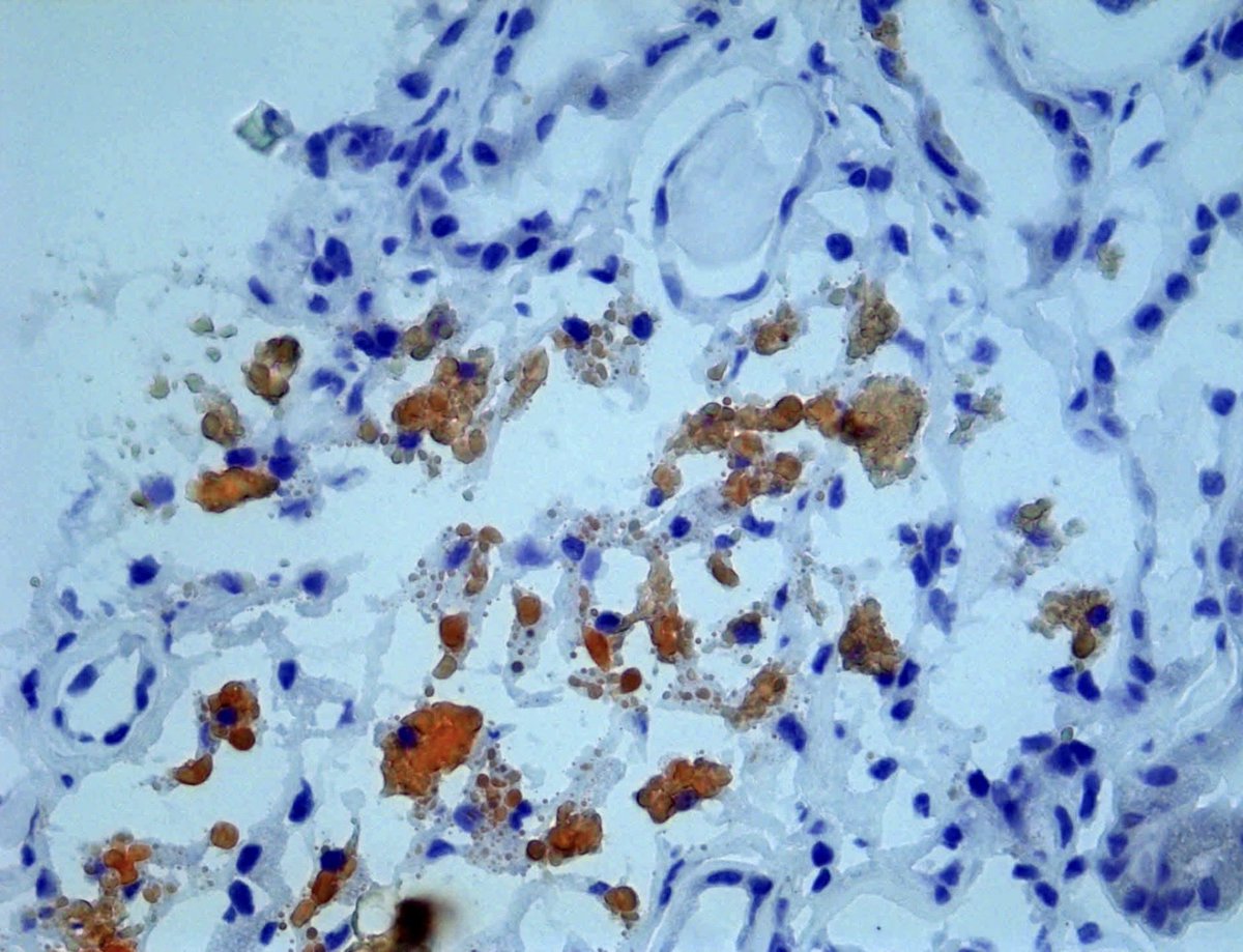
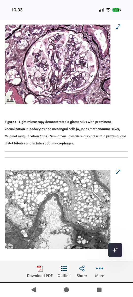
@grace_choung Here in our laboratory, we’ve seen the same changes in two patients who were also receiving pegcetacoplan. We performed Oil Red O staining and found lipid positivity. We had started writing up our cases for publication, but hadn’t finished the manuscript yet.
Tough case.
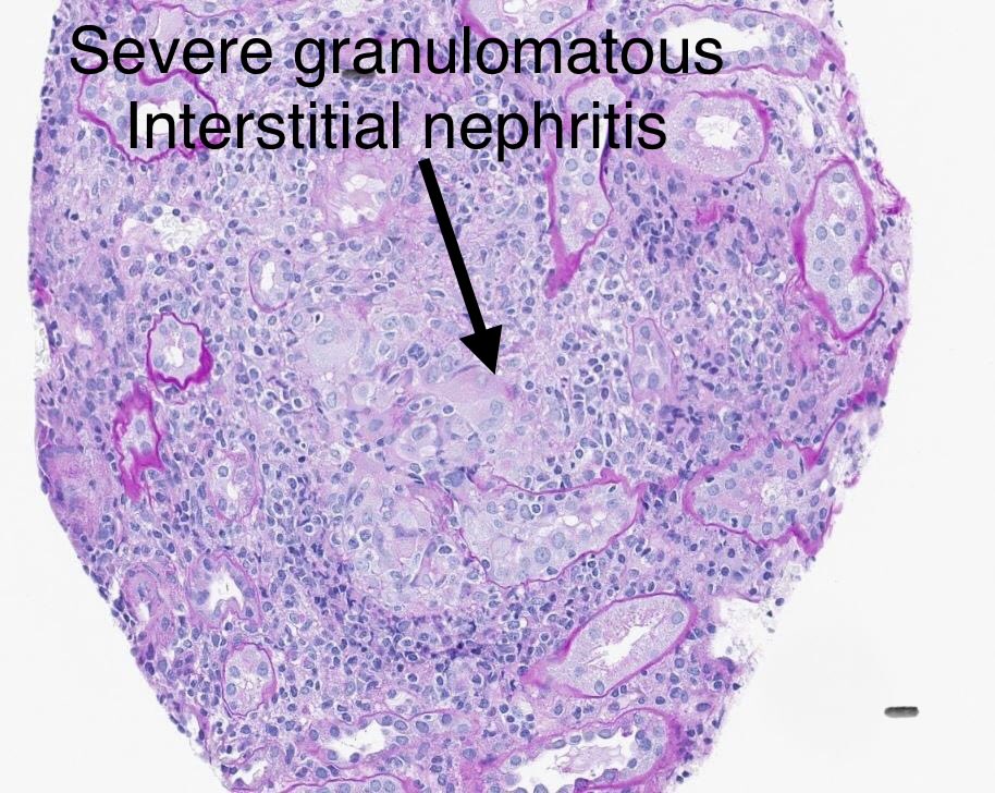
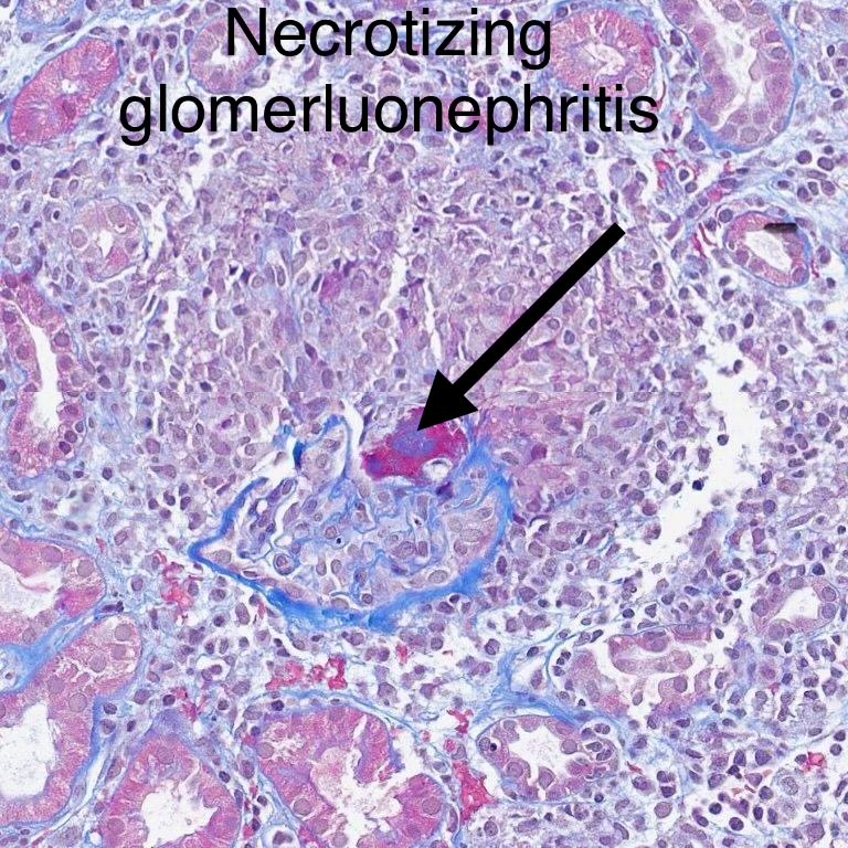
LM: granulomatous interstitial nephritis, extensive.
IF: negative.
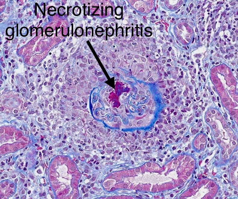
Look carefully and you start seeing small foci of necrosis in glomeruli, surrounded by inflammation/rupture of Bowman’s capsule.
Dx: 1. Granulomatous interstitial nephritis. 2. Necrotizing glomerulonephritis, pauci-immune. (both possibly drug related).
30-yr woman with rash, serum creatinine 10, bipolar disorder-just started treatment. ANCA, other serology negative.
Pathological Diagnostic Criteria for Thrombotic Microangiopathy in the Renal Allograft: Lessons Learned During the Banff Working Group Consensus Process - Kidney Medicine https://t.co/U3wxIUbUxH
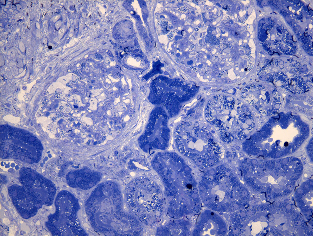
Enjoy these amazing images from this Toluidine blue stain #renalpath#electronmicroscopy
Courtesy of @hawkssox (first time I've ever seen an actual case of this entity)
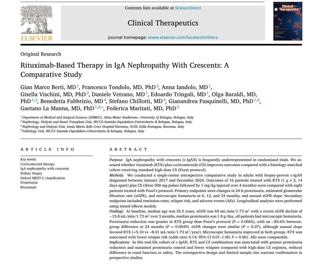
Re-considering Rituximab efficacy in IgA Nephropathy: a new role in patients with crescents?
We explored RTX-based therapy in #IgAN with crescents, showing encouraging signals in proteinuria reduction and relapse prevention.
Great teamwork @Unibo
https://t.co/1IZVlZaHg1
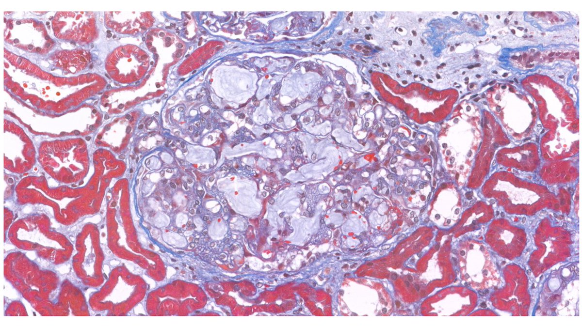
Our teaching case highlights a novel entity of renal toxicity after combination chemotherapy, emphasizing the diagnostic value of kidney biopsy and clinicopathologic correlation.
@gisellavischini @UniversityofBologna
https://t.co/1nbMK4FS7B
@KidneyMed
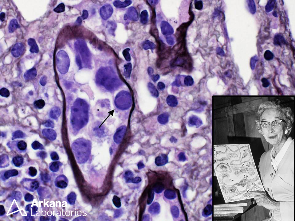
The arrow points to an intranuclear viral inclusion characteristic of BK virus, one species of non-enveloped dsDNA viruses belonging to the polyoma virus family. Sarah Stewart (see inset), a physician and research scientist, studied viral oncogenesis (she was the first woman to earn an M.D. degree from Georgetown Medical School). Dr. Stewart and her collaborator, Dr. Bernice Eddy (Ph.D. virologist), renamed the parotid tumor virus originally discovered by Ludwik Gross “SE polyoma virus” (the “SE” was for “Stewart-Eddy”). Interestingly, Dr. Stewart like the name “omnioma” virus, but Eddy suggested the term “polyoma” virus since not all viral infections caused tumors in all tested animals. The detailed history of this virus family and its discoverers is chronicled in a fascinating article by Gregory J. Morgan (see below).
References
Morgan GJ. Ludwik Gross, Sarah Stewart, and the 1950s discoveries of Gross murine leukemia virus and polyoma virus. Stud Hist Philos Biol Biomed Sci. 2014 Dec;48 Pt B:200-9. PMID: 25223721.
Photograph of Dr. Sarah Stewart, 1971. Public domain (National Library of Medicine)
#TeachingPoints #kidneypath #renal #pathology
We're excited to share that @Tiff_Caza, Dr. Frank Boyd, and Dr. Patrick Walker are featured collaborators on a new research publication titled, "Clinical and histologic predictors of non-diabetic kidney disease in patients with diabetes mellitus."
Read the publication here: https://t.co/xYpHiEdPbT
#renalresearch #kidneyresearch #nephtwitter #pathtwitter #diabetes
Acellular scanning EM from a biopsy with membranous glomerulonephritis, stage I showing numerous small shallow depressions representing the site of immune complexes which were extracted corresponding to “pinholes” viewed on silver stain.
Photo courtesy of Dr. Stephen Bonsib. #renal #nephropathology
A sample from the pathology teaching slide set on Lupus Nephritis, jointly prepared by Arkana Laboratories and @goKDIGO. https://t.co/KwXMfV3nTh
#KDIGO#pathology#renal#kidneypath