David Sinclair, el científico líder en longevidad, tiene un mensaje claro:
El envejecimiento es una enfermedad y la principal causa de cáncer, enfermedades cardíacas y Alzheimer.
Creó un protocolo para revertir el envejecimiento entre 8 y 10 años.
Aquí tienes cómo aplicarlo:🧵
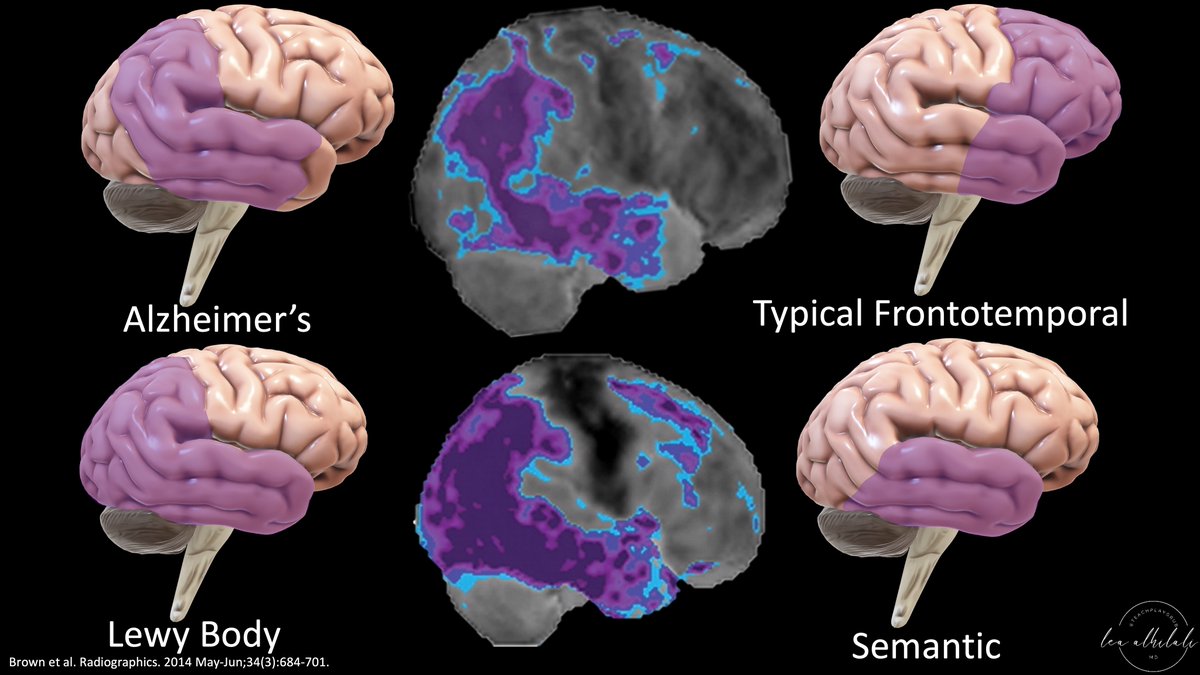
1/Having trouble remembering how to differentiate dementias on imaging?
Is looking at dementia PET scans one of your PET peeves?
Here’s a thread to show you how to remember the imaging findings in dementia & never forget!
Batiendo a Wall Street es uno de los mejores libros de inversión que ha caído en mis manos.
Si no te apetece leerte el libro entero, aquí tienes sus 8 ideas principales.
Dentro hilo 🧵
✨️Sistema de conducción cardíaca.
🫀⚡️El corazón humano late más de 2 mil millones de veces durante una vida promedio, impulsado por despolarizaciones rítmicas iniciadas por células marcapasos cardíacas dentro del nodo SA. 🫀⏱️
✅️La automaticidad de las células de marcapasos se basa en la actividad de osciladores duales para generar potenciales de acción rítmicos ➡️ la actividad sincronizada de estos osciladores, conocidos como reloj de membrana y reloj de calcio, genera el perfil de acción único de las Cels. Marcapasos.
📰Marcapasos biológicos actuales ➡️ Reingenieria funcional, reprogramacion somática, cels. Madre pluripotenciales. @CircAHA 💯
📚⚡️⤵️
https://t.co/AvNH1kUJkd
https://t.co/4z3bYlCKSP
Daniel Kahneman, quien cambió la psicología y la economía para siempre, falleció ayer a los 90 años.
Lo recordamos en 10 grandes lecciones de sabiduría:
Forgetting to look for something on dementia imaging?
Atrophy patterns matter!
How often have you said “diffuse volume loss” but it wasn’t really diffuse?
The 3D images below can be made in just ONE STEP from ANY MRI & can help assessing atrophy patterns
Here's how to do it!
▶️Ideally, every patient for dementia would receive a volumetric study to help assess atrophy patterns & focal volume loss.
▶️But in real life, that is often not the case. So when you have lemons—make lemonade!
▶️Or in this case—if you have a routine study without volumetric imaging, use your diffusion images! It’s as easy as 1, 2, 3!
(1) Diffusion images are already skull stripped, so they are already ready to process
(2) Use the 3D volume tool on any PACS that you would use for CT
(3) Choose the bone 3D volumetric tool—this will pick up the bright signal of brain on diffusion & automatically make a 3D brain volume that can make atrophy patterns pop!
▶️Important patterns to look for:
(1) BILATERAL medial temporal atrophy in Alzheimer’s
(2) ASYMMETRIC temporal atrophy in frontotemporal dementia
(3) Anterior greater than posterior gradient of atrophy in frontotemporal dementia
(4) Anterior temporal volume loss in frontotemporal dementia
(5) Asymmetric or severe frontal atrophy in frontotemporal dementia
🔸The images below give some examples of focal atrophy that can be seen well on these “poor man’s” renderings compared to normal patients.
🔸3D renderings from diffusion images aren’t quantitative or perfect, but they can be helpful to make patterns pop when you don’t have the volumetric imaging you need.
Remember—just because the imaging isn’t the best doesn’t mean you don’t do your best to get the diagnosis for patients
Hopefully, this will help you to make lemonade when you only have routine images for dementia work up! Don’t sour on a study just because it isn’t perfect!
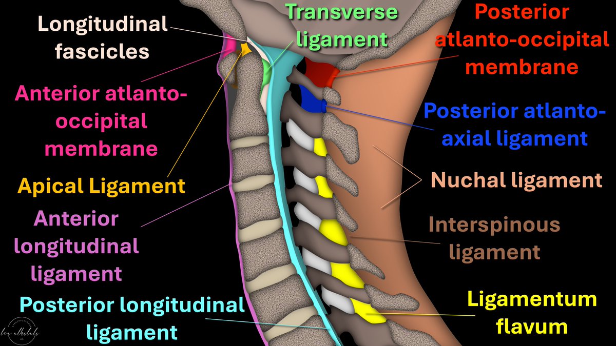
Afraid to stick your neck out when it comes to naming cervical spine ligaments?
Know the anterior longitudinal ligament (ALL) & that’s ALL?
Is the nuchal ligament a NEW CALL for you?
How many ligaments do YOU know?
Here’s are the ligaments to know & how you can remember them
➡️ALL:
🔸As it’s name implies, it runs ALL the way down the spine from the occiput to the sacrum
🔸It’s the ANTERIOR longitudinal ligament so it is ANTERIOR to the vertebral bodies
➡️PLL:
🔸PLL stands for POSTERIOR longitudinal ligament, so it’s posterior to vertebral bodies
🔸I call it “PARTIALLY longitudinal ligament” bc it only runs from C2 inferiorly
🔸Above C2 it’s the tectorial membrane.
🔸I remember this bc it has a different name but is TECT-nically part of the PLL
➡️Cranio-cervical ligaments:
🔸In the sagittal plane C2 looks like a mountain peak
🔸Ligaments here are arranged how you climb a mountain!
▶️AT Last you reach the ANTERIOR mountain (first is ANTERIOR ATLanto-occipital membrane)
▶️You hike up the APEX (next is APICAL ligament)
▶️Finally, you have to TRAVERSE the LONG way down (TRANSVERSE & LONGitudinal fibers)
➡️Posterior ligaments:
▶️Ligamentum flavum:
🔸I remember this bc Flavum & Facets both start w/F = Flavum is by the Facets
🔸F is also for FAR FIVE, so ligamentum flavum is only on the lowest/farthest five cervical vertebral bodies
🔸Above this, membranes are named for what they connect (posterior atlanto-occipital & posterior atlanto-axial membrane)
▶️Nuchal ligament:
🔸This is where we see the nuchal fold & is the equivalent of the supraspinous ligament from the occiput to C7.
Now you know the major cervical ligaments you can see on sagittal images!
Hopefully, now you will be ready when injury to these structures occurs in your neck of the woods!
1/They say form follows function!
Brain MRI anatomy is best understood in terms of both form & function.
Here’s a thread to help you to remember important functional brain anatomy
1/Correlate clinically!
It’s harder than you think in THALAMUS—where its size is small & but the clinical symptoms are large.
Here’s a thread to help you remember the main thalamic syndromes & their locations!
Si te quejas de que la vida es dura...
Lee este hilo sobre la vida de Keanu Reeves.
Fue abandonado por su padre con 3 años y creció con 3 padrastros diferentes.
Es disléxico.
Su sueño de convertirse en jugador de hockey quedó destrozado por un grave accidente.
Su hija...🧵 👇