Msc in Molecular, Cellular Biotechnology and Genetics.
Experienced in molecular biology, drug discovery, cancer research, reproductive medicine, grant writing
Out today in @nature.com: our review on how single cells evolve into multicellular organisms, with Ozan Bozdag, @kai_tong_mc25, Peter Yunker, and Matt Herron.
https://t.co/mJYWOSK3g3
For an overview, check out the video below.
The most important briefings of the day.
Karl Deisseroth, professor of bioengineering and of psychiatry and behavioral sciences, shares the news of his Nobel Prize with his kids.
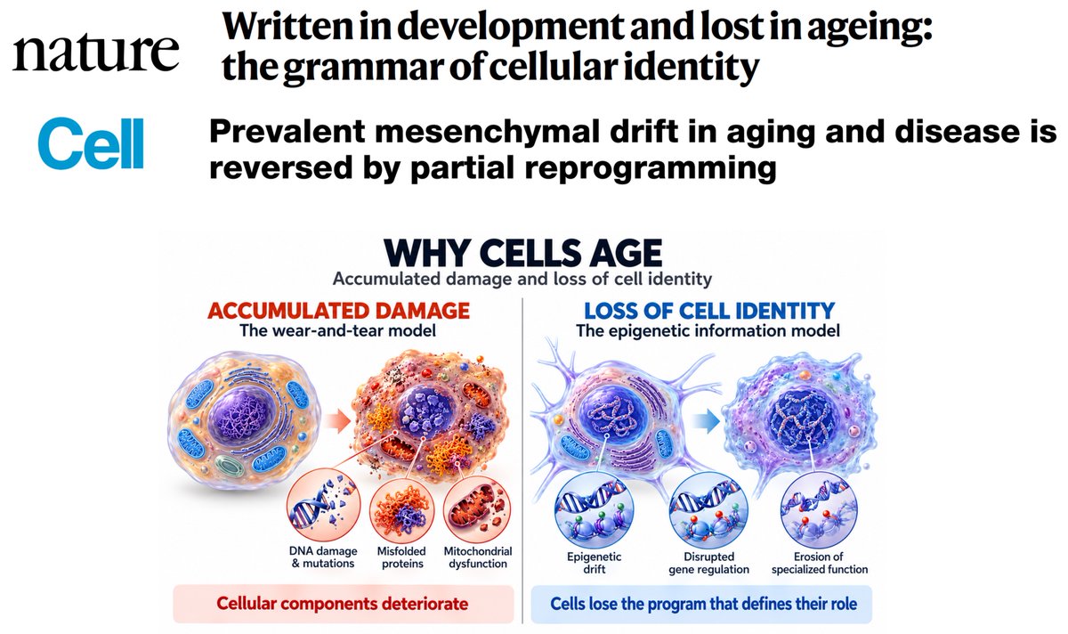
Why do our cells age?
It's not just wear and tear, accumulated damage.
Loss of cell identity takes center stage.
A @Nature review today by @gladyshev_lab and connection with prior work that substantially advances the science of aging.
In the new Ground Truths.
Nobel Prize winner Ada Yonath sadly passed away today. She was a legend for her pioneering work on figuring out the structure of ribosomes.
“Focus on what you love to do, what you want to know, first of all, or what you want to achieve, or what you want to do. If you want to be a performer, a dancer, it's okay with me, as long as this is really built in, and you can do it well. Courage is very important, but for science, curiosity is not enough, it should come with the courage. Really wish to understand something. If there is no wish like this, and they ask me where will I get the best salary, or I become more famous, or then I have no answer for that.”
Ahead of the 20-year anniversary of induced pluripotent stem cells #iPSCs, Joseph Wu & colleagues review how this technology has moved from bespoke experimental models toward standardized, engineered biological medicines.
https://t.co/xx86z0G1Mr
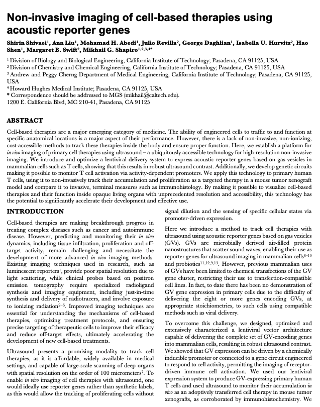
It is possible to image cells (using ultrasound) as they move through the body. This could transform medicine.
Consider a pharmaceutical company that wants to test whether an engineered immune cell, such as those used for CAR-T therapy, can detect and destroy cancer cells. At least six types of CAR-T cells have already passed through clinical trials and earned FDA approval. But during these clinical trials, only a handful of indicators are measured: the number of cells infused into the body, the size of the tumor over time, and some blood marker proxies for the immune system’s activity. Just about everything happening inside the body itself—the number of cells that make it to a tumor, the time it takes for them to reach that tumor, or the route by which they get there—is a mystery. But now there is a technology that enables us to *actually* figure out where those CAR-T cells moved through the body.
The secret is gas vesicles, or little protein shells that trap gas, rather than liquids or solids. If you engineer cells to express these gas vesicles, then they will show up very strongly on ultrasound scans (because everything else in the body is liquid; so the air acts as a contrast agent.)
In one paper, Mikhail Shapiro’s group at Caltech did exactly this. They used a lentivirus to put “acoustic reporter genes” (aka gas vesicles) into T cells, injected those T-cells into mice with cancer, and then watched “their function inside opaque living organs.” Brilliant.
(The image below is an E. coli bacterium with these gas vesicles.)
Please watch and consider that Russia is proudly targeting and murdering Ukrainian children, and that there are real people operating these drones. That they have commanders all the way up to Putin authorizing this terror campaign.
When cells grow together, they often align with their neighbors, forming liquid crystal-like patterns known as “nematic order.” But that order isn’t perfect, and at certain points, the alignment can break down, creating “topological defects”: small regions where cells swirl or fan out instead of lining up cleanly.
And according to research published in Science, what looks like a defect can be used strategically as built-in instructions for shaping larger structures. Instead of growing upward, cells can store and release stress in certain places along a previously flat sheetlike structure, forming a bowl, a ridge, or a fold.
Learn more: https://t.co/OSMGhNzR6d
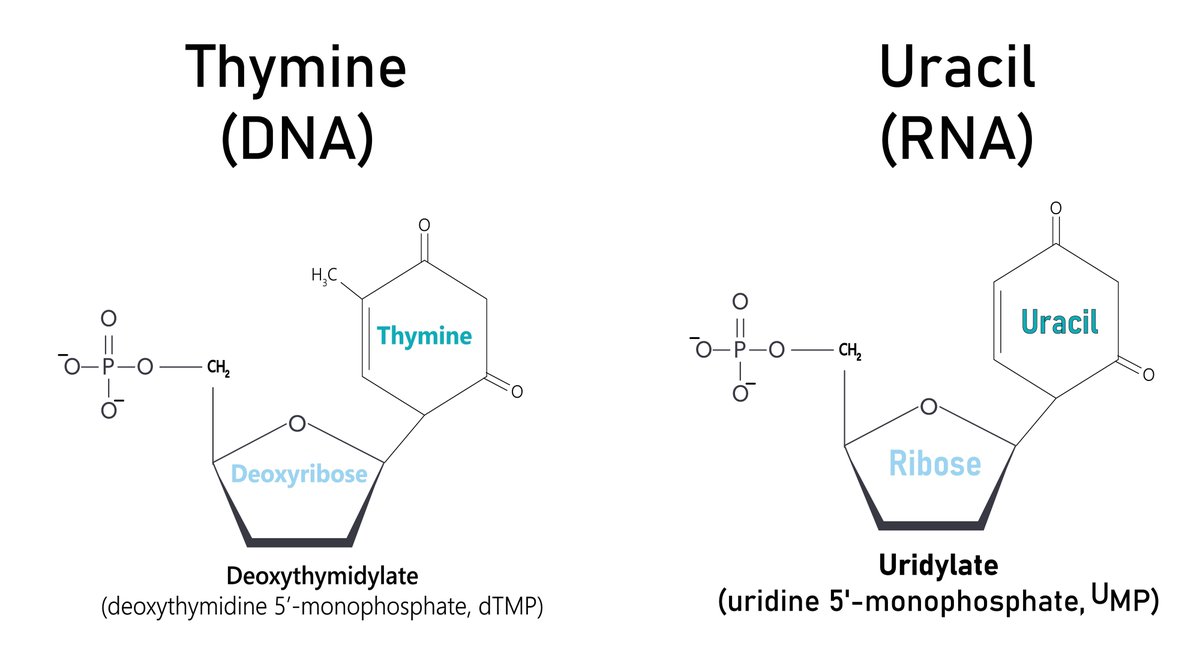
Why does DNA use thymine, whereas the evolutionarily older RNA uses uracil, given thymine’s susceptibility to damage from UV light? A study suggests that although thymine breaks down more easily, its products are less harmful to the DNA molecule. In PNAS: https://t.co/5u5xWyoHPG
Drug candidates that form covalent bonds with their targets were historically avoided because of toxicity concerns. However, covalent drugs with controlled reactivity are now recognized as promising agents that combine manageable safety with substantial advantages, including enhanced potency, prolonged target residence times, and access to challenging targets.
This shift is reflected in recent covalent drug approvals, particularly those targeting protein-phosphorylating enzymes (kinases) and cancer drivers such as the oncogenic protein KRAS. A central feature of these drugs is a “warhead” reacting with an amino acid residue in the target protein to form a covalent bond. Yet the repertoire of drug-like, chemically accessible reactive moieties remains limited.
In a new Science study, researchers report streamlined access to an underexplored warhead type with well-tempered reactivity that can be appended to complex scaffolds late in synthesis, enabling practical use in drug discovery.
Learn more in a new #SciencePerspective: https://t.co/zvnUNfkBNP
Largest cross-tissue map of the human aging epigenome yet: 15,000+ methylation profiles across 17 tissues reveal both shared and tissue-specific DNAm signatures of aging. Nature Aging 2026. 🧬🔬
[1/5]
Today in @Nature we report how AI-guided redesign enhances protein evolution. Integrating ProteinMPNN sequence design with autonomous laboratory evolution, we establish a workflow to engineer enzymes with improved properties over those evolved from natural proteins. Redesigned starting points consistently evolve an expanded fitness landscape, reaching new function with higher activity, specificity, and stability than their natural counterparts.
https://t.co/pNlw6z1zvU
1/14
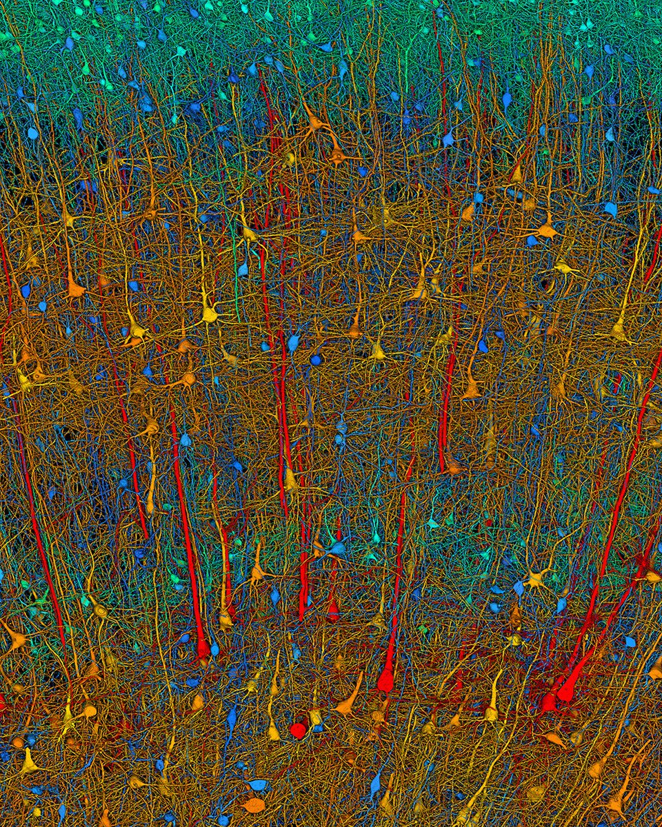
Using more than 1.4 petabytes of electron microscopy (EM) imaging data, researchers generated a nanoscale-resolution reconstruction of a millimeter-scale fragment of human cerebral cortex, providing an unprecedented view into the structural organization of brain tissue at the supracellular, cellular, and subcellular levels.
The human brain is a vastly complex organ and, to date, little is known about its cellular microstructure, including the synaptic and neural circuits it supports. Disruption of these circuits is known to play a role in myriad brain disorders. Yet studying human brain samples in such great detail comes with a host of challenges, ranging from technological limitations to the availability and preservation of tissue samples from healthy individuals.
In a 2024 Science study, researchers performed a high-resolution EM reconstruction of the ultrastructure of a cubic millimeter of human temporal cortex. According to the authors, the reconstruction contains roughly 57,000 cells, about 230 millimeters of blood vessels, and nearly 150 million synapses, comprising 1400 terabytes of data. The authors generated a three-dimensional reconstruction of nearly every cell and process in the cubic millimeter sample and developed a freely available tool for visualizing and analyzing the vast dataset.
Learn more on #WorldBrainDay: https://t.co/ye8JncNkrJ
About 6.2 billion basepairs of DNA are 'packed' into the nucleus of each human cell. Scaled by a factor of 500,000x, this is akin to packing a 1,000-kilometer-long string (roughly the distance from New York City to Detroit) into a small bedroom.
How does all that DNA fit? Well, let’s do some back-of-the-envelope calculations:
The human genome is 6.2 billion basepairs in length, and each basepair is separated by 0.34 nanometers along the helix. (A carbon-carbon single bond is about half that length.) Multiply these numbers together, and we see that the DNA inside of each cell stretches about 2.1 meters in length. This is not a scaled value! A human cell nucleus has a diameter of about 6 micrometers.
So what fraction of that nucleus is 'occupied' by the DNA?
First, we calculate the volume of the cell nucleus. Given a radius of 3 micrometers, and using the regular equation (V = 4/3*pi*r^3), we get a volume of 113 cubic micrometers.
The DNA can be modeled as a cylinder. This cylinder is 2.1 meters long and about 2 nanometers in diameter. Again, using the equation for volume of a cylinder (V=π*r^2*h) we get 6.6 cubic micrometers.
In short: The human genome occupies about 6% of the cell nucleus. This is not tightly packed at all.
The main problem is that once you pack all of this 'string' into the nucleus, you have created a really difficult search problem for yourself. This string is coiled up tightly and, if you are an enzyme, then you might need to find a particular segment of the string located at, say, position 4,284,283,192. Good luck with that! And yet, cells have evolved highly effective search strategies to quickly retrieve information, which I think is one of the great innovations of evolution.
Just published @ScienceMagazine
Impressively broad range of life science research getting done autonomously with agentic AI to accelerate discovery
https://t.co/YpRBoUA5Rw








![biogerontology's tweet photo. Largest cross-tissue map of the human aging epigenome yet: 15,000+ methylation profiles across 17 tissues reveal both shared and tissue-specific DNAm signatures of aging. Nature Aging 2026. 🧬🔬
[1/5] https://t.co/4RtwhE9WcF](https://pbs.twimg.com/media/HOQJwx1WcAEZdHE.jpg)





