Check out my new video of how the nystagmus of vestibular neuritis changes with time, and one way you can avoid missing subtle nystagmus. https://t.co/qFXQOHPJ5M
This is an eye-opening study for me... Apparently, cerebrovascular hyperemia and /or vasospasm can occur in reversible cerebral vasoconstriction syndrome (RCVS). They may even coexist in different vascular territories or at different times in the same patient.
In RCVS, the hallmark is multifocal cerebral arterial vasoconstriction, which can progress and lead to ischemia and infarction.
At the same time, some regions can show vasodilation and impaired autoregulation with vasogenic edema, often overlapping with PRES, reflecting a hyperemic or “leaky” circulation pattern rather than pure spasm.
A mixture of distal vasodilation, proximal vasoconstriction, edema, and ischemia is well described, making it difficult to cleanly separate hyperemia from vasospasm. Indices like the Lindegaard ratio can help distinguish hyperemia from vasospasm rather than assuming only one process is present. A Lindegaard ratio of < 3 suggests hyperemia and > 7 is s/o vasospasm.
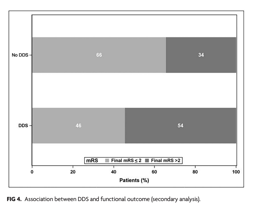
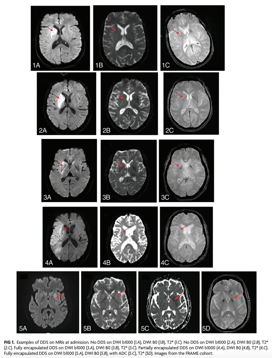
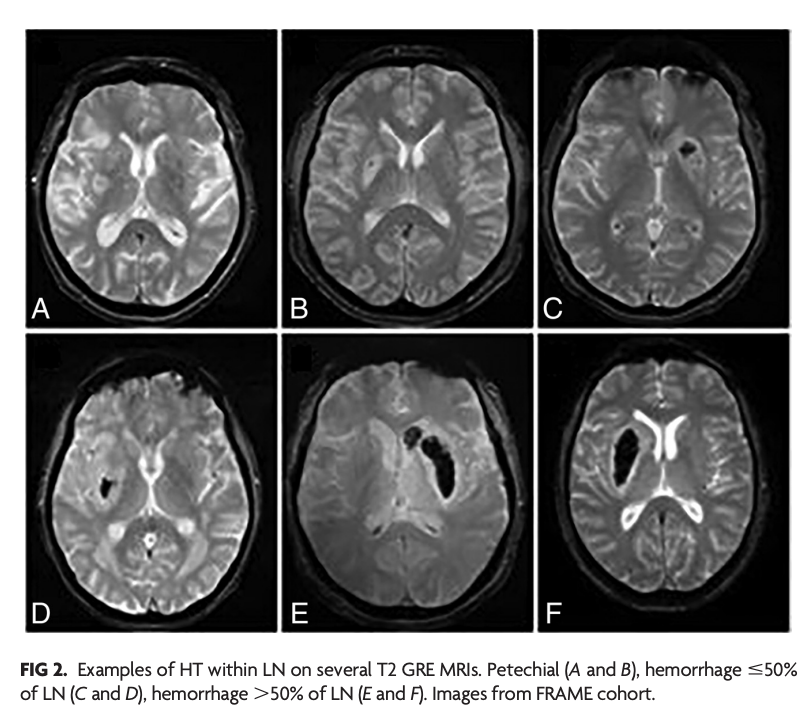
"Dark Diffusion Sign: A Novel MRI Predictor of Hemorrhagic Transformation in Patients with Stroke Treated with Endovascular Therapy?"
https://t.co/mRMc4vaK63
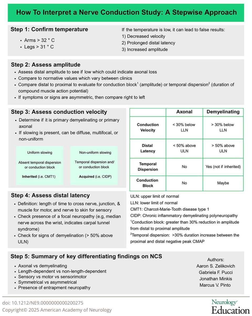
In the latest Neurology Education Teaching NeuroVisual, Zelikovich and colleagues break down interpreting nerve conduction studies into a 5-step guide: https://t.co/TU7v5mCMGY
#NeurologyEd@aszelikovich@gabifpucci@MarcusVPinto
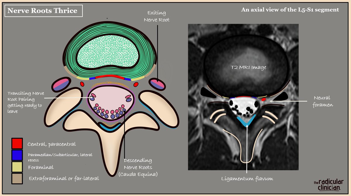
Nerve Roots 'Thrice'
Clinical anatomy applied
Lumbosacral nerve roots can be described in three geographical locations on axial T2 MRI, each with important clinical implications if they are compressed or inflamed. The picture illustrates the L5-S1 level, so we are looking at the L5 disc. Focus on the colour codes in order to appreciate disc location in relation to each geographical nerve root location.
Descending (bilateral S2, S3, S4 and S5 roots as part of the cauda equina).
An acute central or paracentral disc herniation here is rare but can cause cauda equina syndrome if compressed. Also relevant with crowding as seen with LSS.
Transiting/Traversing (here bilateral S1 roots).
As the bilateral transiting nerve roots leave the thecal sac centrally, they remain enveloped by its outer connective tissue layer, referred to as the nerve root sleeve. The lateral recess is the most common site of disc related compression because of its anatomical position and the root being fixed in place. This is why radicular pain is more commonly noted in one leg. Note that the nerve roots do not lie flat when you do.
Exiting (here L5).
As the nerve root enters its respective neural foramen. This is the second most common site of compression, typically from a far lateral disc herniation or from crowding within the foramen.
Tele-stroke rule out!
Acute wrist drop - peripheral- with further wrist flexion on making a fist- characteristic of radial palsy. In a cortical stroke synergy would cause the flexed wrist to remain neutral or extend
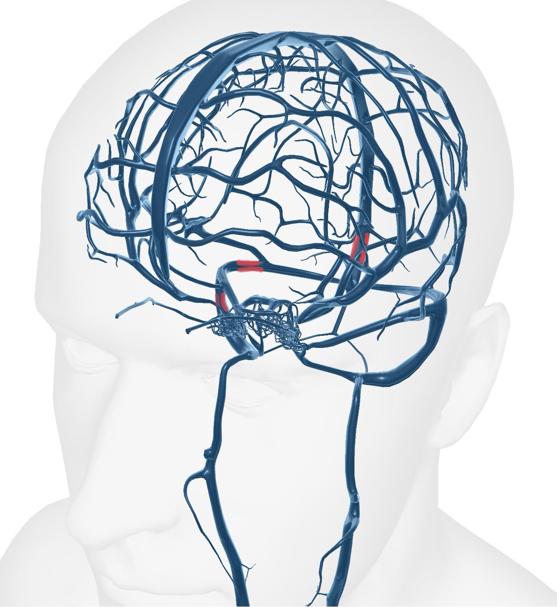
The most common sites of venous stenosis in Idiopathic intracranial hypertension (IIH).- the transverse- sigmoid junction, posterior sigmoid and distal superior sagittal sinus
#STROKE: In a prospective observational cohort study, patients with a Carotid Artery Risk Score ≤20% had a very low risk of recurrent stroke, suggesting that selected patients may be managed conservatively. #AHAJournals https://t.co/mw9j8hlyYm
#MondayTip
😵💫Acute Vestibular Syndrome and Stroke
#AVS👉continuous vertigo/nausea/nystagmus, worse with head motion but present at rest
⚡️~25% are posterior strokes, often missed by CT/MRI.
💡#HINTS exam > early MRI for diagnosis
Libgen, Sci-Hub, and Z-library had millions of pirated academic books and papers.
So, they were shut down. We shouldn't use them anyway.
We should help billion-dollar academic publishers get richer.
Anyway, here's how to access these libraries:
Don't do this!
🧵 “ANA Positive ≠ Lupus: The Art of Interpreting Autoantibodies”
ANA positivity is one of the most misunderstood findings in medicine.
Here’s how to approach an ANA report systematically, clinically, and with confidence.
👇 #Rheumatology#ANA#Autoimmunity#MedEd @DrAkhilX @IhabFathiSulima #MedTwitter
The Notes app on your iPhone is one of the most powerful tools available.
But 99% of People don't know its true potential.
Here are 20 amazing features you must know:⤵️
1/One important aspect to stroke care is well, ASPECTS.
It’s a simple score system—but it’s important to understand all aspects!
Read on for the latest research on ASPECTS in this month’s @theAJNR SCANtastic!
https://t.co/UiExzEzQoD
Libgen, Sci-Hub, and Z-library had millions of pirated academic books and papers.
So, they were shut down. We shouldn't use them anyway.
We should help billion-dollar academic publishers get richer.
Anyway, here's how to access these libraries:
Don't do this!
Knowledge is contagious!
Infectious encephalitis can be a difficult diagnosis, but patterns on MRI can suggest an etiology.
Most know herpes & the temporal lobe
Do YOU know any more?
Here’s a figure to help you remember the most “common” encephalitis patterns for different organisms
Since CSF PCR studies take time, some imaging can provide helpful clues:
Temporal lobe: Although other infectious agents may involve the temporal lobe, statistically this is overwhelming HSV-1
Deep Gray: Think of flaviviruses such as Japanese encephalitis & West Nile encephalitis
Splenium: Think of common infections, especially infections kids get—rotavirus, influenza, COVID.
Brainstem: This is most commonly enteroviruses (hand, foot and mouth disease). Remember, all the motor for the hand, foot, & mouth meet in the brainstem, so this is where the encephalitis will be!
Infarcts: Think VZV. Varicella and Vasculitis both start with V!
White matter: Think of immunocompromised viruses (HIV, PML/JC virus)
Hopefully, you will have an infectious curiosity for the MR findings in encephalitis!