The big takeaway here is that it looks, feels and acts nothing like an Apple product or anything the company is currently planning. OpenAI hasn’t found any evidence they’re violating trade secrets with this device, I’m told.
@TrackYourHeart Plenty of “ICD”s in the comments below. However this is an oversimplification of type 1 Brugada management. For patients symptomatic for syncope or documented TV, that’s the right answer. However in many cases this ECG finding is incidental in asymptomatic patients.
Commotio Cordis is a sudden cardiac arrest caused by a blunt, non-penetrating impact to the chest, often from a baseball, hockey puck, or lacrosse ball.( Football is less common )
The risk is highest when the impact hits the precordium during a 20-millisecond window on the upstroke of the T wave, triggering ventricular fibrillation (VF).
Common in young athletes with a compliant chest wall. Immediate CPR + defibrillation is critical.
A wide QRS tachycardia is ventricular tachycardia (VT) until proven otherwise.
The two most common causes are:
⬜ Ventricular tachycardia (VT)
🟪 Supraventricular tachycardia (SVT) with aberrant conduction
Other less common causes include:
🟦SVT with pre-excitation (WPW syndrome)
🟩SVT with baseline bundle branch block
🟨 Hyperkalemia
🟧 Ventricular pacing
A practical ECG clue:
- LBBB-type wide QRS: the terminal portion of the QRS in V1 is predominantly negative
- RBBB-type wide QRS: the terminal portion of the QRS in V1 is predominantly positive
If the QRS pattern in V1 is atypical (such as an Rs or qRs pattern), the rhythm is much more likely to be ventricular tachycardia.
Key point:
Because VT is the most common and potentially life-threatening cause of a wide QRS tachycardia, it should be the default diagnosis until an alternative is confidently established.
@IM_Crit_@RogerSeheult AFib cardioversion of a septic patient such as this one is always risky, given (above all) the higher thromboembolic risk. However I definitely agree on the decision in this case, and I’m glad he recovered
Coronary artery territories
The main epicardial coronary arteries supply distinct regions of the myocardium, which can be mapped and assessed during an ultrasound examination. For standardization, the left ventricle (LV) is divided along its long axis into four quadrants: anterior, inferior, septal, and lateral.
At the basal and midventricular levels, the septal and lateral walls are further subdivided into anterior and inferior segments. Each wall is then divided in short-axis views into basal, mid, and apical thirds. Beyond the LV cavity, the distal apex forms a cap segment. Altogether, this results in a total of 17 myocardial segments.
Most of the heart’s blood supply comes from the left main coronary artery, which divides into the left anterior descending (LAD) and left circumflex (LCx) arteries. The LAD supplies most of the anterior ventricular wall, while its septal branches supply the anterior two-thirds of the interventricular septum. Its diagonal branches supply the anterolateral wall. In some cases, a large LAD may wrap around the apex and supply the distal portion of the inferior wall.
The LCx travels in the atrioventricular groove, and its obtuse marginal branches supply the inferolateral wall. The right coronary artery (RCA) supplies the inferior third of the septum and the inferior wall, and it also provides blood to the right ventricle.
Adapted from Bulwer BE, Rivero JM, eds. Echocardiography Pocket Guide: The Transthoracic Examination
A dislodged TAVR valve (Transcatheter Aortic Valve Replacement valve embolization or migration) is a rare but serious complication where the implanted prosthetic valve moves from its intended position after deployment.
📊 JAMA Clinical Guidelines Synopsis: The 2025 guideline for #ACS management recommends dual antiplatelet therapy with aspirin and a P2Y12 inhibitor, high-intensity statins, invasive revascularization, and radial access for angiography.
https://t.co/cRfu42LO0s
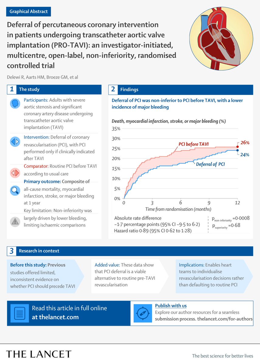
PCI before TAVI—necessary?
In patients with CAD, deferring PCI was noninferior to a PCI-first strategy at 1 year for the composite of death, MI, stroke, and major bleeding.
https://t.co/KwN2tauPyY #ACC26