Pericardial effusions have long been described in millimeters.
But when the clinical question is fluid burden, a single linear dimension may not be enough.
In our new JASE editorial with David Dudzinski, we discuss the practice-changing work by Kane et al. and the accompanying tutorial, which challenge us to rethink how we quantify pericardial effusions by echo.
The key shift:
From a single maximal width
to a more integrated assessment of volume, distribution, phenotype, and physiology.
Milliliters may bring us closer to the anatomic truth.
But tamponade remains a physiologic diagnosis.
Free access:
https://t.co/R2UgItJ6B3
#EchoFirst #CardioTwitter #PericardialDisease #JASE
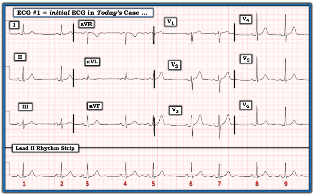
The ECG in the Figure is from a man in his 60s who is aware of his “irregular heart beat”. — Why is this rhythm irregular? — What is your differential diagnosis? — GO TO — https://t.co/CuTZVMvalV — :)
I was Going through Af management - changes and nuances for a presentation
And some change that I read which are noteworthy #MedTwitter#CardioTwitter#NeuroTwitter#Epeeps
( updates from the new ESC / European Heart Rhythm Association guidelines)
1. CHA₂DS₂-VASc → CHA₂DS₂-VA
• Female sex ❌ no longer a risk factor
• It’s a risk modifier, not an independent predictor
2. Some AF cases = anticoagulate anyway (don’t calculate scores)
• HCM→ OAC irrespective of score
• Cardiac amyloidosis → OAC strongly favoured
• Significant valvular disease (esp. MS) → treat as high risk
“high-risk AF phenotypes”
3. DOACs
• No clear superiority of one DOAC over another
• Switching without reason = no benefit
• Dose correctly
4. Rhythm control is back favoured over rate ctrl except in PERMANET t AF
• Early rhythm control > rate control in:
– Paroxysmal AF
– Persistent AF
👉 Now Class I recommendation
5. Ablation early, preferred
• Early referral for catheter ablation
– Paroxysmal AF
– Persistent AF
👉 First-line strategy in selected patients (Class I)
6. AF + ACS (antithrombotics simplified) #cardiotwitter
• DOAC + DAPT → very short (≈1 week, up to 1 month)
• Then DOAC + single antiplatelet → up to 12 months
• Then DOAC alone lifelong
👉 Bleeding reduction is the priority
7. Transient / secondary AF
(e.g. sepsis, post-surgery, pericarditis)
• OAC ~ 4 weeks
• Reassess with monitoring (Holter/ELR)
• If no recurrence → stop OAC
8. No AF documented = no DOAC
• Even in cryptogenic stroke
• Need ECG evidence (Holter / ILR)
👉 No more “empirical anticoagulation”
9. Risk reassessment is dynamic
• Stroke risk ≠ static
• Re-evaluate periodically (age, DM, BP, etc.)
10. Lifestyle is now guideline-level therapy
• Weight loss
• Exercise
• Alcohol reduction
• Treat OSA
👉 Risk factor control = AF control
11. AF burden matters (but not fully there yet)
• Device-detected AF → still evolving
• Not all subclinical AF = anticoagulate
Bottom line
AF management is shifting from:
👉 “Rate control + late decisions”
to:
👉 “Early rhythm control + early ablation + precise anticoagulation”
Any other points?!
Don’t wait for disease. Know your numbers.
Most heart attacks, strokes & diabetes are predictable years in advance, if you track the right markers.
For Indian adults, these cut-offs matter more than ever:
1. Blood Pressure (BP)
Target: <130/80 mmHg
Even “borderline” BP in Indians carries higher cardiovascular risk due to earlier vascular aging.
2. Fasting Blood Sugar (FBS) <100 mg/dL
Indians develop insulin resistance earlier, even at “normal” BMI.
3. HbA1c <5.7%
Prediabetes starts silently. By the time glucose rises, damage has already begun.
4. Insulin Resistance (HOMA-IR)
Ideal: <2
Insulin resistance (IR) is not routinely measured, but it is extremely useful in high-risk individuals. IR is a hidden driver of diabetes, fatty liver & PCOS.
5. Waist Circumference (Most important for Indians)
Men: <90 cm
Women: <80 cm
🔸“Thin outside, fat inside” (TOFI) phenotype is common.
🔸Waist circumference is better than BMI for risk prediction.
6. LDL Cholesterol
Target
<100 mg/dL (general)
<70 mg/dL (high-risk)
🔸Lower is better; there is no “safe high LDL.”
7. HDL Cholesterol>50 mg/dL
Low HDL is extremely common in Indians and amplifies risk.
8. Triglycerides (TG)<150 mg/dL
High TG is an insulin resistance marker, not just a lipid issue.
9. TG : HDL Ratio<2
🔸TG:HDL ratio is one of the simplest markers of metabolic health.
🔸A ratio of 2 or more is insulin resistance until proven otherwise.
10. hs-CRP (inflammation marker)<2 mg/L
Atherosclerosis is an inflammatory disease, not just high cholesterol.
11. ApoB (the underrated one)
Target: <80 mg/dL (general)
<65 mg/dL (high-risk)
ApoB is better than LDL at predicting risk, as it counts actual atherogenic particles.
12. Sleep Duration
Optimum: 7–9 hours/night
Less sleep is associated with ↑ insulin resistance, ↑ BP, ↑ appetite, ↑ CV risk.
▶️What makes this crucial for Indians?
🔸Higher risk at younger age
🔸More visceral fat at lower BMI
🔸Greater tendency for diabetes & heart disease
🔸Normal-looking person does not mean low risk
▶️If you are above these thresholds, do not panic. Start with exercise, healthy diet, optimum sleep, and consistency. Consult your physician for individual opinion.
Dr Sudhir Kumar @hyderabaddoctor
(Disclaimer: This is not a medical consultation. The information provided here is general in nature, and applies to healthy people, who are not on medications. In any case, consult your doctor for individual opinion)
Back to Basic Electrophysiology
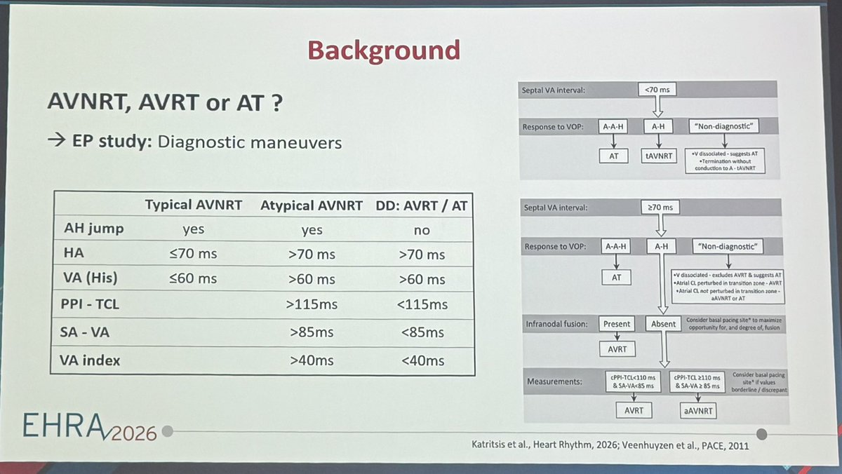
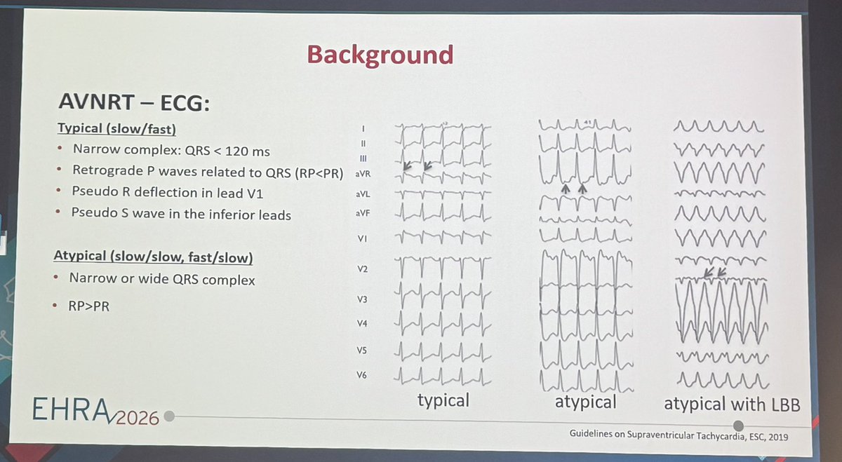
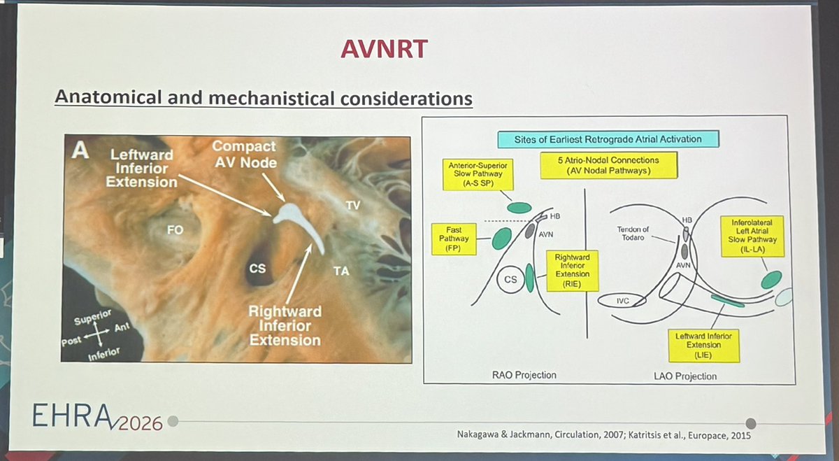
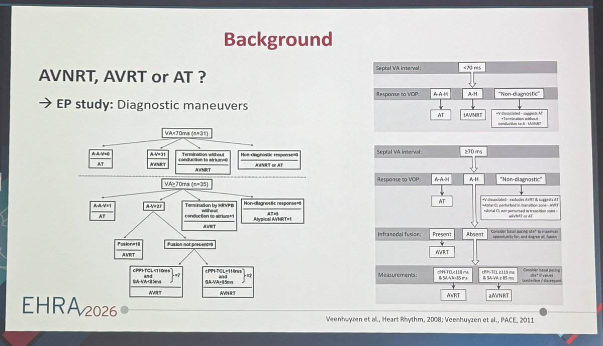
Nice educational case and discussion of different types and rare forms of AVNRT by Dr Christiane Jungen at #EHRA26#EPeeps
For fellows bookmark this tweet for exams #cardiotwitter#MedTwitter
🧵
First of all What is a significant step-up?
👉remember 7-5-5-7 rule
•RA to mixed venous sats ≥7%
•RV to RA ≥5%
•PA to RV ≥5%
•. At any level >7%
{Mixed venous sats = (3* SVC + IVC)/4 )
Normally IVC sat is higher than SVC - please don’t say svc is higher (read up why )
1/n
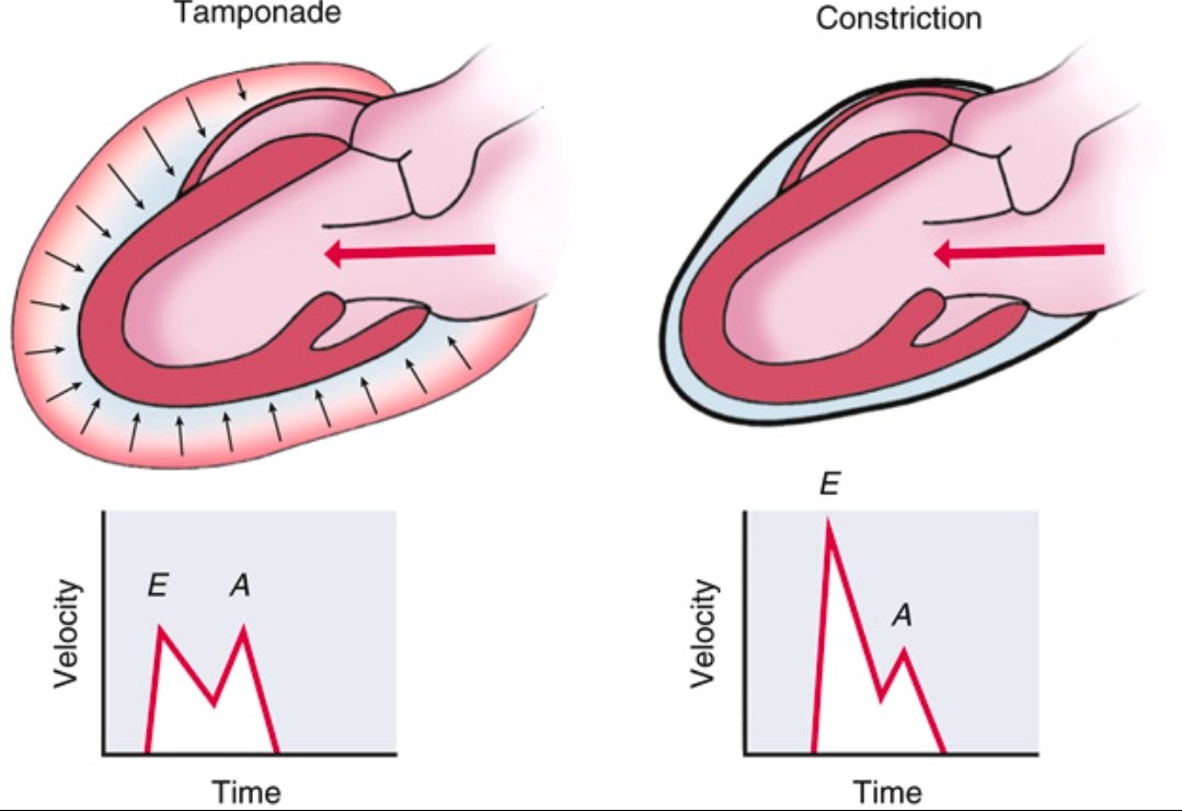
Pericardial Tamponade vs Constrictive Pericarditis ➡️Spot the Difference
Tamponade
⬇️
Accumulation of fluid in the pericardial space
⬇️
↑ intrapericardial pressure
⬇️
impairs diastolic filling throughout the cardiac cycle (early and late diastole).
Doppler shows blunted E and A waves due to restricted filling.
Constrictive Pericarditis
⬇️
Thickened, non-compliant pericardium
⬇️
rapid early diastolic filling that abruptly halts once volume limit is hit.
Doppler shows prominent E wave, reduced A wave, classic for constriction.
Recognizing the echocardiographic filling patterns is essential for diagnosis.
Ref: Catherine M. Otto, Textbook of Clinical Echocardiography
#CardioNugget:
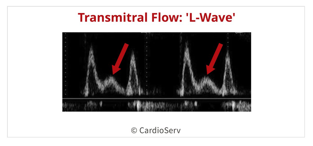
L wave = extra filling wave between E & A (mid-diastole)
Not normal → suggests ↑ LV filling pressures + abnormal relaxation
Seen in advanced diastolic dysfunction
E–L–A pattern = abnormal filling
#CardioNuggets #EchoBoards #Diastology
🩻Contrast-induced AKI:
one of the biggest myths still shaping clinical decisions
For decades we were taught:
👉 “Contrast damages the kidneys”
👉 “Avoid CT with contrast in CKD”
👉 “Hydrate, protect, delay imaging if needed”
But what if… most of this is wrong?🤔
->The uncomfortable reality
Modern evidence shows:
👉 Low-osmolar contrast rarely causes true nephrotoxicity
👉 Even in CKD, AKI, and ICU patients
👉 The risk is often overestimated—or nonexistent
So where did the fear come from?
📍 1950s high-osmolar contrast (actually toxic)
📍 Poorly controlled observational studies
📍 “Creatinine rise = contrast injury” assumption
👉 Correlation became causation
👉 And the dogma stayed
⚠️What recent data tells us
✔ No difference in AKI rates with vs without contrast
✔ No benefit from bicarbonate, NAC, or aggressive hydration
✔ Even ICU and AKI patients show no worsening outcomes
->Translation to real life
👉 The patient was going to develop AKI anyway...Not because of contrast!!
->The real problem: “Renalism”
👉 Avoiding necessary imaging
👉 Delaying diagnosis
👉 Choosing inferior tests
And that leads to:
❌ Missed PE
❌ Delayed sepsis source control
❌ Worse outcomes
->Clinical mindset shift
Instead of asking:
👉 “Will contrast harm the kidneys?”
We should ask:
👉 “Will NOT doing the scan harm the patient?”
->Who still deserves caution?
✔ eGFR <30
✔ Severe hemodynamic instability
✔ Multiple nephrotoxins
Even then:
👉 Optimize volume
👉 Minimize dose
👉 Don’t delay critical imaging
🤓Bottom line
✔ Contrast nephrotoxicity exists… but is rare
✔ The fear is bigger than the risk
✔ The harm of NOT imaging is often greater
In critical care
👉 We don’t treat creatinine
👉 We treat patients
And sometimes…
👉 The most dangerous thing is NOT the contrast
👉 It’s hesitation.
📃Reference
Florens N, Demiselle J.
Kidney360 7: 445–449, 2026. doi: https://t.co/CWzi7WC9Wx