🎯Join the upcoming ESHNR webinar!
📅 April 28 | ⏰ 18:00 CEST
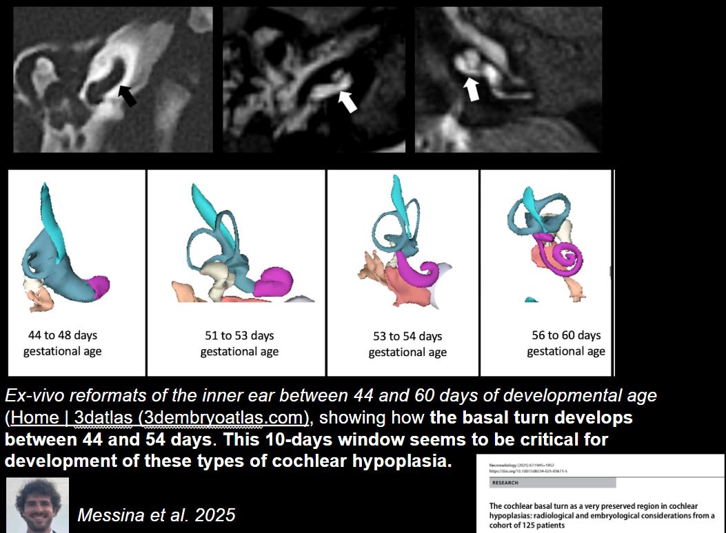
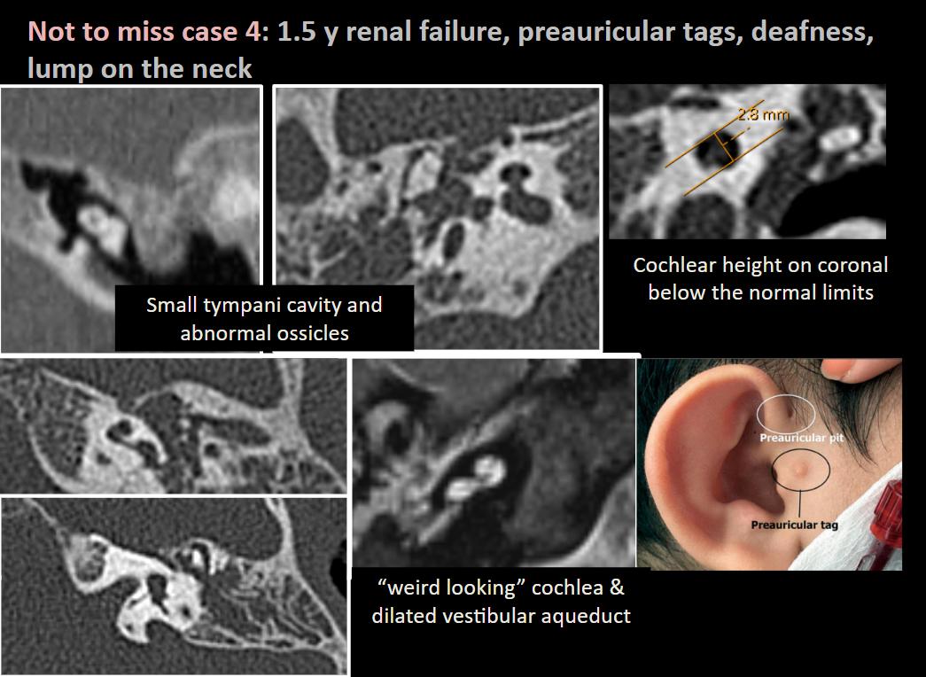
🦻Advanced Imaging of inner ear malformation
▶️Speaker: @FeliceDArco
▶️Moderator: @Bert_DeFoer
🔗 Register: https://t.co/9kVZA8qj42
📖 Related reading: https://t.co/3d2nxxYns3
👀 A sneak preview of the talk!
Congenital Posterior Fossa Malformations: Imaging Patterns and Diagnostic Approach. An amazing learning experience putting it together. Thank you for the help, guidance and mentorship @elka_miller@DrNeetikaGupta Drs. Krishnan and Shinar @RadioGraphics
https://t.co/clTLHhZiuS
Important review of a common abnormality in clinical workflow!
Congenital Posterior Fossa Malformations: Imaging Patterns and Diagnostic Approach | RadioGraphics @RadioGraphics@RadG_Editor
https://t.co/oB9q82oUcI
This is the value of expert neuroradiology
I am pretty sure this would not have been done in most centers
Even I would have missed it
A treatable cause of myelopathy
Great teaching point by @SharathKumarGG7 sir
This is why when people tell me that the MRI is normal
I always take it with a pinch of salt
This is also why I always prefer a neuroradiologist whenever I can
#MedTwitter
#NeuroTwitter
#RadTwitter
#dAVF
A sobering reminder of why sequence selection matters in unexplained myelopathy. 🛑
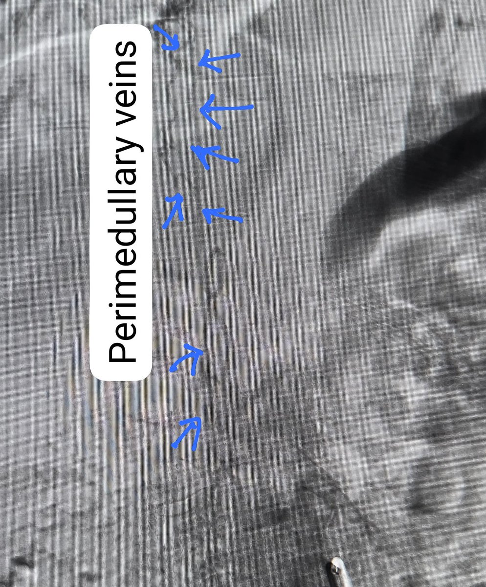
Case: Patient suffered 18 months of progressive lower limb weakness. Routine T2 sagittal MRI was falsely reassuring, entirely missing the subtle flow voids of a Spinal Dural Arteriovenous Fistula. This missed diagnosis resulted in prolonged delays, irreversible cord damage, and severe morbidity.
The turning point? Running a heavily T2-weighted 3D sequence. The high-resolution CSF contrast beautifully delineated the tortuous perimedullary vessels that routine T2 missed. ⚡️
Takeaway: Never settle for standard T2s if you suspect an SDAVF. 3D CISS is absolutely essential to catch these flow voids early and prevent devastating clinical delays!
#MedTwitter #Neuroradiology #FOAMrad #SDAVF #SpineSurgery #NeuroIntervention
"Brain CT for Diagnosis of Intracranial Disease in Ambulatory Patients with Cancer: Assessment of the Diagnostic Value of Scanning without Contrast Prior to with Contrast"
https://t.co/564pgKQjmE
👋Do you know about my website with almost 150 pediatric neuroradiology cases? Searchable database by diagnosis. Classic cases. Free resource for anyone. Hope to add more of cases soon! #MedEd
https://t.co/fVAgtTqwvA
Join us in this case-based review session curated by Dr. Burce Ozgen! Perfect for R3s and R4s preparing for the Core exam and looking to refresh their knowledge on Head and Neck topics.
Thank you @burceozgen!
Thrilled to share our article on interrater reliability studies! Huge thanks to everyone who made this possible, especially my senior co-resident @umontreal and future co-fellow @UofTMedIm@AnassBenomarMD
The idea that doctors need to become prompt engineers for ChatGPT-like models of the future is not new. Doctors now need to be better prompt engineers for your radiologists. Provide 1) relevant history/exam findings and 2) your diagnostic query.
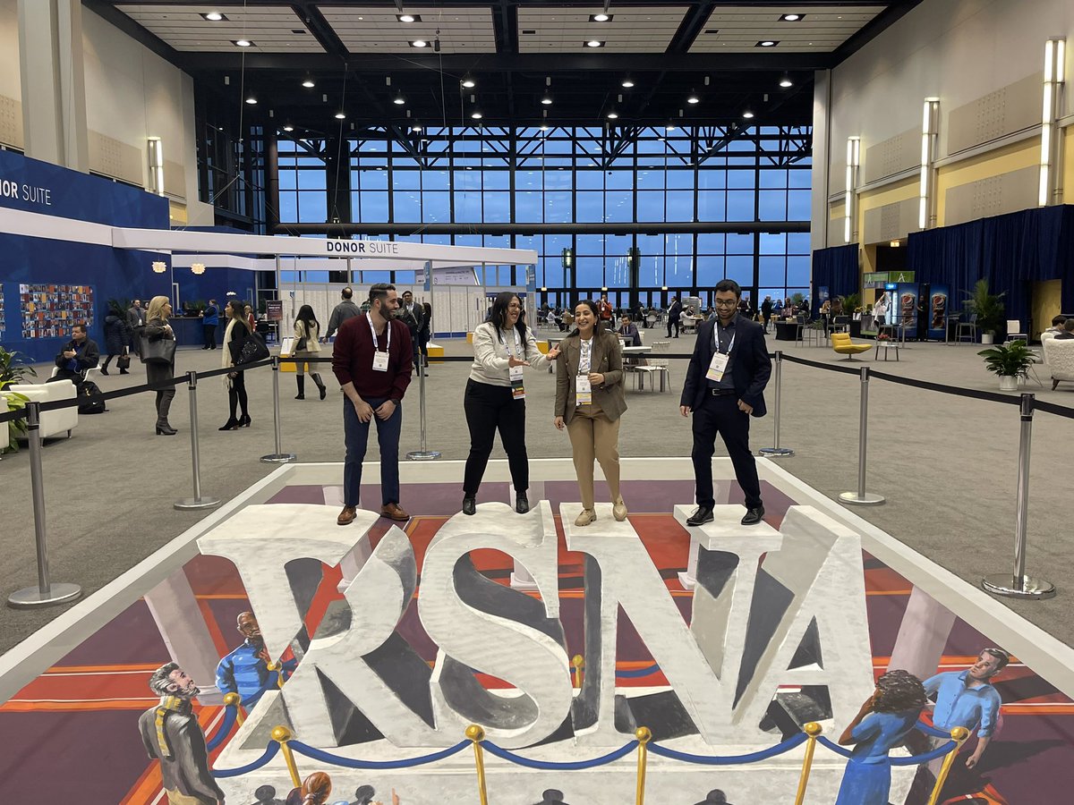
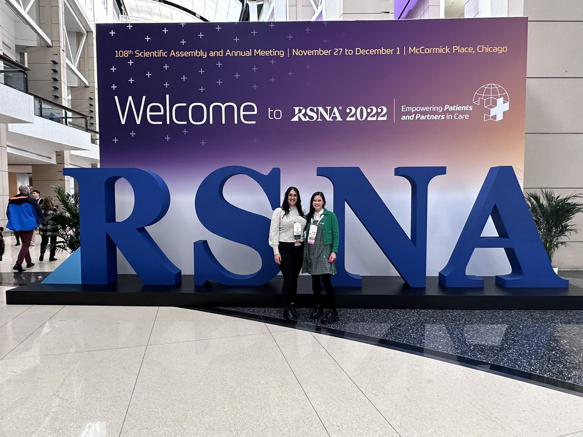
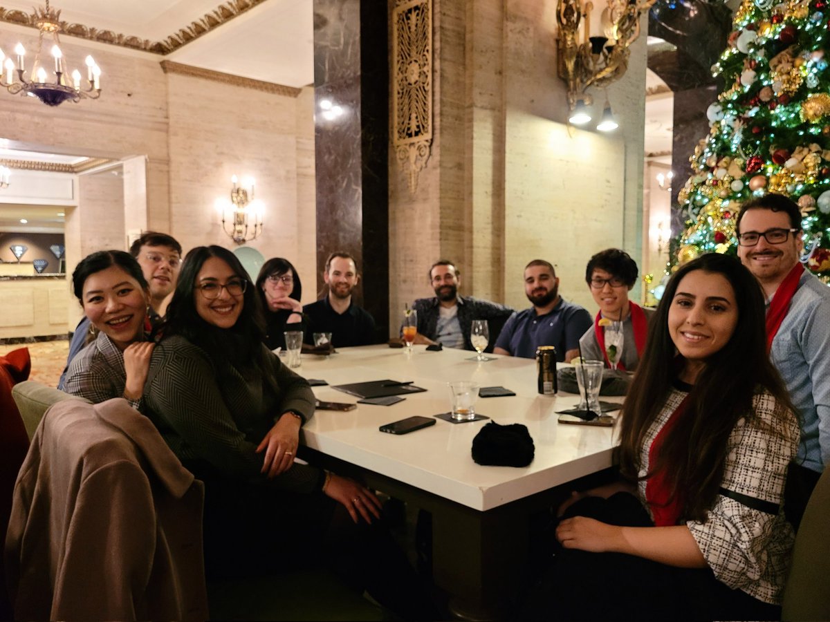
An incredible week @RSNA#RSNA22 spent with friends old and new! I’m grateful to have met my mentors and @canadaradwomen, connect with new colleagues across 🇨🇦 and 🇺🇸, as well as work with the fantastic #RGTEAM at @RadioGraphics. Looking forward to 2023!
#RSNA22, a fantastic first @RSNA meeting. Grateful to have spent time with this crew of wonderful Cdn #radres, met so many of the rads community from across the world, and learned from the best. 🙏 Looking forward to representing @CARadiologists on the RSNA RFC--and to #RSNA23!
Such an honour to learn from leaders in Academic Radiology like @SilviaChangMD! Her lecture on goal setting and career development geared towards trainees was practical and insightful. Looking forward to putting these nuggets of wisdom into action back home in 🇨🇦