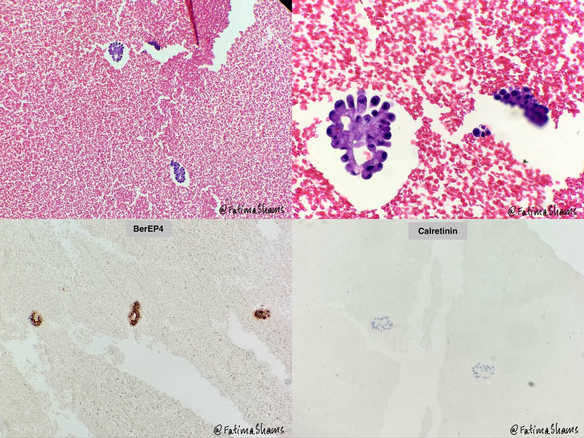
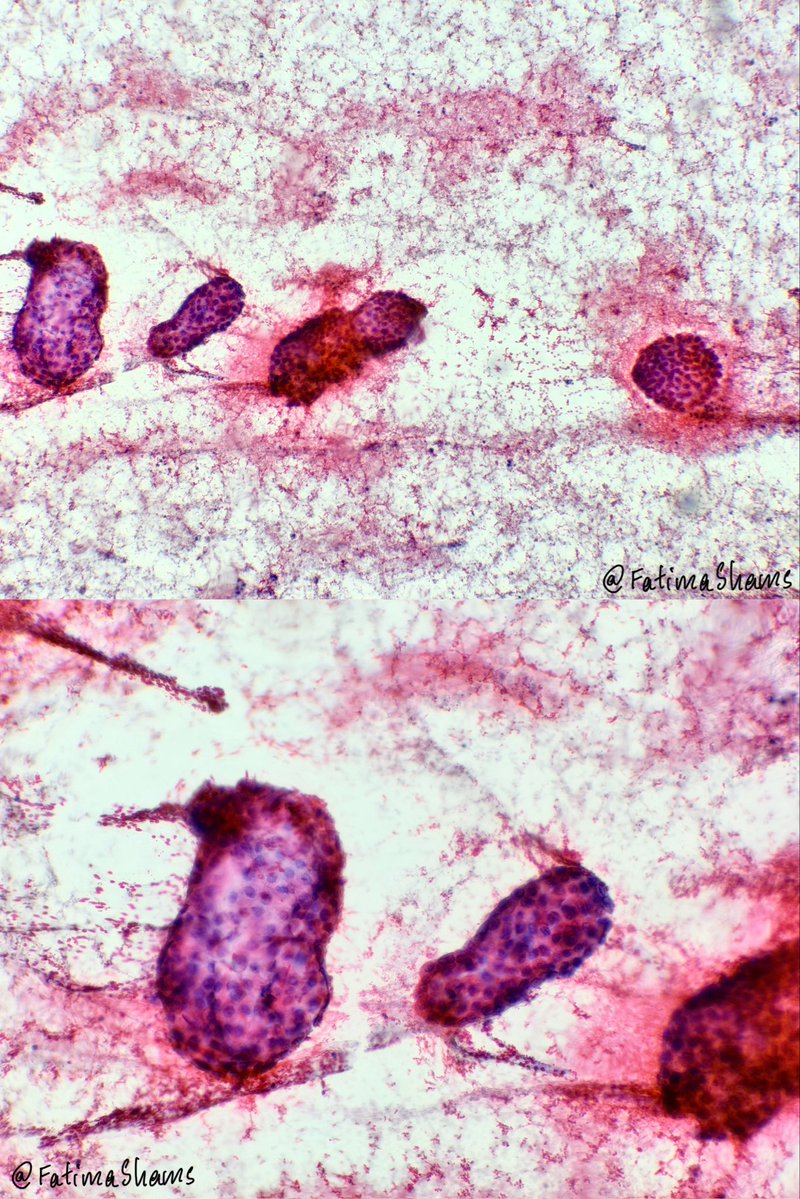
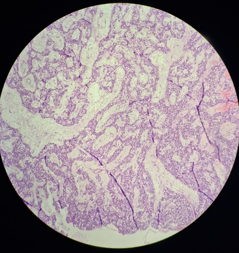
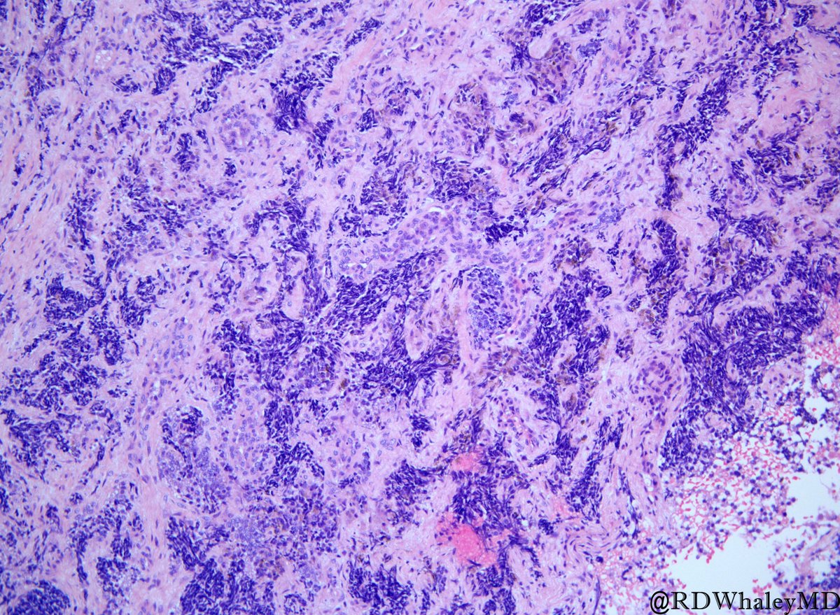
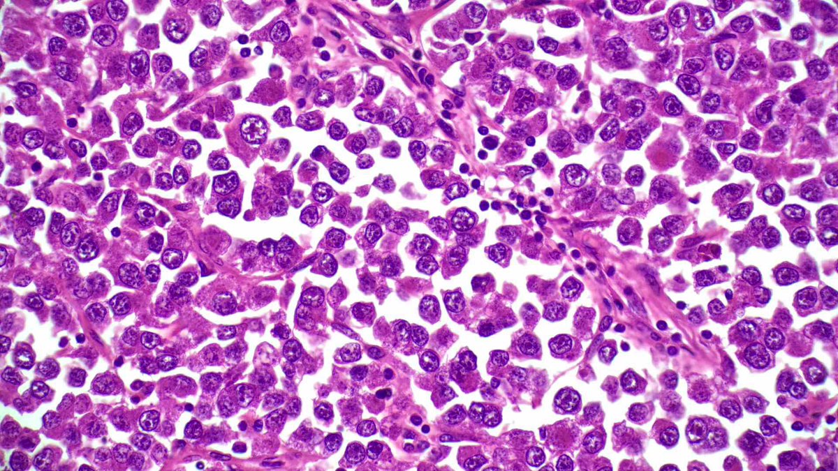
📣 CYTOLOGY - PERICARDIAL EFFUSION
👉 64 YO male presenting with seizures, massive pericardial effusion and cardiac tamponade.
DQ, pap, cell block and IHC provided.
What is your diagnosis ⁉️
#Cytology#PericardialEffusion#EffusionCytology#Pathology#PathTwitter #ThoracicPathology
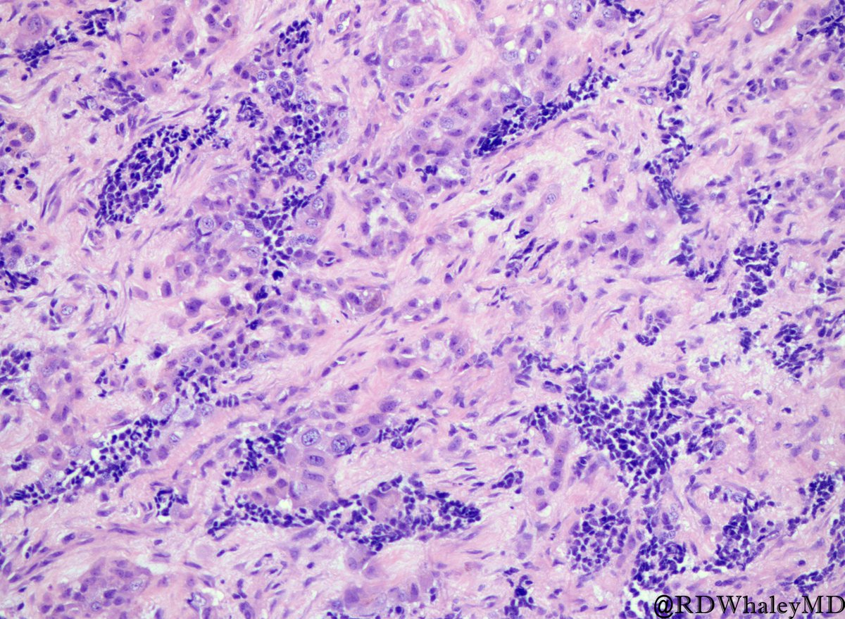
Melanotic Neuroectodermal Tumor of Infancy: Biphasic alveolar proliferation composed of small neuroblast-like cells and larger melanin-producing epithelioid cells separated by fibroblastic stroma. Presentation - first year of life. #Pedipath#TSPSpecialEdition@RDWhaleyMD
Epithelioid AML it is. 😊
By definition, ≥80% of the tumor is composed of epithelioid cells ( polygonal with abundant cytoplasm, vesicular nuclei, and prominent nucleoli)
Epithelioid AML has malignant potential. Worth paying attention to atypia, mitotic activity/atypical mitoses, necrosis, large size , and extrarenal renal/renal vein involvement; different series have proposed different risk parameters.
Sarcomatoid RCC https://t.co/MXnAVIyN1U and sarcomatoid urothelial carcinoma can be important mimickers.https://t.co/r6dx5z5fWp
📚 Brimo et al. AJSP. 2010.https://t.co/HUy8PbMOoh
📚 Nese et al. AJSP. 2011.https://t.co/otwdXZSxUr
📚 WHO Classification of Urinary and Male Genital Tumours, 5th ed.
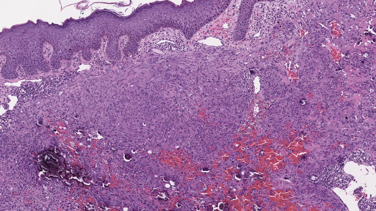
The missing history was immunosuppresion for a solid organ transplantation.
In contrast to the unconvincing CD34, there was clear ERG expression.
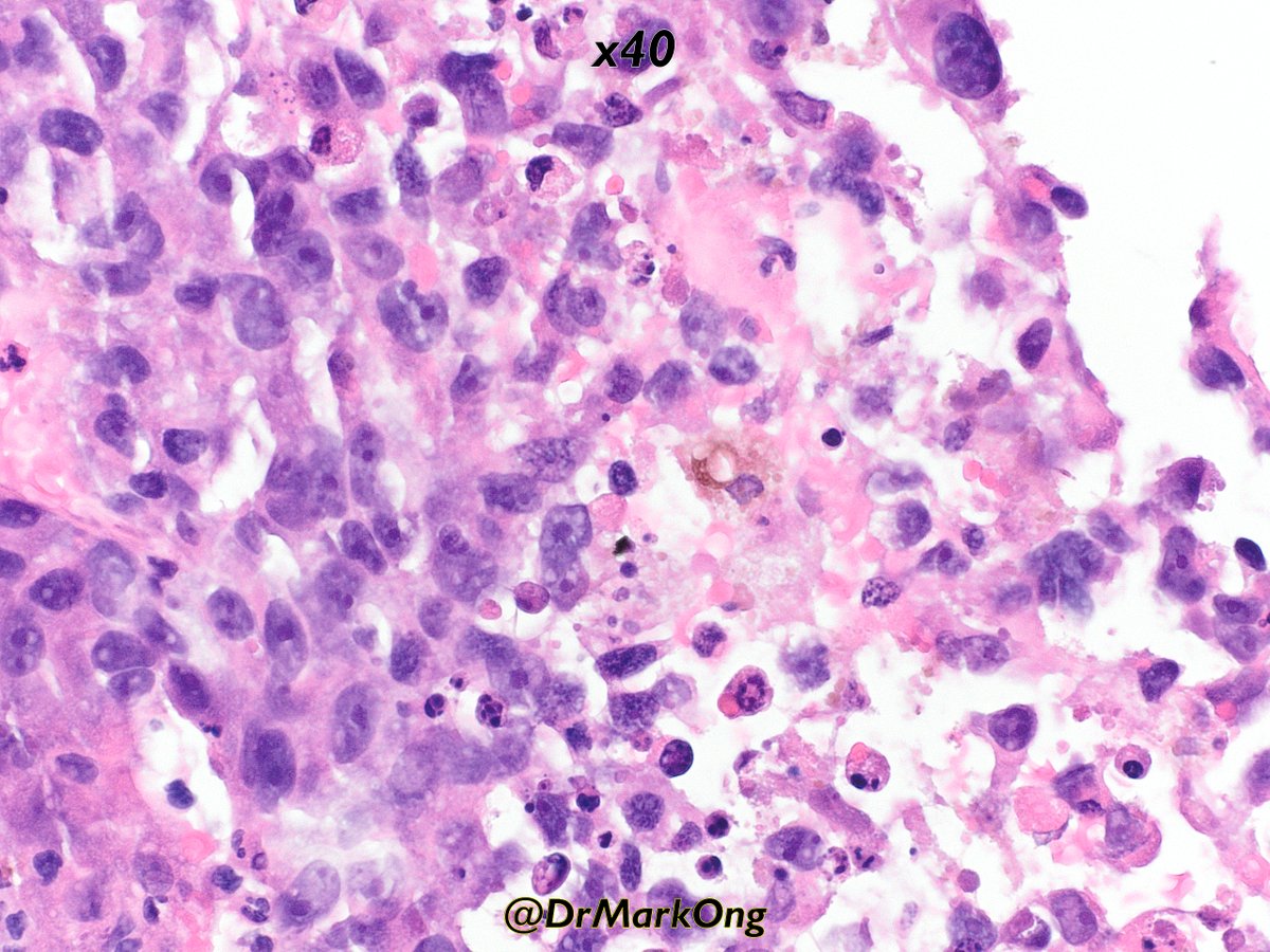
HHV8 was also positive (the dot-like pattern is easier to appreciated on the x40 picture).
Final diagnosis: Kaposis' sarcoma
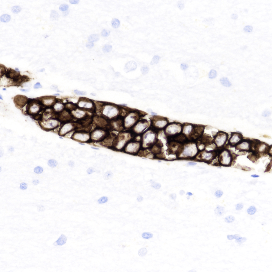
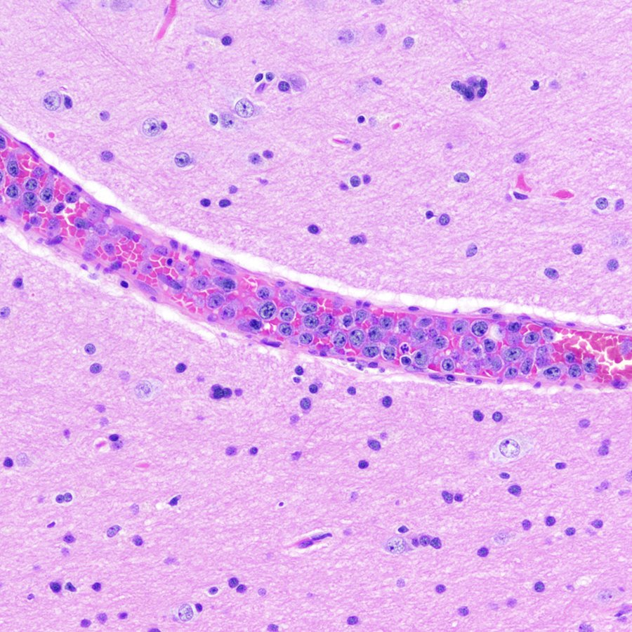
To kick off the month of #February, this might at first glance look like an innocuous section of cerebral cortex, but look more closely. What's the mystery immunostain? What's the diagnosis? #pathology#neuropath#PathTwitter
Let’s review atypical apocrine lesions of the breast this week! 💄
Atypical Apocrine Adenosis 🔬
A non-expansile proliferation of atypical apocrine cells within sclerosing adenosis, showing cytologic atypia that falls short of apocrine DCIS.
#PathX#PathTwitter#breastpath
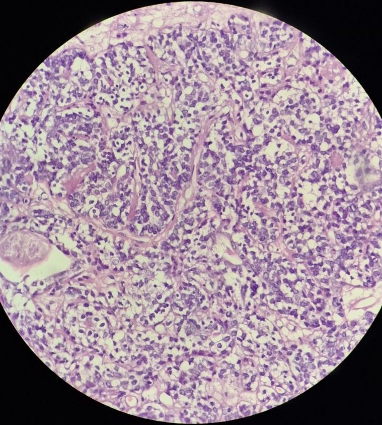
Biopsy of abdominal mass in elderly male with disseminated disease on radiological imaging.
Immunohistochemistry in next post.
What's your preferred diagnosis?
#GIpath#pathology#pathX#pathtwitter
Epithelioid features can be seen in a variety of lesions- let's review:
What is your interpretation?
A. Epithelioid sarcoma
B. Epithelioid histiocytoma
C. Epithelioid angiosarcoma
D. Epithelioid hemangioendothelioma
#PathPearls#PathQuiz
Our Gynecologic Pathology cases @UMiamiPathology are fascinating! Here’s a striking example of pilomatrix-like high-grade endometrial carcinoma (PiMHEC), a recently described entity mostly driven by CTNNB1 mutations. Look for solid basaloid growth with conspicuous central tumor cell necrosis, and pilomatrix-like keratinization with the hallmark ghost cells. This case also had a focal conventional FIGO grade 1 endometrioid component. IHC shows diffuse nuclear β-catenin with loss of PAX8 and ER in the pilomatrix-like component. These tumors are believed to behave aggressively.