

Top Tweets for #Peripheralnerveblock

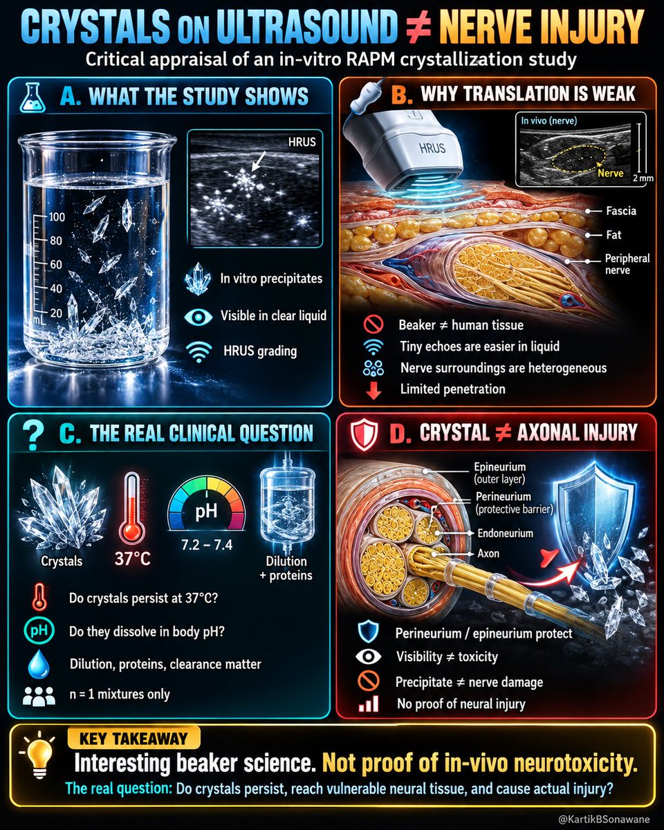
🧊 𝐂𝐑𝐘𝐒𝐓𝐀𝐋𝐒 𝐎𝐍 𝐔𝐋𝐓𝐑𝐀𝐒𝐎𝐔𝐍𝐃 ≠ 𝐍𝐄𝐑𝐕𝐄 𝐈𝐍𝐉𝐔𝐑𝐘
#RegionalAnesthesia #LocalAnesthetics #Neurotoxicity #Crystallization #Ultrasound #PeripheralNerveBlock #NerveInjury #InVitro #CriticalAppraisal #EvidenceBasedMedicine #RAPM
𝗖𝗥𝗜𝗧𝗜𝗖𝗔𝗟 𝗔𝗣𝗣𝗥𝗔𝗜𝗦𝗔𝗟:
🔵 𝐀. 𝐖𝐇𝐀𝐓 𝐃𝐈𝐃 𝐓𝐇𝐄 𝐒��𝐔𝐃𝐘 𝐀𝐂𝐓𝐔𝐀𝐋𝐋𝐘 𝐒𝐇𝐎𝐖?
▪️ LA/adjuvant mixtures can form visible precipitates.
▪️ 24–33 MHz ultrasound can detect them in a clear liquid environment.
▪️ A semiquantitative sonographic crystallisation score was proposed.
▪️ This demonstrates visibility of precipitates, not biological toxicity.
🟠 𝐁. 𝐁𝐄𝐀𝐊𝐄𝐑 ≠ 𝐇𝐔𝐌𝐀𝐍 𝐓𝐈𝐒𝐒𝐔𝐄
▪️ Hyperechoic particles are easy to identify against an almost anechoic liquid background.
▪️ Around nerves, fascia, fat, muscle, epineurium and connective tissue already create heterogeneous echogenicity.
▪️ Distinguishing a tiny crystal from normal tissue echoes may therefore be extremely difficult.
▪️ UHF imaging also has limited penetration - approximately 1–2 cm in this setting.
▪️ The authors themselves acknowledge that in-vivo visibility remains uncertain.
🔴 𝐂. 𝐂𝐑𝐘𝐒𝐓𝐀𝐋𝐋𝐈𝐒𝐀𝐓𝐈��𝐍 𝐈𝐒 𝐍𝐎𝐓 𝐓𝐇𝐄 𝐂𝐋𝐈𝐍𝐈𝐂𝐀𝐋 𝐄𝐍𝐃𝐏𝐎𝐈𝐍𝐓
▪️ Mixing two solutions may alter pH, ionisation and solubility → precipitation.
▪️ The real question is what happens after injection.
▪️ Do crystals remain undissolved at 37°C and physiological pH?
▪️ What happens after dilution by extracellular fluid, buffering, protein interaction and vascular clearance?
▪️ Do they dissolve, fragment, aggregate or persist?
▪️ Persistence was not studied.
▪️ Temperature was not even controlled in this experiment.
🟣 𝐃. 𝐂𝐑𝐘𝐒𝐓𝐀𝐋 ≠ 𝐀𝐗𝐎𝐍𝐀𝐋 𝐈𝐍𝐉𝐔𝐑𝐘
▪️ Axons are not lying freely exposed beside the needle tip.
▪️ They are protected within endoneurial, perineurial and epineurial compartments.
▪️ Therefore, direct access of relatively large particulate material to vulnerable neural structures cannot simply be assumed.
▪️ The important question is not merely “Did crystals form?”
▪️ It is:
Formation → Persistence → Neural localisation → Cellular interaction → Structural injury → Functional neurological deficit
▪️ None of these downstream steps was demonstrated here.
▪️ Importantly, nerve injury does not necessarily require direct crystal entry into an axon; vascular, inflammatory or perineural mechanisms are theoretically possible, but these also remain unproven.
⚠️ 𝐀𝐃𝐃𝐈𝐓𝐈𝐎𝐍𝐀𝐋 𝐌𝐄𝐓𝐇𝐎𝐃𝐎𝐋𝐎𝐆𝐈𝐂𝐀𝐋 𝐂𝐎𝐍𝐂𝐄𝐑𝐍𝐒
▪️ Only n=1 preparation per formulation was reported.
▪️ Multiple images from one sample ≠ independent experimental replication.
▪️ Samples underwent gravitational settling and were subsequently stirred before later measurements.
▪️ Stirring itself could have influenced the apparent increase in crystallisation grade.
▪️ The proposed ultrasound grading score was not microscopically validated.
▪️ Therefore, excellent observer agreement confirms agreement between readers, not necessarily biological validity of the grading system.
���� 𝐊𝐄𝐘 𝐓𝐀𝐊𝐄𝐀𝐖𝐀𝐘𝐒
▪️ Crystal formation ≠ crystal persistence.
▪️ Crystal persistence ≠ neural exposure.
▪️ Neural exposure ≠ neurotoxicity.
▪️ Ultrasound visibility in liquid ≠ reliable identification in living tissue.
▪️ A laboratory precipitate should not become a surrogate marker for nerve injury.
The clinically meaningful question is therefore not:
“𝐂𝐚𝐧 𝐰𝐞 𝐬𝐞𝐞 𝐋𝐀 𝐜𝐫𝐲𝐬𝐭𝐚𝐥𝐬?”
It is:
“𝐃𝐨 𝐭𝐡𝐞𝐬𝐞 𝐜𝐫𝐲𝐬𝐭𝐚𝐥𝐬 𝐬𝐮𝐫𝐯𝐢𝐯𝐞 𝐢𝐧 𝐯𝐢𝐯𝐨, 𝐫𝐞𝐚𝐜𝐡 𝐚 𝐛𝐢𝐨𝐥𝐨𝐠𝐢𝐜𝐚𝐥𝐥𝐲 𝐯𝐮𝐥𝐧𝐞𝐫𝐚𝐛𝐥𝐞 𝐧𝐞𝐮𝐫𝐚𝐥 𝐜𝐨𝐦𝐩𝐚𝐫𝐭𝐦𝐞𝐧𝐭, 𝐚𝐧𝐝 𝐜𝐚𝐮𝐬𝐞 𝐝𝐞𝐦𝐨𝐧𝐬𝐭𝐫𝐚𝐛𝐥𝐞 𝐢𝐧𝐣𝐮𝐫𝐲?”
Until that chain is demonstrated, this remains an interesting proof-of-concept imaging experiment, rather than evidence of clinically relevant nerve toxicity.
💥 𝗔𝗡𝗞𝗟𝗘 𝗕𝗟𝗢𝗖𝗞: 𝗜𝗧’𝗦 𝗡𝗢𝗧 𝗔𝗕𝗢𝗨𝗧 𝗧𝗛𝗘 𝗡𝗨𝗠𝗕𝗘𝗥
#RegionalAnesthesia #AnkleBlock #FITBlock #SciaticBlock #SaphenousBlock #MotorSparing #FootSurgery #PeripheralNerveBlock #RegionalAnaesthesia #CriticalAppraisal #EvidenceBasedPractice #ClinicalReasoning
𝗖𝗥𝗜𝗧𝗜𝗖𝗔𝗟 𝗔𝗣𝗣𝗥𝗔𝗜𝗦𝗔𝗟:
🧠 𝗦𝗛𝗢𝗥𝗧 𝗔𝗡𝗔𝗧𝗢𝗠𝗬
🔹 Sciatic nerve → Tibial nerve + Common peroneal/fibular nerve
🔹 Distally → Tibial, superficial & deep peroneal/fibular + sural nerves supply most of the foot
🔹 Saphenous nerve adds the femoral contribution
💉 𝗛𝗢𝗪 𝗠𝗔𝗡𝗬 𝗣𝗥𝗜𝗖𝗞𝗦?
🔹 Conventional ankle block → 5
🔹 FIT ankle block → 2
🔹 Popliteal sciatic + saphenous block → 2
🔹 SPEDI block → 1
🔹 Neuraxial → 1
🎯 𝗦𝗢, 𝗪𝗛𝗔𝗧 𝗥𝗘𝗔𝗟𝗟𝗬 𝗠𝗔𝗧𝗧𝗘𝗥𝗦?
🔹 Not merely how many times we prick
🔹 But where we block, what we block and why we block
🔹 Surgical anesthesia? Analgesia? Motor preservation? Early ambulation?
🔑 𝗞𝗘𝗬 𝗧𝗔𝗞𝗘𝗔𝗪𝗔𝗬𝗦
🔹 Fewer pricks ≠ automatically better block
🔹 Distal block ≠ completely motor-free block
🔹 Ankle block mainly preserves proximal/extrinsic leg motor function, while intrinsic foot motor function may still be affected
🔹 Choose the strategy according to the desired clinical outcome - not the needle count.
𝗧𝗵𝗲 𝗯𝗲𝘀𝘁 𝗯𝗹𝗼𝗰𝗸 𝗶𝘀 𝗻𝗼𝘁 𝘁𝗵𝗲 𝗼𝗻𝗲 𝘄𝗶𝘁𝗵 𝘁𝗵𝗲 𝗳𝗲𝘄𝗲𝘀𝘁 𝗽𝗿𝗶𝗰𝗸𝘀; 𝗶𝘁 𝗶𝘀 𝘁𝗵𝗲 𝗼𝗻𝗲 𝘁𝗵𝗮𝘁 𝗯𝗲𝘀𝘁 𝗺𝗮𝘁𝗰𝗵𝗲𝘀 𝘁𝗵𝗲 𝗽𝘂𝗿𝗽𝗼𝘀𝗲.

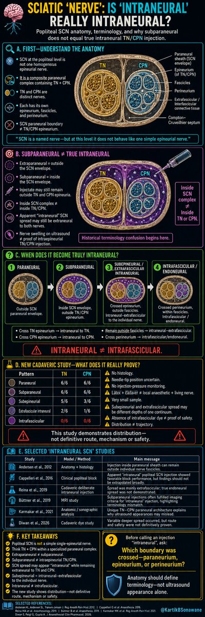
🧠 𝗦𝗖𝗜𝗔𝗧𝗜𝗖 “𝗡𝗘𝗥𝗩𝗘”: 𝗜𝗦 “𝗜𝗡𝗧𝗥𝗔𝗡𝗘𝗨𝗥𝗔𝗟” 𝗥𝗘���𝗟𝗟𝗬 𝗜𝗡𝗧𝗥𝗔𝗡𝗘𝗨𝗥𝗔𝗟?
#RegionalAnesthesia #SciaticNerve #SciaticNerveBlock #PoplitealBlock #PeripheralNerveBlock #NerveAnatomy #Sonoanatomy
#GrayZonesInRA #GrayAreasInRA
𝐆𝐑𝐀𝐘 𝐙𝐎𝐍𝐄𝐒 𝐈𝐍 𝐑𝐀:
🔬 𝗔. 𝗙𝗜𝗥𝗦𝗧 𝗨𝗡𝗗𝗘𝗥𝗦𝗧𝗔𝗡𝗗 𝗧𝗛𝗘 𝗔𝗡𝗔𝗧𝗢𝗠𝗬
▫️ Calling SCN, a nerve, is itself is MISNOMER.
▫️ It is a composite neural complex containing TN + CPN within a paraneural envelope.
▫️ TN & CPN are distinct nerves.
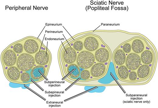
▫️ Each has its own epineurium, fascicles, perineurium.
▫️ Therefore:
SCN paraneural boundary ≠ TN/CPN epineurium.
🎯 𝗕. 𝗦𝗨𝗕𝗣𝗔𝗥𝗔𝗡𝗘𝗨𝗥𝗔𝗟 ≠ 𝗧𝗥𝗨𝗘 𝗜𝗡𝗧𝗥𝗔𝗡𝗘𝗨𝗥𝗔𝗟
▫️ Extraparaneural → outside the SCN paraneural envelope.
▫️ Subparaneural → inside that envelope.
▫️ Yet injectate may remain outside both TN & CPN epineuria.
���𝗻𝘀𝗶𝗱𝗲 𝗦𝗖𝗡 𝗰𝗼𝗺𝗽𝗹𝗲𝘅 ≠ 𝗶𝗻𝘀𝗶𝗱𝗲 𝗧𝗡/𝗖𝗣𝗡.
▫️ Thus, spread may look “intraneural” relative to the composite SCN while remaining extraneural to its component nerves.
▫️ Nerve swelling on ultrasound ≠ proof of intraepineurial TN/CPN injection.
▫️ Previous MRI/sonographic studies have already demonstrated this important terminology–anatomy mismatch.
“Intraneural” means little unless we specify WHICH boundary has been crossed.
🧩 𝗖. 𝗪𝗛𝗘𝗡 𝗗𝗢𝗘𝗦 𝗜𝗧 𝗕𝗘𝗖𝗢𝗠𝗘 𝗧𝗥𝗨𝗟𝗬 𝗜𝗡𝗧𝗥𝗔𝗡𝗘𝗨𝗥𝗔𝗟?
▫️ Cross TN epineurium → intraneural to TN.
▫️ Cross CPN epineurium → intraneural to CPN.
▫️ Remain outside fascicles → intraneural–extrafascicular.
▫️ “Subepineurial” therefore belongs within this broad extrafascicular domain.
▫️ Cross perineurium → intrafascicular/endoneural.
Paraneural → Subparaneural → Subepineurial/Extrafascicular → Intrafascicular
𝗜𝗡𝗧𝗥𝗔𝗡𝗘𝗨𝗥𝗔𝗟 ≠ 𝗜𝗡𝗧𝗥𝗔𝗙𝗔𝗦𝗖𝗜𝗖𝗨𝗟𝗔𝗥.
🧪 𝗗. 𝗡𝗘𝗪 𝗖𝗔𝗗𝗔𝗩𝗘𝗥𝗜𝗖 𝗦𝗧𝗨𝗗𝗬 - 𝗪𝗛𝗔𝗧 𝗗𝗢𝗘𝗦 𝗜𝗧 𝗥𝗘𝗔𝗟𝗟𝗬 𝗣𝗥𝗢𝗩𝗘?
▫️ Paraneural/subparaneural spread was universal.
▫️ Deeper subepineurial and extrafascicular spread occurred variably.
▫️ No intrafascicular spread was demonstrated.
But important limitations remain:
⚠️ No histology → exact microscopic boundary crossing unconfirmed.
⚠️ Needle-tip position uncertain → deeper dye cannot automatically prove sequential inward migration.
⚠️ No pressure monitoring → mechanism of tissue separation remains unknown.
⚠️ Latex + cadaver ≠ LA + living nerve.
⚠️ Very small sample → TN–CPN differences remain hypothesis-generating.
⚠️ “Subepineurial” & “extrafascicular intraneural” may represent different depths of the same continuum, not completely separate compartments.
⚠️ Absence of fascicular dye ≠ proof of clinical safety.
Most importantly:
The study demonstrates DISTRIBUTION, not necessarily TRAJECTORY.
📚 𝗘. 𝗪𝗛𝗬 𝗢𝗟𝗗 “𝗜𝗡𝗧𝗥𝗔𝗡𝗘𝗨𝗥𝗔𝗟 𝗦𝗖𝗡” 𝗦𝗧𝗨𝗗𝗜𝗘𝗦 𝗡𝗘𝗘𝗗 𝗥𝗘𝗜𝗡𝗧𝗘𝗥𝗣𝗥𝗘𝗧𝗔𝗧𝗜𝗢𝗡
▫️ Much of the reassuring “intraneural” literature comes from the popliteal SCN.
▫️ Prior clinical, MRI, sonographic & cadaveric studies have shown that:
apparent SCN intraneural spread may actually be subparaneural or extrafascicular.
▫️ Their findings therefore cannot be automatically extrapolated to ordinary single-epineurial peripheral nerves.
The “safe intraneural SCN” narrative may partly be an ANATOMICAL NOMENCLATURE problem.
💡 𝗞𝗘𝗬 𝗧𝗔𝗞𝗘𝗔𝗪𝗔𝗬𝗦
▫️ Popliteal SCN ≠ simple single-epineurial nerve.
▫️ Think TN + CPN within a specialised paraneural complex.
▫️ Extraparaneural ≠ subparaneural.
▫️ Subparaneural ≠ intraepineurial TN/CPN.
▫️ SCN spread may appear “intraneural” while remaining extraneural to TN & CPN.
▫️ Subepineurial = intraneural–extrafascicular to the individual nerve.
▫️ Intraneural ≠ intrafascicular.
🎯 𝗕𝗘𝗙𝗢𝗥𝗘 𝗖𝗔𝗟𝗟𝗜𝗡�� 𝗔𝗡 𝗜𝗡𝗝𝗘𝗖𝗧𝗜𝗢𝗡 “𝗜𝗡𝗧𝗥𝗔𝗡𝗘𝗨𝗥𝗔𝗟”, 𝗔𝗦𝗞:
Which boundary was crossed : PARANEURIUM, EPINEURIUM or PERINEURIUM?
Anatomy should define the terminology, not ultrasound appearance alone.
🔥 RA & ANS: A. Knowing the Sympathetic System Before Blocking It
#RegionalAnesthesia #SympatheticNervousSystem #AnesthesiaEducation #NeuraxialAnesthesia #PeripheralNerveBlock #PainMedicine #RAandANS
#PerioperativeMedicine #StressResponse #AdrenalMedulla #AnatomyMatters
#TipOfTheDay #MyRATips
Tip of the Day:
Why regional anesthesia does more than just numb pain?
The sympathetic nervous system quietly travels alongside our nerves and vessels and when we perform RA, we often block it without even trying.
🧠 The Sympathetic Highway
🔹 🚦 Origin: Starts in the spinal cord from T1–L2 (thoracolumbar outflow)
🔹 🛣️ Exit route: Leaves via ventral root → spinal nerve → white ramus
🔹 🔗 Relay station: Enters the sympathetic chain. May synapse at the same level, go up/down, or pass through as splanchnic nerves
🔹 🌿 Final journey: Returns via gray ramus. Travels with peripheral nerves and blood vessels to skin, muscles, joints, and organs
🎯 One-line rule👇
White ramus = exit from spinal nerve (T1–L2 only)
Gray ramus = entry into spinal nerve (all levels)
💉 What Happens During Regional Anesthesia?
🟢 Sympathetic fibers are small, superficial, and highly sensitive to local anesthetics
➡️ They get blocked earlier and more easily than motor fibers
🌡️ Clinical Signs
✨ 🔥 Warm limb after block → sympathetic vasodilation
✨ 💧 Dry skin → sudomotor block
✨ 📉 Drop in BP after spinal/epidural → sympathetic blockade
✨ 👁️ Ptosis, miosis, warm arm → Horner’s syndrome (after stellate spread)
⛔ But Important Reality Check
⚠️ Not all pain is sympathetic pain
❌ Tourniquet pain
❌ Ischemic pain
❌ Compartment syndrome pain
👉 These persist because they involve:
🔹 Deep C-fibers & A-delta fibers 🧬
🔹 Central sensitization🧠
🔹 Multi-segmental spinal input 🔄
🔹 Ischemic chemical mediators 🧪
🔹 Blocking sympathetic efferents ≠ blocking all pain afferents🔹
🎯 Why This Matters to Us (RA Perspective):
🧩 Perfect block ≠ pain-free tourniquet
🧩 Large-volume proximal blocks feel “better”
🧩 Vasodilation happens before sensory loss
🧩 Sympathetic block is a feature, not a complication
🏁 Take-home
RA always blocks the sympathetic system but pain relief depends on much more than just sympathetic blockade.

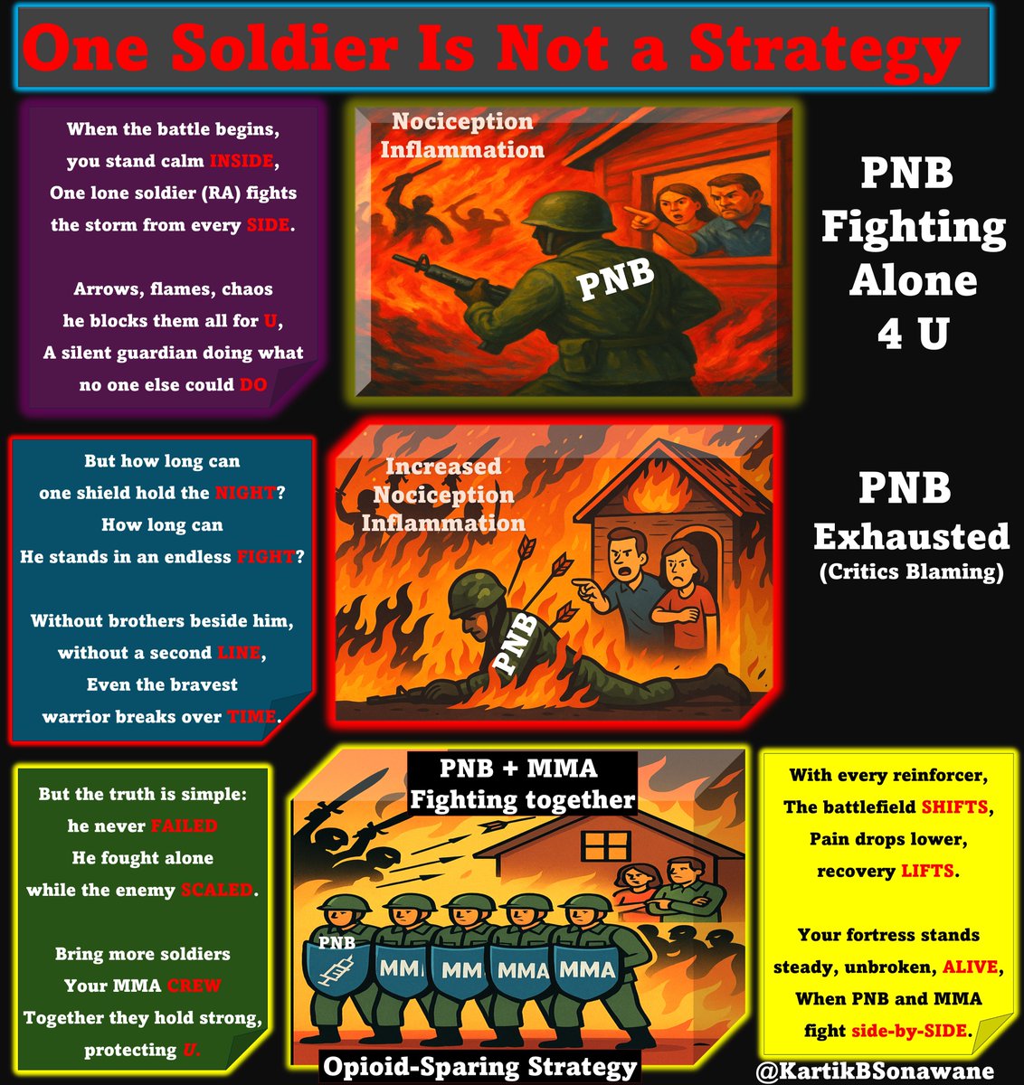
🥅 The Great Regional Anesthesia ‘Self-Goal’: Blaming the Protector 🪖
#RegionalAnaesthesia #PeripheralNerveBlock
#ReboundPain #MultimodalAnalgesia
#OpioidSparing #AcutePainManagement
#Anaesthesia #EvidenceBasedPractice
#TipOfTheDay #MyRATips
Tip of the Day:
👁️Imagine This…
👉Your home is under constant attack (the nonstop barrage of postoperative nociception).
👉At the front gate stands one brave soldier (your Peripheral Nerve Block (PNB).
🪖 One Soldier… Fighting for You
He:
🛡️ Blocks every strike
🔥 Absorbs every hit
🤫 Keeps your home quiet
⏳ Protects you for hours
He fights alone… but he fights brilliantly.
💀When the Soldier Falls…
Every warrior has limits.
When exhaustion takes him down (the block wears off):
💥 The attackers surge
🏚️ Damage occurs
😱 Chaos breaks loose
And what happens?
You turn around and say: “The destruction started when he fell - so he must be responsible.”
That, right there, is the SELF GOAL.
Blaming the only thing that protected you.
🎯 The Correct Logic
The soldier saved you for hours.
The real problem is that the walls were never reinforced.
You never sent in:
💊 Paracetamol
💊 NSAIDs / COX-2 inhibitors
💊 Pre-emptive dosing
💊 Scheduled MMA
When the soldier fell, the unprepared system collapsed.
👉 The enemy didn’t get stronger.
Your defenses were incomplete.
🔍 What Is Rebound Pain?
⭐Rebound pain = the normal return of surgical nociception once a single-shot PNB wears off.
⭐It is:
🧠 Physiological, not pathological
🕒 Transient, not persistent
💊 Preventable, not inevitable
✅Rebound pain only becomes dramatic when block offset meets weak MMA.
✅If the systemic analgesia is poor, the rebound looks like a “complication.”
✅But it's really a system-created phenomenon.
💥Rebound Pain Doesn’t Increase Opioids - Bad MMA Does
Many claim:
❌ PNB → Rebound Pain → More Opioids
But the true sequence is:
🛡️ PNB provides exceptional early analgesia
🔄 Block wears off
⚠️ Weak MMA creates an analgesic gap
💉 Rescue opioids shoot up
So the correct chain is:
❌ PNB → Opioids
✔ Poor MMA → Opioids
🚨The Real Problem in the BMJ Paper
📌 “Rebound pain” was a label, not an outcome - the 0.17/10 NRS difference is clinically negligible.
📉 Raw data show PNB patients used fewer opioids; the ‘22.7% increase’ appears only after heavy modelling.
💊 PNB patients received more pre-op opioids and weaker MMA - yet the block was blamed instead of the analgesic plan.
🔔 Pain spikes after block regression only when MMA is inadequate - that’s a system failure, not a block failure.
🩺 The same study shows lower chronic pain at 12 months in PNB patients - clear proof that PNB is the protector.
⭐Final Take-Home Message
PNBs are NOT the problem - poor perioperative analgesia is.
✔ PNBs reduce early pain and early opioid use
✔ PNBs reduce chronic post-surgical pain
✔ PNBs improve recovery & shorten hospital stay
✔ Rebound pain is fully preventable with good MMA
❌ PNBs do NOT inherently increase opioids
❌ Rebound pain is NOT a block complication
❌ Blaming RA is a clinical self-goal
💡PNB is the protector — not the culprit.
💡Rebound pain reflects a gap in MMA, not a flaw in RA.

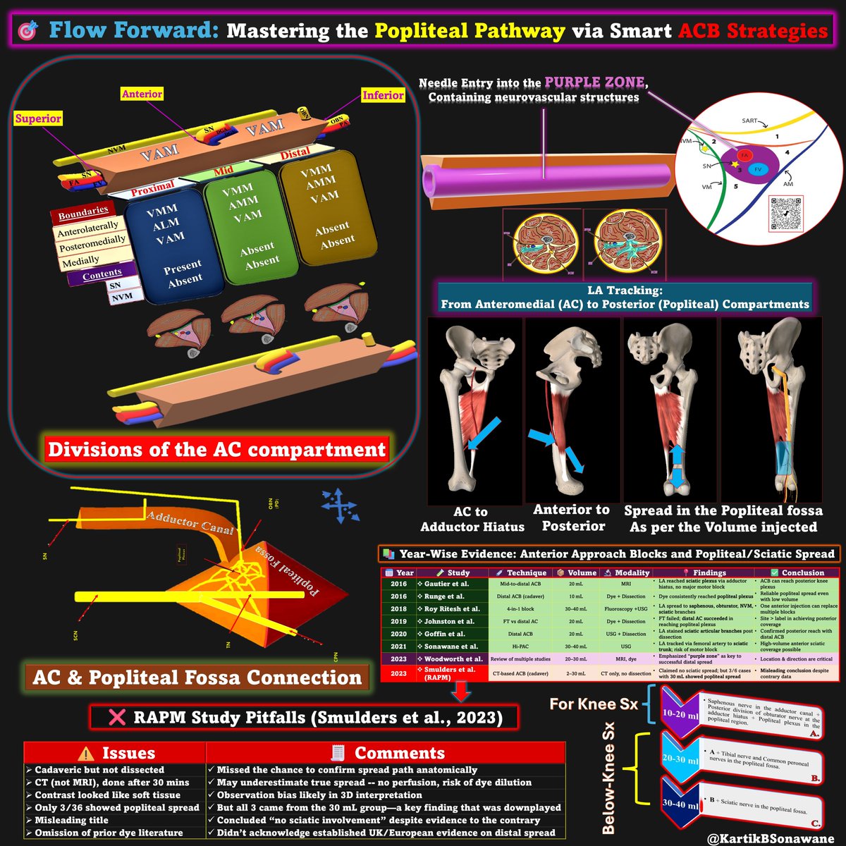
🎯 🚀 Forward Flow Forward: Mastering the Popliteal Pathway via Smart ACB Strategies 🛠
#RegionalAnesthesia #NerveBlocks #UltrasoundGuided #PeripheralNerveBlock #AnesthesiaEducation #AdductorCanalBlock #PoplitealPlexus #AnteriorApproach #DSB #HiPAC #4in1Block #KneeAnalgesia #MotorSparing #TipOfTheDay #MyRATips
Tip of the Day:
🧠The Adductor Canal: Nature’s Passageway to the Popliteal Fossa
👉The AC is a musculoaponeurotic tunnel, bounded by two muscles (VMM & ALM/AMM) and one aponeurosis (VAM).
👉It naturally connects the anterior thigh to the posterior thigh through the adductor hiatus.
👉 Using this anatomical passageway, local anesthetic can be directed from an anterior approach to reach posterior targets like the popliteal plexus and even sciatic nerve branches.
📢Volume Dynamics & Direction in ACB-Based Techniques
🔍 When performing ACBs for knee or below-knee surgeries, understanding how LA moves and how we can guide that movement is critical for:
🦵Targeting the popliteal plexus (for knee analgesia),
🦵Or reaching the sciatic nerve trunks (for foot/ankle surgery).
💡 Popliteal Plexus = Enough for Knee Surgery
✨LA spread to posterior capsule & intra-articular branches (tibial & obturator articular twigs) is usually sufficient for TKA and arthroscopy.
✨Achievable with 15–20 mL when injected in the PURPLE perivascular zone at the proximal AC (just distal to FT apex).
💡 Sciatic Trunk Coverage = Needed for Below-Knee Surgery
✨Full coverage of the TN & CPN requires: High-volume LA (30–40 mL) in the AC.
✨The drug follows the femoral artery through the adductor hiatus into the popliteal fossa and around the sciatic nerve.
📢📚 What Did Dye Studies Show?
📝Multiple dye and contrast studies have confirmed spread to the popliteal plexus when large volumes (20–30 mL) are injected perivascularly in the distal AC.
📝In the landmark Gautier et al. series, 20 mL of contrast injected into the AC showed dye around the sciatic nerve in all cases, with limited sensory or motor impact.
📝Similarly, Runge et al. and Goffin et al. (cadaveric work) demonstrated consistent spread into the popliteal fossa, especially when dye was injected 1–2 cm above the adductor hiatus.
📝Most importantly, Johnston et al. confirmed that dye injected into the distal AC could spread to the popliteal plexus, while distal FT injections did not.
📝Hi-PAC and 4-in-1 innovated further by extending the anterior spread to the sciatic trunk.
��The only contradictory claim (RAPM, 2023) lacks dissection and underreports its own positive data.
💉 Where to Inject? The “Purple Zone”
🎯 Optimal target = deep to sartorius, lateral to femoral artery, and under the VAM.
🟣 This is the perivascular PURPLE zone, a conduit that sends LA through the adductor hiatus → popliteal fossa.
📏 How Much? Volume Matters
📍<10 mL: Often insufficient to reach the popliteal plexus or include major articular branches.
📍15-20 mL: Ideal for proximal AC – to achieve popliteal spread without sciatic involvement.
📍≥20 mL: Increases likelihood of sciatic spread, especially if injected distally in the AC.
⚙️ DSB Technique = Dual Injection for Strategic Spread
🔑Volume Efficiency + Direction Control
🔹 1st injection: 10-20 mL in distal FT pre-fills the proximal tract, restricting retrograde spread of the second injection.
🔹 2nd injection: 10–20 mL in AC moves distally, pushing the drug toward the popliteal plexus
✅ Key Advantage: DSB achieves popliteal plexus involvement with less total volume than needed in a single-shot ACB alone.
🧠 Concept: Blocking the Backdoor, Opening the Front - By “sealing off” the proximal path with the first bolus, DSB converts the second bolus into a distally driven column, minimizing leakage and maximizing effect.
🛑Why the RAPM (Smulders et al.) Study Missed the Mark?
🧟Cadaveric study, but no dissection❌.
🕓CT after 30 min - not real-time, no vascular flow, potential underestimation.
🧪Used 2 mL groups (!), only 30 mL showed popliteal spread in 3 cases - yet still concluded “no sciatic spread.”
❌ No comparison with prior positive studies
❌ Ignored key findings in 30 mL group
📛Titled with a strong claim despite contradictory findings: misleading, non-neutral, and goal-directed bias.
📉Contrast in CT can resemble soft tissue = interpretation bias.
🔄 Why Modify ACB Instead of Adding Blocks?
🚫 You don’t always need iPACK or posterior sciatic blocks.
✅ A single smart ACB (or DSB) can:
🔳Cover posterior capsule of the knee
🔳Reach articular sciatic branches
🔳Sometimes even spread to sciatic trunk (if needed, with higher volume)
🙌 Minimizes motor block, preserves function, reduces number of injections.
🟥Achieve foot analgesia from the front in below-knee surgeries when using Hi-PAC or 4-in-1.
���🔑 Final Take-Home Message
✅ Site trumps name: Where and how you inject matters more than the block label
✅ 15–20 mL in the purple zone = safe & effective posterior knee coverage
✅ DSB enhances forward flow, limiting volume while boosting effectiveness
✅ 30–40 mL directed distally (Hi-PAC/4-in-1) can reach sciatic trunk from anterior thigh
✅ One modified ACB can replace multiple blocks -if you respect anatomy and guide your volume!
✅Popliteal-plexus spread rarely weakens ankle; Hi-PAC/4-in-1 almost certainly will factor this into rehab plans. So, always use diluted LA for selective sensory blockade.
✅Document your plan - write “Popliteal-plexus target (20 mL)” or “Hi-PAC (35 mL) for below-knee surgery” so PACU and physio teams know what to expect.
🚀 One clever anterior injection can replace multiple blocks - if you know what you're doing.💡
"🧬 Vastoadductor Membrane (VAM): Gateway to Smart Analgesia in TKA 💡"
#VastoadductorMembrane #VAM #DualSubsartorialBlock
#MotorSparingAnesthesia #RegionalAnesthesia #AdductorCanalBlock #UltrasoundGuidedBlock #AnatomyMatters #FascialPlanes #PeripheralNerveBlock
#AnesthesiaEducation #SmartBlocking #AnatomicalPrecision #TKAPainManagement #EnhancedRecoveryAfterSurgery #FastTrackSurgery
#KneeSurgeryCare #TipoftheDay #MyRATips
Tip of the Day:
🧠Anatomical Role of the VAM
🧵The VAM is a dense, collagen-rich fascial septum forming the anteromedial boundary of the adductor canal.
🛖It lies deep to the sartorius, separating the vastus medialis muscle (VMM) from the adductor longus and magnus muscles.
🧱It acts as a physical barrier and anatomical tunnel wall, crucial for directing local anesthetic (LA) flow.
📍Attachments and Boundaries
🔹Laterally: Attaches to the posteromedial border of vastus medialis.
🔹Posteromedially: Anchors to the anterior surfaces of adductor longus (proximal) and adductor magnus (distal).
🧩Forms the anteromedial fascial boundary of the adductor canal, opposite the posteromedial muscle wall.
📏Extent and Dimensions
📐Length: Approximately 7.6 cm from the apex of the femoral triangle to the adductor hiatus.
📏Width: Varies - about 2.2 cm proximally, 1.7 cm midsection, and 0.5 cm distally.
📍Proximal origin: Around 28 cm distal to ASIS.
📍Distal termination: About 10 cm above the adductor tubercle.
🌬️Fenestrations in the VAM
🔍Present in ~75% of cadaveric specimens.
🔄Allow limited cross-compartment communication between the STM–VMM plane and the adductor canal.
🟢Fenestrations are physiological gaps in the VAM that allow passage of:
🟩Nerve to vastus medialis (NVM)
🟦Saphenous nerve branches (31%)
🟧Obturator nerve branches (12.5%)
🟪Perforating veins (22%)
💧Local Anesthetic Flow Dynamics
💥When LA is injected in the STM–VMM interfascial plane, it prefers to spread:
🔽Beneath the VAM into the adductor canal - the path of least resistance.
🔁Along the natural musculoaponeurotic tunnel, facilitating broader spread and nerve coverage.
⚠️While fenestrations exist, they offer greater fascial resistance than the canal tunnel, hence most LA flows under the VAM, not through it.
💡Clinical Relevance in Regional Anesthesia
❌ACB targets the saphenous nerve, but often misses the NVM and other terminal femoral branches.
✅ DSB uses:
🩺A proximal injection in the STM–VMM plane to directly block NVM and SN.
💉A second injection into the adductor canal to reinforce distal coverage.
💉Why ACB Alone is Not Enough for TKA?
❌ACB only targets the saphenous nerve in the adductor canal.
❌ It misses:
💡NVM (often outside the canal in the STM–VMM plane)
💡Peripatellar plexus and Subsartorial plexus branches
💡Obturator nerve contributions and
💡Posterior knee nerves (if volume <15ml)
🔄 While fenestrations help some spread, they are not sufficient alone to ensure full knee coverage.
🧠Take-Home Messages
🛠️The VAM is not the roof of the AC - that’s the sartorius. Instead, VAM forms the medial fascial wall that governs how LA spreads.
✅ The VAM is not just a fascial boundary - it’s an anatomical gateway that determines the success or failure of motor-sparing regional anesthesia techniques.
📦LA injected beneath the VAM into the adductor canal flows easily along the low-resistance tunnel, not primarily through fenestrations.
🌀Its fenestrations allow cross-compartmental spread.
❌ACB alone, despite VAM fenestrations, is not sufficient for TKA.
🧩Mastering the VAM's anatomy and spread mechanics is essential for maximizing regional block efficacy while preserving quadriceps strength.


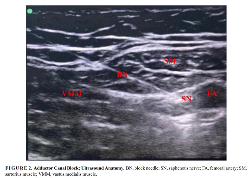
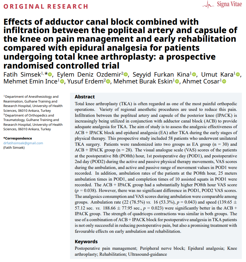
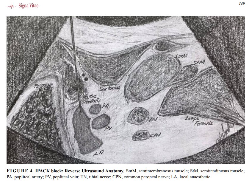
📷【Click】https://t.co/kbYr7P6bGV
#Postoperativepainmanagement;#Peripheralnerveblock;#Kneearthroplasty;#Epiduralanalgesia;#Rehabilitation;#Rehabilitation;

Data suggest periph nerve blocks provide analgesic benefit when intrathecal morphine can't be used or as rescue technique for refract pain. ASRA Pain Medicine News: https://t.co/RrcC497j4Z @MKwesiKwofie @DElmofty @NarouzeMD @ruthi_landau #Cesarean #Analgesia #PeripheralNerveBlock

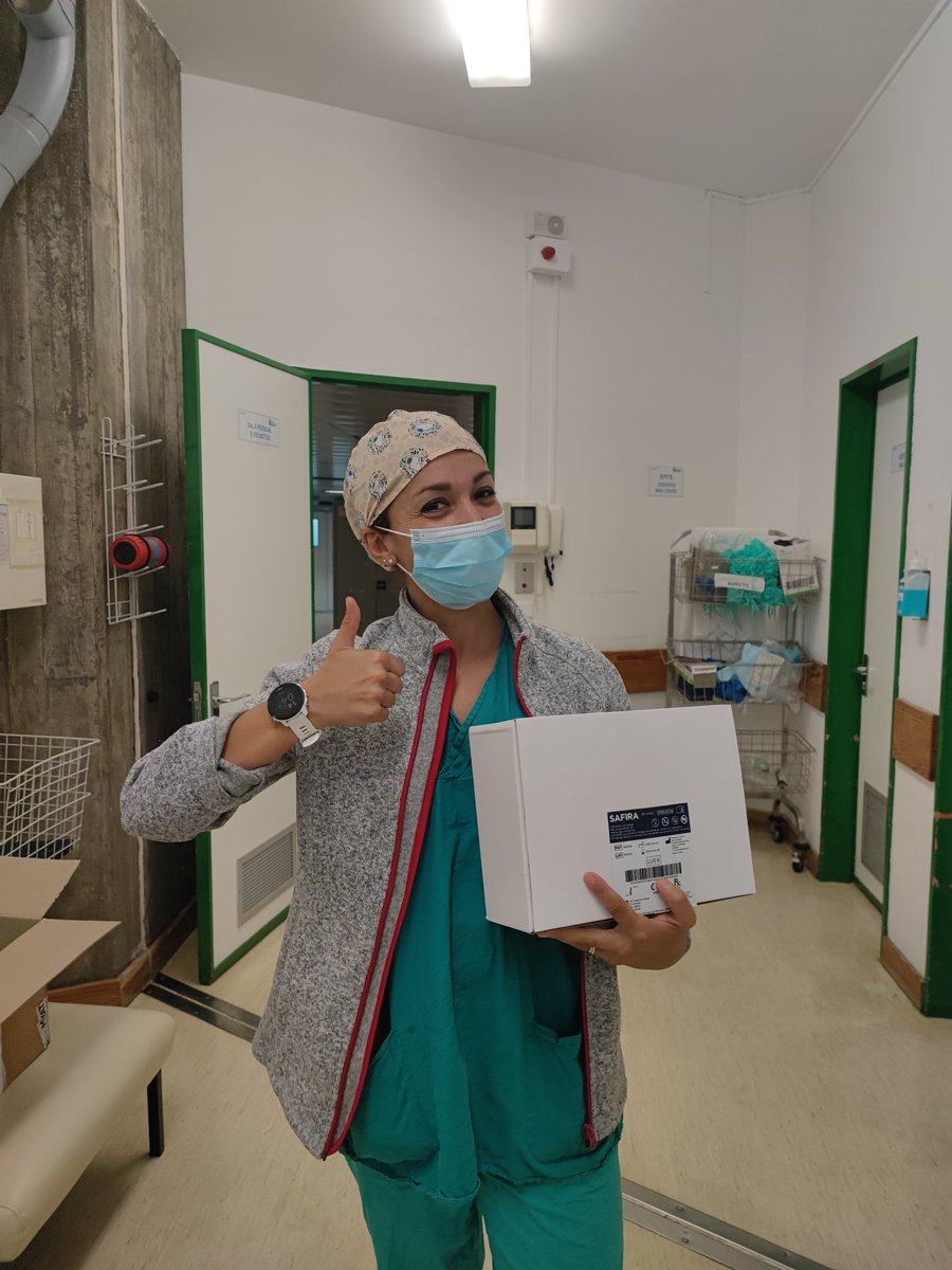
Delivered today! A #SAFIRA evaluation pack to @JoanaMMagalhae👍 Looking forward to hearing the results of the evaluation!
#regionalanaesthesia #innovation #UGRA #nerveblock #anesthesiology #nerveblocks #anaesthesia #USGRA #peripheralnerveblock

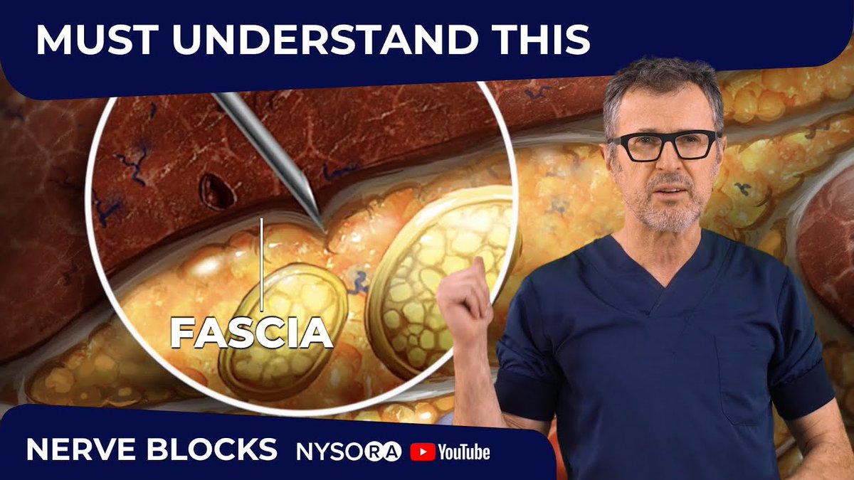
#Fascia plays an essential role in #PeripheralNerveBlock. Check our newest video here to learn the tricks to make your practice safer and more successful. https://t.co/olisHOBaiE

@NaveenEipe @Jerryblocksthat @Nadia_Hdz_MD @docmatter @OttAnesthesia @OttawaHospital @OttawaTrauma Many thanks @Jerryblocksthat for organizing this opportunity to discuss #RibFracture management with #PeripheralNerveBlock Catheters.
See how SAFIRA® can be integrated into #peripheralnerveblock procedures such as this #ankleblock
Watch the 2 minute video https://t.co/pZ7aS2HJWX
#regionalanaesthesia #anaesthetist #SAFIRA #footsurgery
What challenges face #anaesthetists when carrying out a #peripheralnerveblock & how can #SAFIRA® help overcome them?
Dr Peter Merjavy shares his thoughts in this short video https://t.co/glSZki1QOY
#regionalanaesthesia #anaesthesia

#regionalanesthesia #jedi #peripheralnerveblock Is Jedi Grip efficient and effective in ultrasound-guided peripheral n... https://t.co/TaxYg8OO3r

@NarouzeMD That block was so beautiful it inspired me to do a Regional Anesthesia Fellowship. #peripheralNerveBlock

#morningglory !!! one of @PennAnesthesia residents who rotated through #regionalanesthesia made the #peripheralnerveblock a primary anesthetic @PennMedicine #HUP #culturalshift #heretheycome @nelkassabany @VeenaGraffMD @jochrochmd @shuang821 @ibadiolamd @James22kim

Are you joining us in #Asheville? It's going to be beautiful that time of year! This is our last #PeripheralNerveBlock course for 2018! Check it out and grab your seat now.
@terasonUS

Editorial: What are the risks and benefits of intraneural local anesthesia injection? #RegionalAnesthesia #PeripheralNerveBlock #Anaesthesia #Anesthesia #RegionalAnaesthesia #localAnesthesia https://t.co/yyX8iooyjU
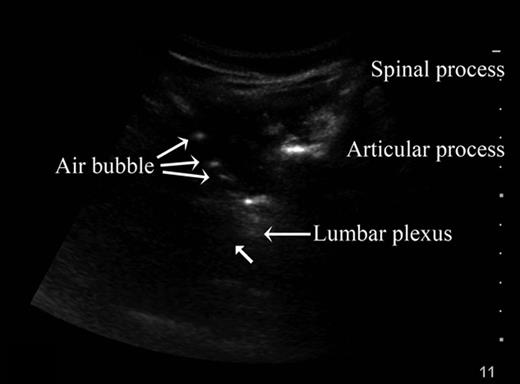
Images in #Anesthesiology: Ultrasound-guided #regionalAnesthesia micro air bubble contrast to visualize the needle. #peripheralNerveBlock #anesthesia #anaesthesia https://t.co/DO5lTAmoRB
Last Seen Hashtags on Sotwe
虐阳
Seen from Germany
pussy contractions
Seen from Indonesia
thick #mexican
Seen from Turkey
ctlartists
Seen from United States
ekjbthesla
Seen from United States
anxiouspanda(*******)*********** filter:videos
Seen from Netherlands
นัดเย็ดโคราช
Seen from Thailand
ديرتية
Seen from Switzerland
CureDApp
Seen from United States
mama短剧
Seen from Turkey
Most Popular Users

Elon Musk 
@elonmusk
241.7M followers

Barack Obama
@barackobama
119M followers

Cristiano Ronaldo
@cristiano
114.5M followers

Donald J. Trump
@realdonaldtrump
111.9M followers

Narendra Modi
@narendramodi
107.2M followers

Rihanna
@rihanna
98.7M followers

NASA
@nasa
92.4M followers

Justin Bieber
@justinbieber
91.8M followers

KATY PERRY
@katyperry
90M followers

Taylor Swift
@taylorswift13
84M followers

Lady Gaga
@ladygaga
75.5M followers

Virat Kohli
@imvkohli
73.4M followers

Kim Kardashian
@kimkardashian
70.9M followers

YouTube
@youtube
68.8M followers

Neymar Jr
@neymarjr
66.4M followers

Bill Gates
@billgates
65.2M followers

Selena Gomez
@selenagomez
63.1M followers

The Ellen Show
@theellenshow
62.3M followers

CNN
@cnn
61.8M followers

X
@x
60.7M followers