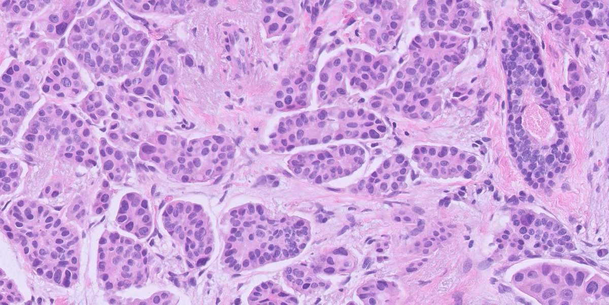
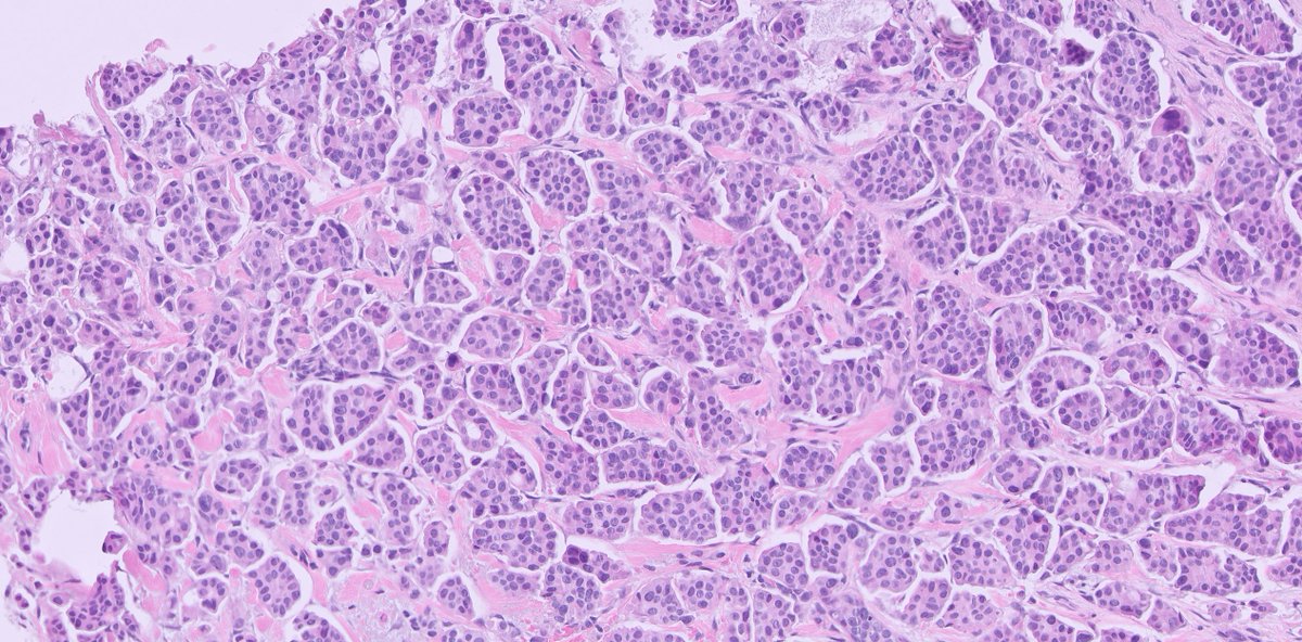
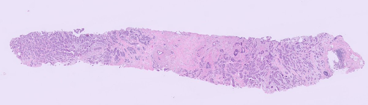
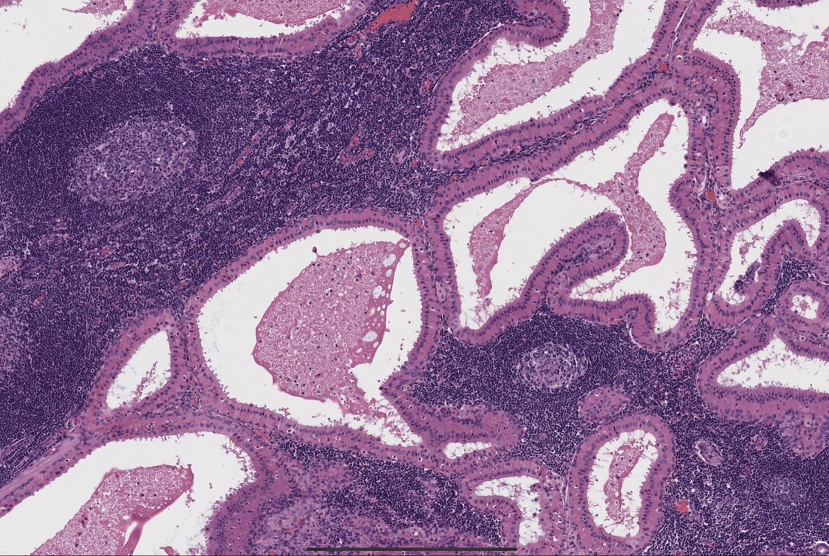
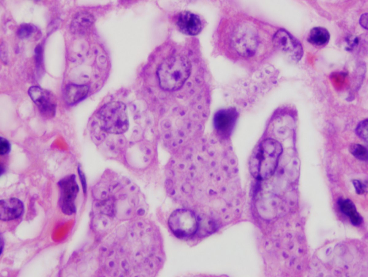
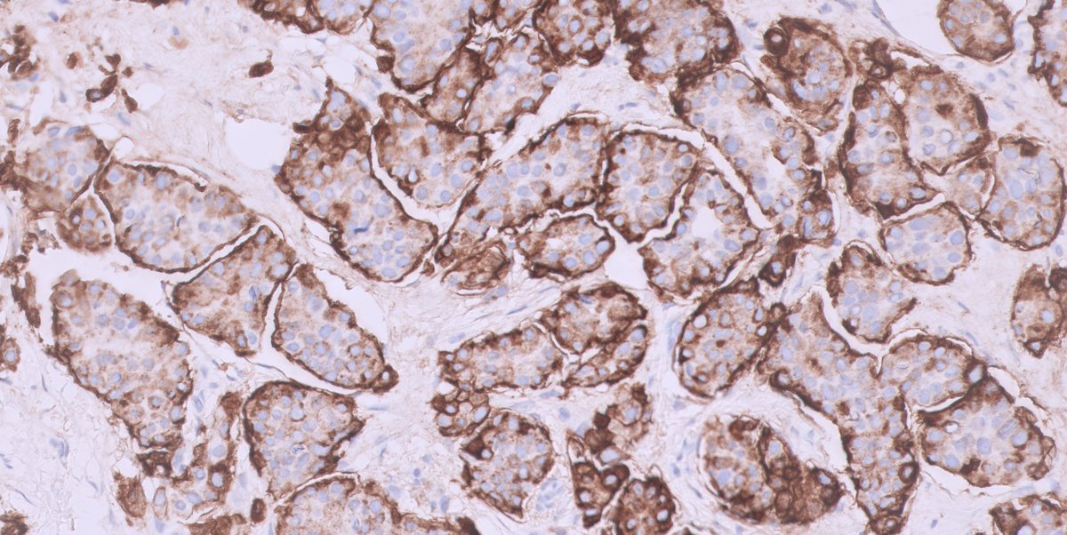
The foundation of histopathology is being able to accurately describe morphological appearances. In simple terms what words would you use to describe the image below? #histohandbook#pathology
51 year-old female presented with extensive microcalcification in left breast. The morphology of the partial mastectomy showed in figures. ER was positive in 50% of the lesional cells.
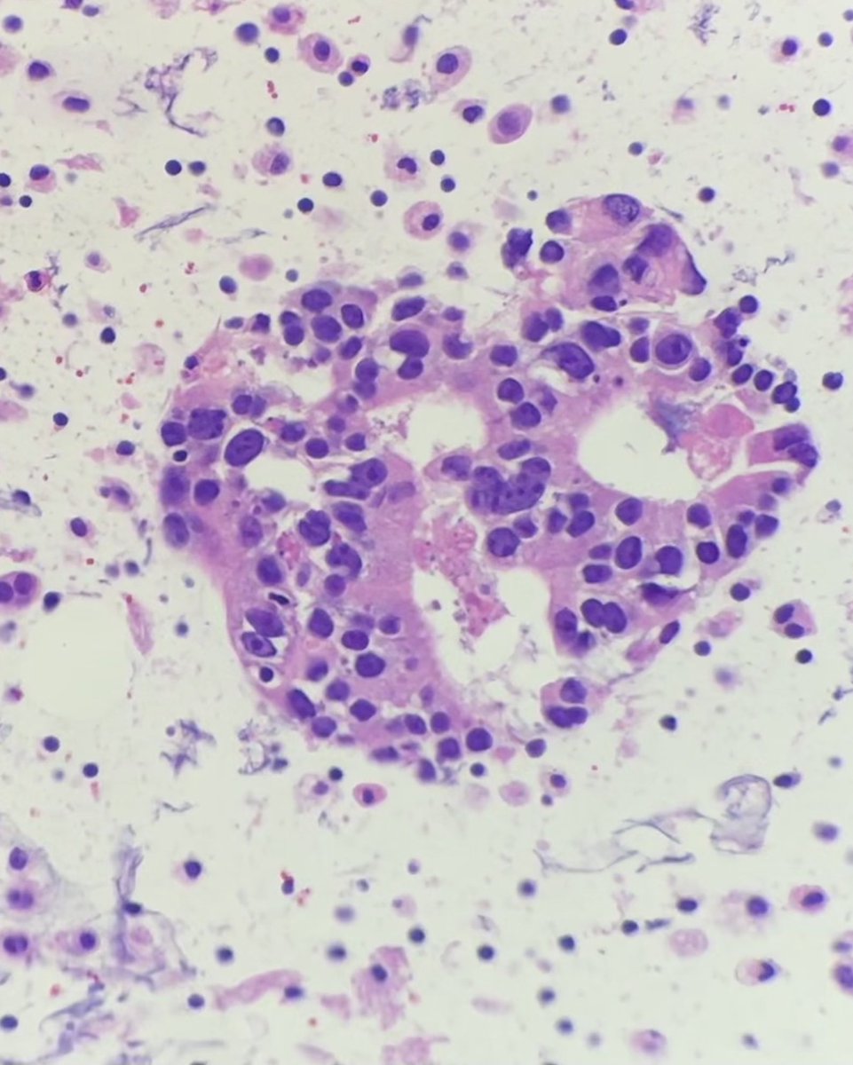
Cytology #fluidfriday 💉
Pleural fluid: PAP and cell block 🔬
How would you call this?
What stains would you order to work up tumor of unknown origin?
#cytopath#PathTwitter
@DrBMcGinn@MAHoureih Maybe we need to start one?
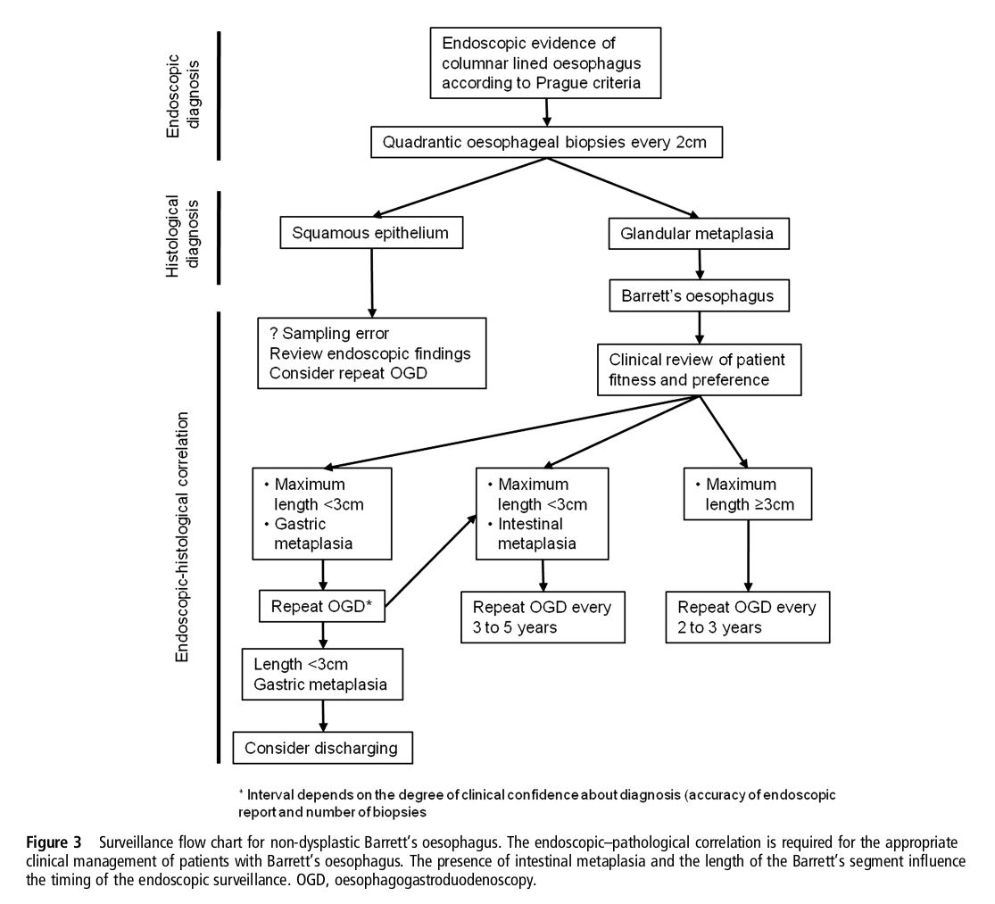
BSG guidelines on polyp follow-up.
Useful to know as no point chasing levels on a tiny polyp if it isn't going to change management.
#GIpath
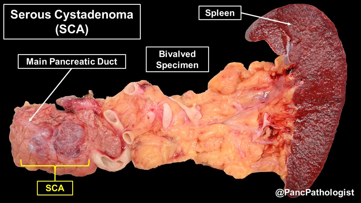
Although benign, a subset of Serous Cystadenomas (SCAs) can grow & occlude the #pancreatic duct. In addition to VHL, these SCAs have mutations in TP53 & the hTERT promoter. Pic#3 in formalin to bring out their architecture. #Pathology#GIPath#Pancreas#PrecisionMedicine
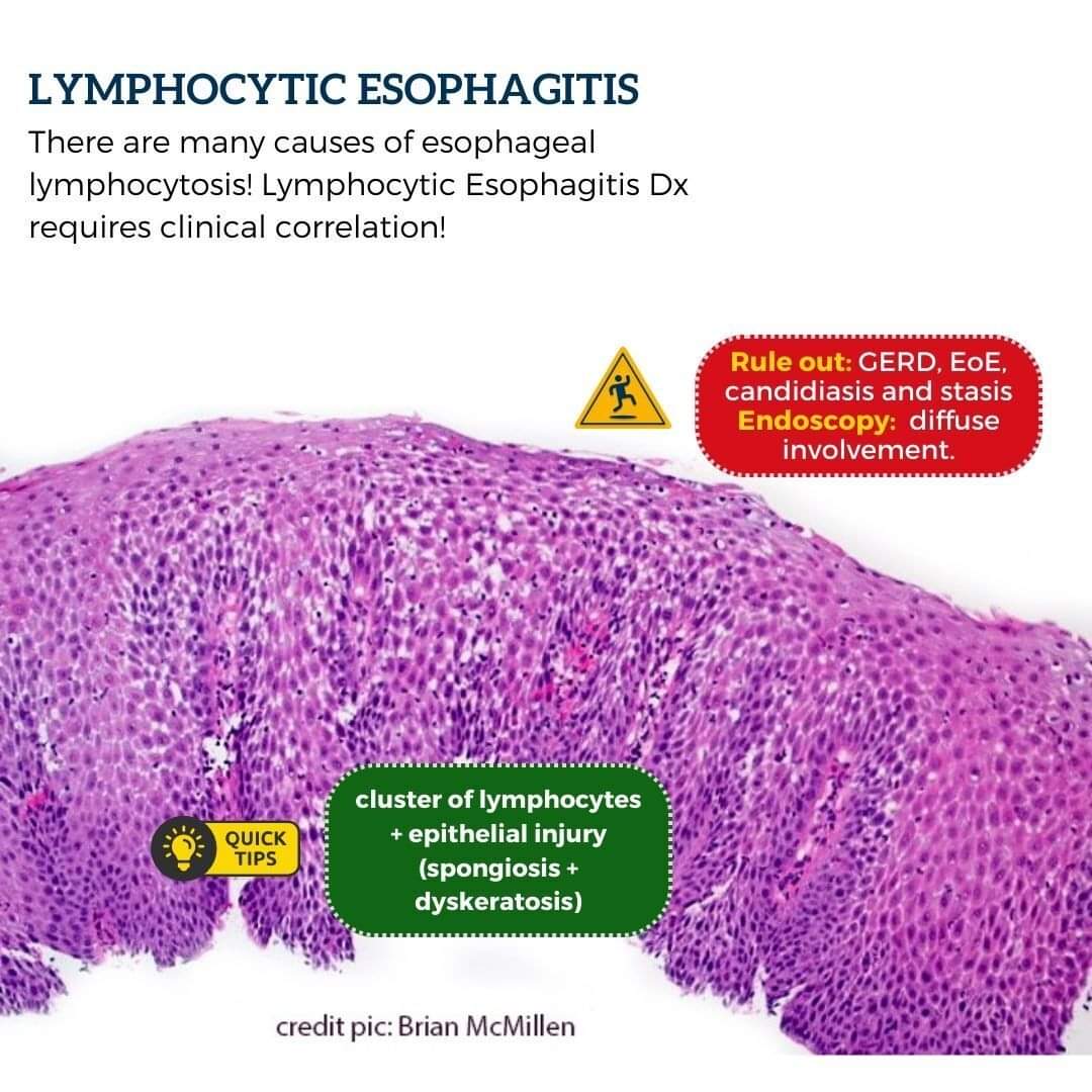
Stay tuned! Do the lymphocytic esophagitis dx if u see cluster of lymphocytes + signs of epithelial injury (spongiosis + dyskeratosis) + excluded GERD, EoE, candidiasis, stasis and endoscopy showing diffuse involvement.
In your report, mention associations.
#gipath#pathology