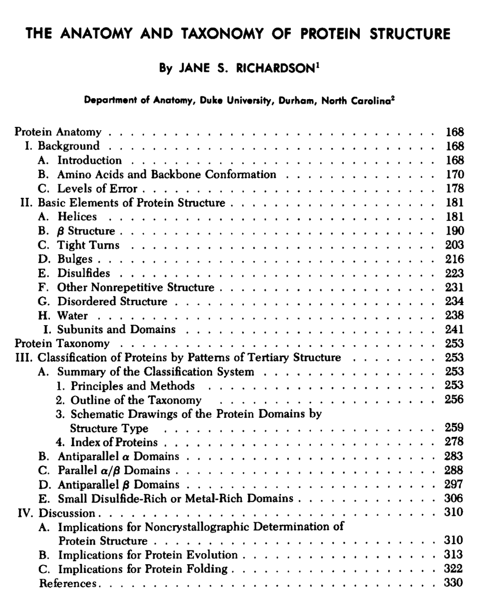
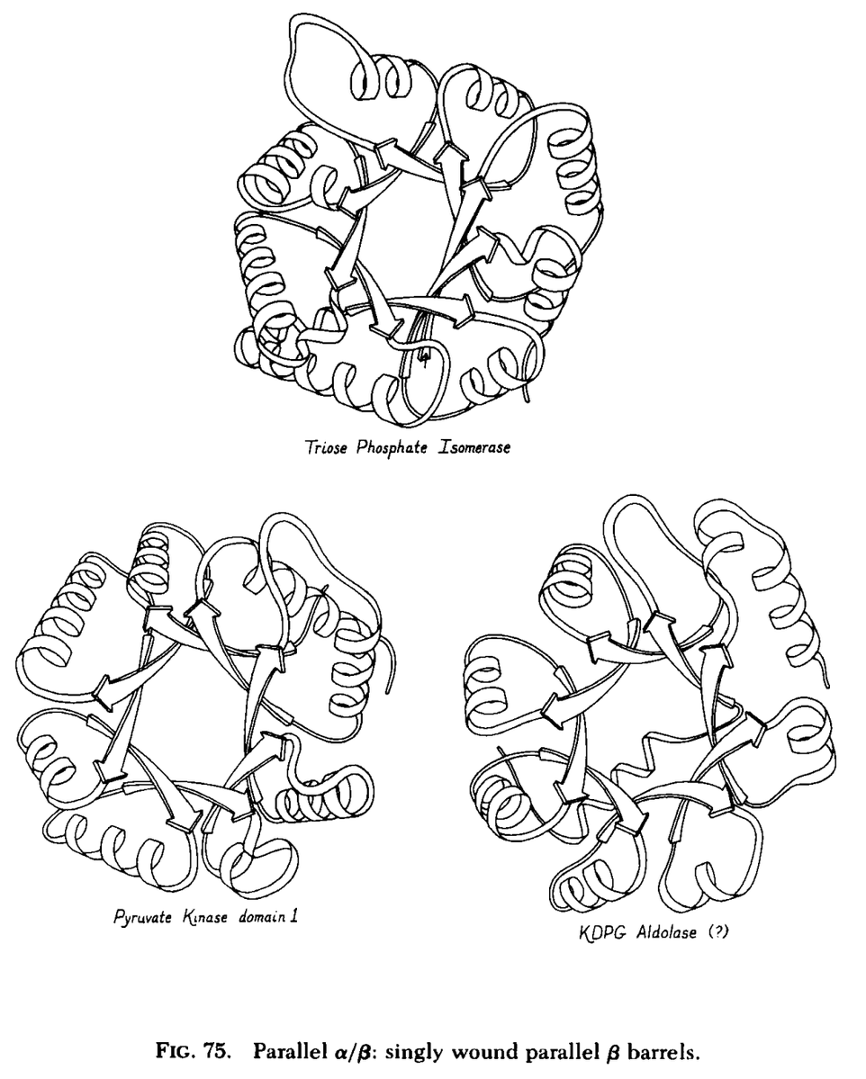
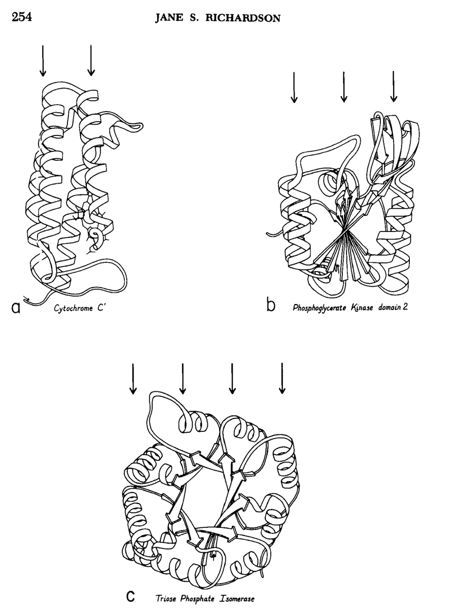
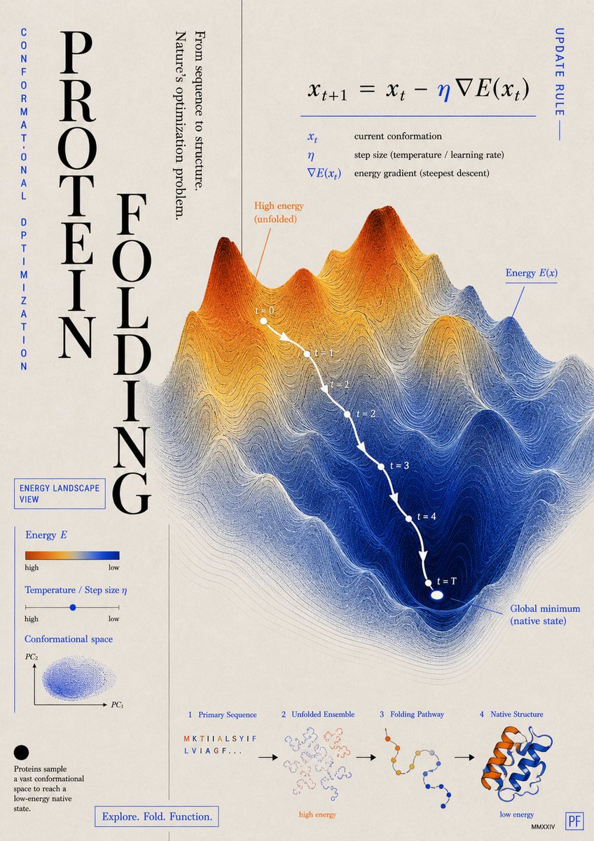
Mother of the ribbon diagram
One paper in 1981.
Changed how we see proteins.
If you haven’t read it, you’re late.
https://t.co/5OIR6riN3K
#compchem#phd
The world’s first AI-designed virus has officially landed in Science.
Using genome language models, researchers generated complete bacteriophage genomes—and experimentally validated 16 functional phages capable of infecting, replicating, and killing bacteria.
The study was first released as a preprint in 2025. Now, it has passed peer review and entered Science.
AI biology is moving from designing proteins to designing entire genomes.
Researchers in Science report the development of a general-purpose biomedical AI agent that can help automate biomedical research workflows.
The authors say their results point “toward a future in which AI agents work alongside human researchers to accelerate biomedical discovery from basic research to translation.”
Learn more: https://t.co/UdLl5uxMQe
1/10
🚨Preprint! One RING to rule them all (phages)👑💍⚔️
Bacteria build giant DNA-scaffolded RNase rings (larger than the ribosome!) to shut down viral infection🧬🧵👇
🔗https://t.co/NNvYtwvtjA
Research update: Distinct #RNA engagements define genome import (with the help of nsP2 #helicase) and replication elongation (of course by nsP4 #RNA#polymerase) in #alphaviruses#chikungunya
https://t.co/Zaj4rcSlZD
AlphaFold3 is no longer the best model.
Over the past year, we've seen many (mostly companies) claim that their newly released models matched or outperformed AlphaFold3 in some important structure prediction task. A substantive amount of these also underperformed their self-professed benchmarks under independent scrutiny.
This is why I was initially skeptical on the results from the ESMFold2, Protenix-v2, and OpenDDE teams' respective releases. Within a few weeks of each others, these groups each released benchmarks showing meaningful outperformance over AF3.
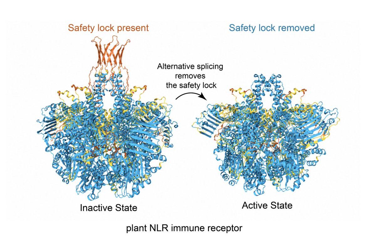
AlphaFold-based modelling lets us visualise these N-terminal “safety caps” and predict how they interfere with formation of the pore-forming resistosome. https://t.co/Ws1mU9fym7
David Baker & Veesler labs "building viruses - Institute for Protein Design (IPD)
-Two new Nature papers - "building viruses, at University of Washington" if you believe the headlines. What they've actually done is more interesting than that.
https://t.co/EjXP70iwQX
🧵1/9 If you're in peptide design, one static snapshot can make a candidate look like a sure thing. Lock it into the receptor in a frozen model, the scores light up. Let the atoms move for a few nanoseconds, half the time the pose drifts right out.
That gap is where most early-stage peptide programs lose money.
PeptAI is a built to catch it before any candidate hits synthesis, and accelerate open R&D at agentic velocity
Golgi–Mitochondria Contact Sites: The Hidden Lipid–Stress Interface
Mitochondria are no longer “stand-alone powerhouses.” They operate as networked organelles, physically coupled to the ER, Golgi, lysosomes, lipid droplets, and plasma membrane through membrane contact sites.
One emerging axis is especially underexplored: Golgi–mitochondria communication.
Recent reviews and mechanistic studies suggest that Golgi-derived membranes and lipids may participate in mitochondrial remodeling, respiratory adaptation, and stress signaling—not simply through vesicular trafficking, but via direct or three-way ER–Golgi–mitochondria contact platforms. The Golgi contact-site review The Fast and the Furious: Golgi Contact Sites highlighted Golgi–mitochondria contacts as one of the most experimentally supported new Golgi contact interfaces, especially in lipid exchange and mitochondrial dynamics.
The key concept:
Golgi-derived PI4P/lipid-enriched vesicles may be recruited near ER–mitochondria contact sites, creating a three-organelle signaling hub that coordinates lipid supply, mitochondrial fission, and metabolic adaptation. This shifts the Golgi from “post-ER cargo station” to an active regulator of mitochondrial state.
A second layer comes from COPI biology. COPI is classically known for Golgi–ER retrograde trafficking, but a 2023 Cell Reports study showed that COPI disruption reduces mitochondria–ER contact sites, impairs Ca²⁺ handling, increases mitophagy, decreases respiratory capacity, and accelerates axonal degeneration. Restoring MERCS partially rescued mitochondrial and neuronal phenotypes.
This suggests a causal chain:
Golgi–ER traffic → MERCS integrity → mitochondrial Ca²⁺/respiration → cell survival
For aging, neurodegeneration, cancer metabolism, and fibrosis, this is a rich hypothesis space. Golgi–mitochondria/MERCS dysfunction could act as a hidden “organelle logistics failure” linking lipid imbalance, mitochondrial fragmentation, impaired respiration, and chronic stress signaling.
The field now needs better tools: split-FP proximity reporters, EM tomography, proximity labeling, lipidomics, and perturbation screens to distinguish true contact-site biology from nearby organelle crowding. A 2023 methods review provides a useful technical roadmap for studying membrane contact sites.
Working hypothesis:
Golgi–mitochondria contact is not a minor cell-biology curiosity. It may be a tunable metabolic control node—where lipid trafficking, mitochondrial dynamics, and disease stress programs converge.
Key references
David Y, Castro IG, Schuldiner M. The Fast and the Furious: Golgi Contact Sites. Contact. 2021. DOI: 10.1177/25152564211034424.
Maddison DC et al. COPI-regulated mitochondria-ER contact site formation maintains axonal integrity. Cell Reports. 2023. DOI: 10.1016/j.celrep.2023.112883.
Diokmetzidou A, Scorrano L. Mitochondria–membranous organelle contacts at a glance. J Cell Sci. 2025. DOI: 10.1242/jcs.263895.
Sarhadi TR, Panse JS, Nagotu S. Mind the gap: Methods to study membrane contact sites. Experimental Cell Research. 2023. DOI: 10.1016/j.yexcr.2023.113756