A new cellular atlas of atherosclerosis based on scRNAseq, snATACseq, and spatial transcriptomics across 216 human samples.
A useful resource, including associations with histological levels of atheroprogression.
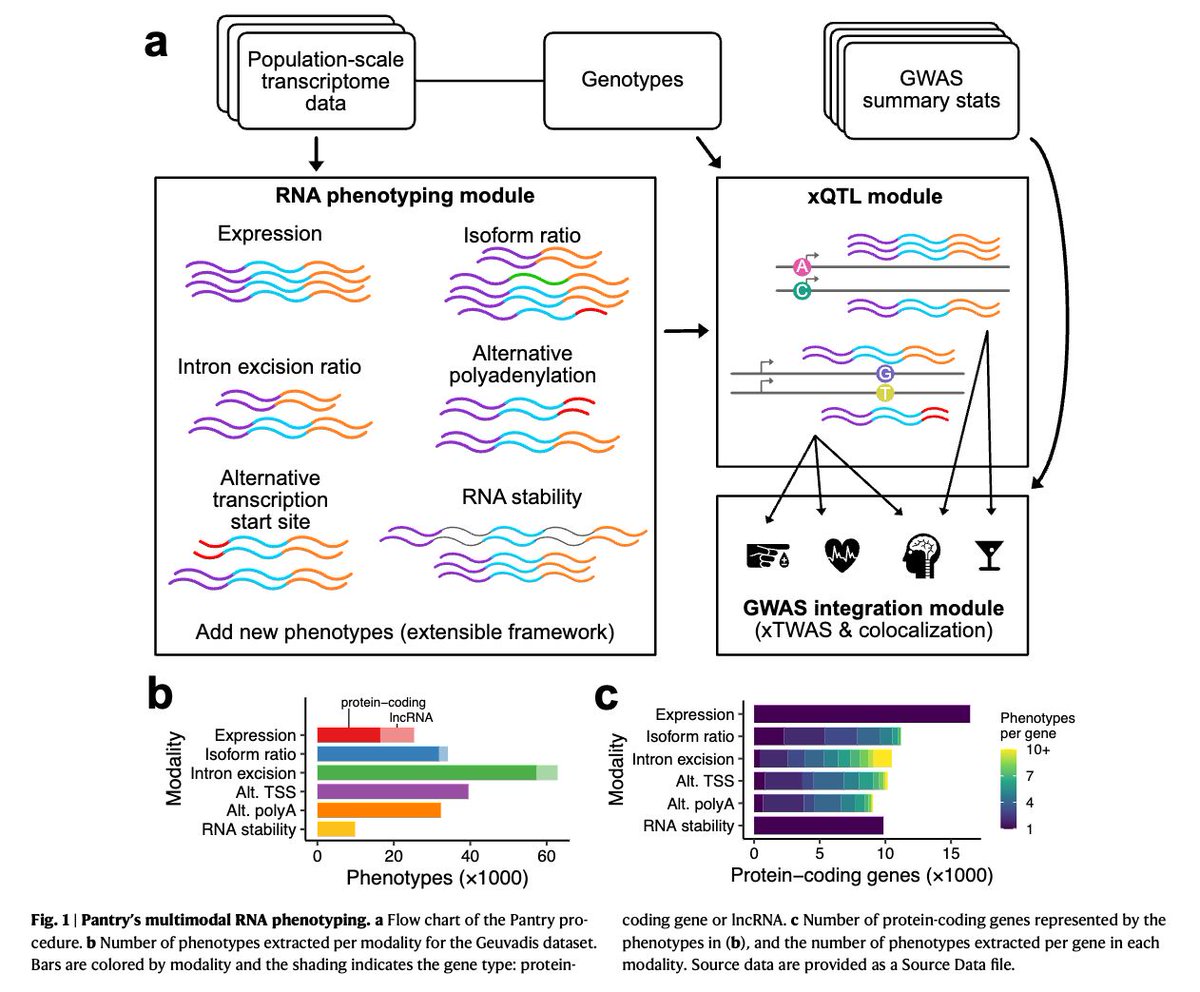
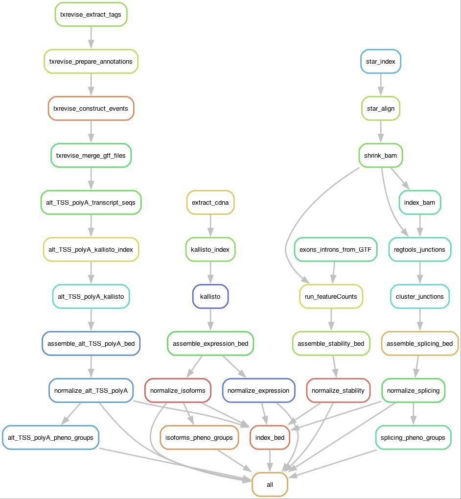
I am happy to share the updated preprint of our TenK10K multiome project. https://t.co/MQcxknvB8m. The full caQTL summary from 922 donors and 3.5M nuclei is now available for download, plus many key summary results! Lots of new analyses since v1. Quick tour of what's changed 👇
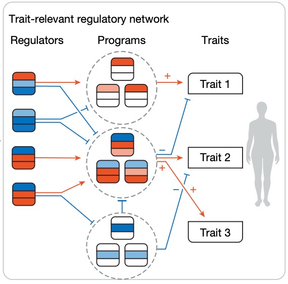
Modern GWAS can identify 1000s of significant hits but it can be hard to turn this into biological insight.
I'm excited to share our new work combining genetic associations and Perturb-seq to build interpretable causal graphs, out today in @Nature:
The 9p21.3 Coronary Artery Disease Risk Locus Modulates Vascular Cell-State Transitions via Enhancer-Driven Regulation of MTAP https://t.co/hRQCzgdRa6 #biorxiv_genomic
Excited to share Nona: a unifying multimodal masking framework for functional genomics.
Models for DNA have evolved along separate paths: sequence-to-function (AlphaGenome), language models (Evo2), and generative models (DDSM).
Can these be unified under a single paradigm? 1/15
We’re thrilled to share that our MERFISH+ preprint is now live on bioRxiv!👉https://t.co/M1SIyfnzMa
In this work, the Bintu and Zhu labs (UCSD) developed MERFISH+, a next-generation spatial genomics platform that combines genome-wide RNA and epigenetic imaging over a large field of view. By introducing acrydite-modified probes covalently anchored to hydrogels, MERFISH+ achieves remarkable imaging stability and enables >1,800-gene, multi-modal, and multi-month experiments.
With this platform, they, together with the Chi lab at UCSD, profiled a whole developing human heart at 12 post-conception week with merely two slides, resulting in a total of 53 slides, 3.1 million single cells and more than 30 cell types. Building upon our previous 3D reconstruction and modeling framework, Spateo (https://t.co/a0BC0Cf3Ec), we reconstruct the 3D human heart that nicely captures the anatomical structure of the heart, including the intricate vasculature network. Sophisticated analyses provide a holistic view of an entire organ and enable systematic characterization of 3D cellular neighborhoods and transcriptional gradients of substructures such as the descending arteries. Furthermore, using a generative integration framework for spatial multimodal data (Spateo-VI), we harmonized these MERFISH+ transcriptomic and chromatin data to reconstruct a 3D spatially-resolved multi-omics atlas of the developing human heart, shared at https://t.co/Jby4Pppzbr and https://t.co/s33SS7jvYL. MERFISH+ thus sets a new standard for large-format, multi-omic spatial profiling, enabling holistic, 3D characterization of organs at subcellular resolution.
Huge congratulations to first authors Colin Kern, @qingquanZhang2, @YifanLu2024 , and Jacqueline Eschbach, and to all collaborators from the Bintu, Zhu, Chi, and Qiu labs for this amazing team effort. Thanks for your diligence, creativity, and hard work on this project. We’re grateful for support from @arcinstitute and our generous donors.
Our lab is expanding—if you’re excited about building the next generation of single-cell and spatial genomics techniques and predictive single cell and spatial foundation models, we’re hiring! If you are interested, please reach out to me via direct message or email at [email protected].
We are excited for any potential collaborations along this line of research in Stanford, UCSF and Berkeley and other labs as well.
🔗link to the caQTL preprint: https://t.co/H7fe0QDKKX
🔗link to the previous single-cell eQTL preprint from TenK10K: https://t.co/2v8fcbXSv2
📊summ stats to become available with publication
Terrific preprint by @hartjackson using imaging mass cytometry (IMC) & matched genomics for building multimodality prediction models:
Integrated spatial proteomics of human PDAC uncovers an expanded tumour–immune– stroma spectrum with genomic associations
https://t.co/zb4njVsBId
@rkramann@NatureCVR @sikandhayat @novonordisk@ivivek87 You're absolutely right, sampling variability is always an important consideration with human specimens. I truly appreciate you bringing this up—thank you and happy holidays😃!
@sikandhayat @NatureCVR Great work! Quick question though—why is the advanced stage vessel's diameter so much larger than the control and early stages? It seems unusual for a human coronary artery to reach such a size.
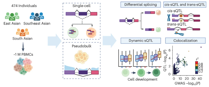
A great resource of single-cell splicing QTLs from the Asian Immune Diversity Atlas❗️
👉474 PBMC samples
👉1M cells analyed with scRNAseq
👉11,577 independent cell-specific cis-sQTLs
📜paper: https://t.co/Q1G6bqfKSl
📊sQTL summ stats: https://t.co/mVGActAE88
We are thrilled to share an HCA Collection of 40+ peer-reviewed papers in @NaturePortfolio shedding light on human development, healthy and disease biology and vital analytical tools. #HCA2024NatureCollection#HumanCellAtlas https://t.co/TKWn8CjXRd
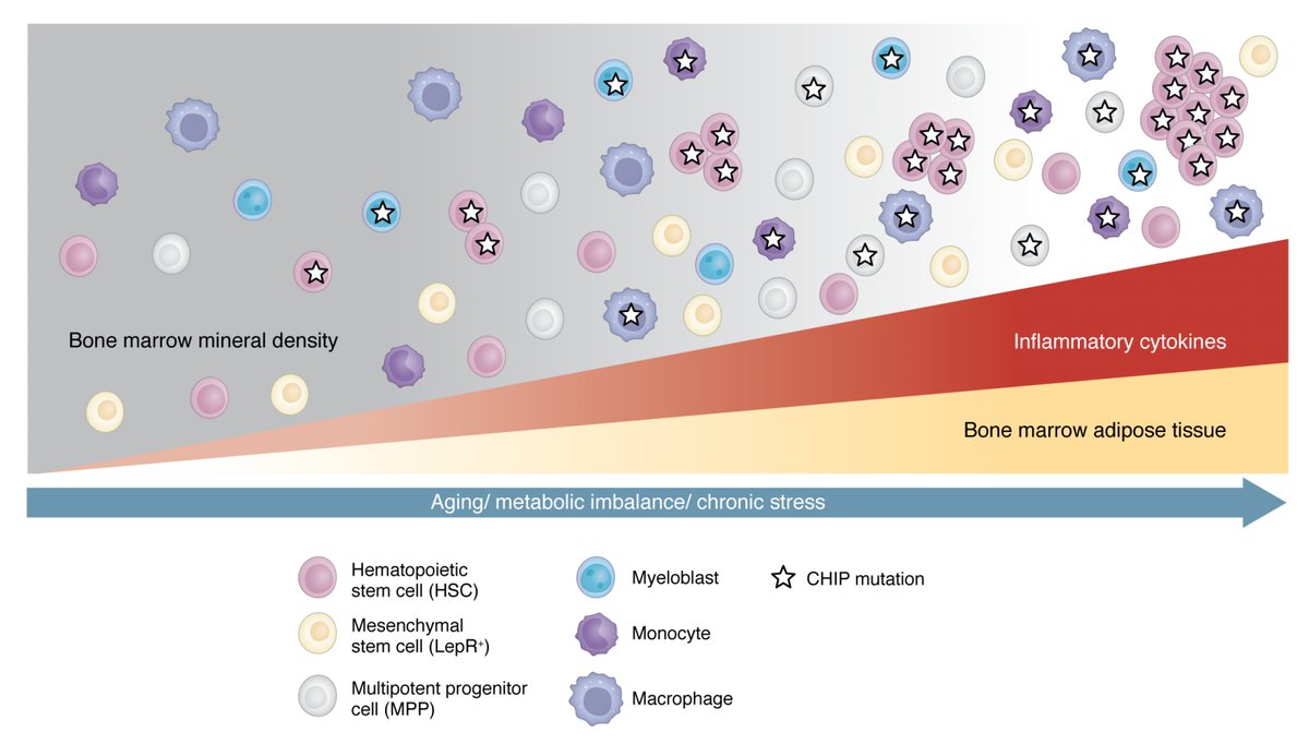
We keep ignoring CHIP (clonal hematopoiesis of indeterminate potential= mutations in our blood stem cells) even though it is no longer "indeterminate." A key marker for risk of cardiovascular events and blood cancers. It should be routinely assayed in older adults.
https://t.co/HpMfany1pi @jclinicalinvest@pietras_eric