🧵 10 HIGH-YIELD ICU PEARLS #002
Temporary Mechanical Circulatory Support in Cardiogenic Shock:
I’ve seen patients die despite temporary Mechanical Circulatory Support (tMCS). I’ve seen others die from complications related to it. And I’ve seen several who, in my opinion, are alive today because of it.
That has made me a believer in temporary MCS devices but deeply respectful of what happens when we get them wrong.
The first rule:
Define the shock phenotype before choosing the device.
Don’t put the cart before the horse.
1️⃣ Phenotype first
LV failure? RV failure? Bi-ventricular failure? Mechanical complication? Obstructive physiology?
The “strongest” device isn’t necessarily the right one.
PHENOTYPE → DEVICE
Not the other way around.
2️⃣ Impella = direct LV unloading
Impella moves blood: LV → ascending aorta
It reduces LV filling while providing forward flow.
The key concept isn’t simply: “More cardiac output.”
It’s: "Unload the failing LV".
3️⃣ Think in pressure-volume loops
As Impella support increases, the pump assumes more of the work of moving blood forward.
The LV PV loop generally shifts leftward:
↓ LV filling pressure/volume
↓ native LV stroke work
↓ pressure-volume area
↓ wall stress
A smaller native LV PV loop ≠ less systemic flow.
The LV does less work because the pump is doing part of it.
That’s LV unloading.
4️⃣ VA-ECMO is different
VA-ECMO provides powerful circulatory + respiratory support.
But peripheral VA-ECMO can bite you:
↑ LV afterload
↑ LV distension
↑ pulmonary congestion
Hemodynamic support ≠ LV unloading.
5️⃣ More support ≠ better support
Temporary MCS trades hemodynamic benefit for potential harm:
Bleeding. Vascular injury. Hemolysis. Thrombosis. Device complications. And I may be missing some...
The goal isn’t the biggest pump. It’s the right level of support for the shock phenotype.
6️⃣ Know DanGer Shock
In selected patients with STEMI-related cardiogenic shock, Impella CP + standard care reduced 180-day mortality: 45.8% vs 58.5% (the number needed to treat was 8)
But:
⚠️ Safety events: 24.0% vs 6.2% (the number needed to harm was 6)
⚠️ Renal replacement therapy: 41.9% vs 26.7%
The lesson isn’t: “Impella saves lives.”
It’s: Benefit comes with complications. Patient selection matters. A lot.
7️⃣ Don’t forget the RV
An LV support device cannot directly support an isolated failing RV. Impella also needs adequate blood reaching the LV.
Ask:
Can the RV provide enough preload for effective LV support? Ignore the RV and you may choose the wrong device.
8️⃣ Find the lesion
Think beyond “low EF.”
Before initiating LV support, ask whether the underlying lesion or physiology makes unloading ineffective or potentially harmful.
Find the lesion → understand the physiology → choose the support.
Cardiogenic shock is a hemodynamic syndrome, not an EF diagnosis.
9️⃣ Timing matters
Escalating vasoactives. Persistent hypoperfusion. Failure to clear lactate. Evolving end-organ dysfunction.
Ask: “Is the support actually working?”
A better cardiac index ≠ reversal of shock. If lactate isn’t clearing and end-organ dysfunction keeps worsening despite apparently adequate support, ongoing hypoperfusion may still be present.
The transition from hemodynamic → hemometabolic shock is ominous and increasingly difficult to reverse.
🔟 Have an EXIT strategy
Where are we going?
Recovery? Bridge to decision? Durable LVAD Transplant? Another temporary support strategy?
Temporary MCS should have a destination.
If none is realistic, that matters before support is initiated.
After years of watching temporary MCS succeed spectacularly -and fail miserably- my framework has become pretty simple:
SHOCK → PHENOTYPE → DEVICE → EXIT STRATEGY
Not:
SHOCK → BIGGER PUMP
#FOAMcc #FOAMed #MedEd
When an MI Tears the Heart
One of the most dangerous complications of myocardial infarction is left ventricular free-wall rupture.
A large transmural infarct can weaken the ventricular wall. If the wall tears completely, blood rapidly enters the pericardial space, causing hemopericardium, cardiac tamponade and often sudden death.
But sometimes the rupture is contained.
That creates an important distinction:
🟥 True aneurysm vs pseudoaneurysm
A true LV aneurysm forms when infarcted myocardium becomes thinned and scarred, but the ventricular wall remains intact.
A pseudoaneurysm occurs when the ventricular wall ruptures and the rupture is contained by pericardium and organized thrombus.
Think of it this way:
True aneurysm: the wall itself balloons out.
Pseudoaneurysm: the wall has ruptured, but the rupture is temporarily contained.
Key imaging clues:
True aneurysm: broad connection with the LV and myocardial wall forming the aneurysm.
Pseudoaneurysm: usually a narrow neck with a sac formed by pericardium and thrombus, without normal myocardial layers.
A pseudoaneurysm is particularly dangerous because it represents a contained rupture and can progress to free rupture.
Echocardiography is often the first-line imaging tool, while CT or CMR can provide additional anatomical detail when the diagnosis is uncertain.
The 2026 ESC clinical consensus emphasizes that these are rare but life-threatening mechanical complications of MI. Early recognition, multimodality imaging and urgent multidisciplinary management are essential. Surgical repair remains the standard approach for many patients, while transcatheter strategies may be considered in selected high-risk or inoperable patients.
Source: Braunwald's Heart Disease, 12th ed.
Updated with contemporary ESC consensus and recent literature.
ICU Stories:
An 80 yo woman is about to go home when she suddenly develops profound shock
SBP: 40s–50s mmHg
Cold. Clammy. Diaphoretic. Weak
The ECG is interpreted as:
“Anterolateral and septal infarct”
iv fluids + norepinephrine aren't working...
What would you do next?
ICU Pharmacology Secrets:
If you work in an ICU of a hospital with busy cardiology and cardiothoracic programs, keep this on your radar:
Ticagrelor (Brilinta) can cause false-negative results in functional laboratory tests for heparin-induced thrombocytopenia (HIT), such as the serotonin release assay (SRA) and heparin-induced platelet activation (HIPA) test
Why this happens
Ticagrelor is a reversible P2Y12 receptor antagonist commonly used after coronary interventions or acute coronary syndrome. Unlike clopidogrel, prasugrel, or aspirin, its reversible binding leaves active drug circulating in the blood sample. HIT antibodies activate platelets through the FcγRIIA receptor, but this pathway relies heavily on P2Y12 co-signaling. Because ticagrelor blocks that receptor, it inadvertently shuts down the lab assay, giving you a clean result on a patient who actually has HIT
Clinical significance
While HIT isn’t everyday occurrence, it carries massive risk, especially in cardiac patients. The incidence of HIT after cardiac surgery is 1-2%, and it is ∼0.5% after transcatheter aortic valve replacement. Even if ticagrelor is held pre-op, we routinely restart it shortly after surgery in high-risk patients
Another high-stakes scenario is managing surgical patients with a recent history of HIT. Heparin remains the standard for cardiopulmonary bypass. Guidelines allow us to use heparin intra-operatively, once functional tests turn negative -even if the antibody ELISA stays positive- while keeping them on non-heparin agents before and after. If ticagrelor is masking that functional test, you could re-expose a patient to heparin under a false sense of security
Take-home messages
1. Never rule out HIT on a negative functional assay alone if the patient is taking ticagrelor
2. Trust the full clinical picture: Re-evaluate the 4Ts score and look closely at the anti-PF4/heparin ELISA. A strongly positive optical density (e.g., OD > 1.5) should raise red flags regardless of a negative SRA
3. Consider workarounds: Send alternative testing like the PF4-dependent P-selectin expression assay (PEA), if available
4. Stay the course: If clinical suspicion remains high, maintain non-heparin anticoagulation (bivalirudin is our go-to), remove all heparin from the lines, and re-test once the drug clears or alternative panels are back
Useful reference:
Blood (2020); 135(11): 875-8
@MrMikeZappulla @IM_Crit_ Well, I tried to emphasise the importance of checking and correcting electrolytes first. But yeah, I cannot argue with the original post. 🙂
@IM_Crit_ New-onset AF is not a ‘give diltiazem first, ask questions later’ diagnosis. Check potassium and magnesium, look for congestion, and use POCUS when you can, because a patient with systolic dysfunction may crash after AV nodal blockade.
Beta blockers and their unique advantage in certain conditions 🙌
📍 Propranolol - Thyroid storm - non-selective β blockade controls adrenergic surge (tachycardia, tremor, anxiety) + uniquely blocks peripheral T4→T3 conversion (no other beta-blocker does this) - directly reduces the most active thyroid hormone; lipophilicity + non-selectivity makes it the only beta-blocker with dual sympatholytic + antithyroid action
📍 Labetalol - Preeclampsia - combined α1 + β1 blockade - α1 blockade reduces peripheral vascular resistance (addresses the vasospasm driving hypertension in preeclampsia) + β1 controls maternal tachycardia; does not reduce uteroplacental blood flow unlike pure beta-blockers (β2 blockade avoided - no uterine vasoconstriction); safe fetal profile - preferred over other antihypertensives in acute severe hypertension of pregnancy
📍 Landiolol - AF/AFL rate control - half-life 3–4 minutes - instantaneous titratability; extreme β1 selectivity (255:1) - rate control without hypotension - critical in AF with sepsis/LV dysfunction where haemodynamic compromise is already present; inactive metabolites - no accumulation; heart rate returns to baseline within 30 minutes if stopped - safety net unavailable with any other beta-blocker
Reading transmitral Doppler
The transmitral inflow pattern is one of the most fundamental windows into left ventricular diastolic function, filling pressures, and relaxation physiology.
After aortic valve closure, the LV begins to relax, but both valves remain closed during the isovolumic relaxation time (IVRT). During this phase, LV pressure falls rapidly without any change in ventricular volume.
Once LV pressure drops below left atrial (LA) pressure, the mitral valve opens and ventricular filling begins.
The first peak is the E wave (early diastolic filling).
This represents passive LV filling, driven by the pressure gradient between the LA and the relaxing LV. In healthy young adults, E velocity is typically dominant because ventricular relaxation is efficient and suction is preserved.
After the E-wave peak, flow slows as the LA-LV pressure gradient declines. This creates the deceleration slope, and the time from peak E to baseline is the deceleration time (DT), an important marker of LV compliance and filling pressure.
A brief phase of minimal flow may follow: diastasis, where LA and LV pressures nearly equalize.
The second peak is the A wave, generated by atrial contraction (atrial kick), which contributes to late ventricular filling.
Key quantitative parameters from transmitral Doppler:
🛑E velocity: peak early filling velocity
🛑A velocity: peak atrial filling velocity
🛑E/A ratio: quick assessment of diastolic filling pattern
🛑IVRT: time from aortic valve closure to mitral valve opening; reflects LV relaxation
🛑DT (deceleration time): reflects LV compliance and filling pressure
🛑VTI (velocity-time integral): total blood flow contribution during filling
🛑DFP (diastolic filling period): duration of ventricular filling
Clinical interpretation:
- A reduced E wave with dominant A wave (E/A <1) often suggests impaired relaxation.
- A very tall E wave with short DT may indicate elevated LV filling pressures and restrictive physiology.
- A seemingly normal E/A ratio can be deceptive (pseudonormal pattern), this is why tissue Doppler and LA assessment matter.
Transmitral Doppler is not just a waveform, it is a hemodynamic conversation between the left atrium and left ventricle.
Ref: Otto Catherine clinical textbook
Your "normal" magnesium lab range was set in 1974. Updated data in the Journal of Nutrition estimates how many US adults fall below the magnesium threshold linked to chronic disease risk (serum Mg <2.06 mg/dL):
- 67.8% of all adults
- 66.6% of metabolically healthy adults
- 68.5% with hypertension
- 71.1% with CKD
- 78.3% with diabetes
The reference range hasn't been updated in 50 years.
📝Entendiendo el Acoplamiento Ventrículo-Arterial 🫀.
📜Notas cafeteras ☕️
🔰El acoplamiento ventrículo-arterial es la traducción fisiológica entre contractilidad y perfusión: cuando se pierde, el corazón puede latir, la presión puede existir, pero la circulación deja de ser eficiente.
🫀El acoplamiento ventrículo-arterial (AVA) describe qué tan bien el ventrículo transforma su contractilidad en flujo efectivo frente a la carga arterial.📝La contractilidad del corazon está acoplada a una vasculatura capaz de recibir y distribuir el volumen sistólico con eficiencia.🫀
🔎La forma clásica de expresarlo es:
🫀VAC = Ea / Ees
📍➡️ Ea es la elastancia arterial efectiva ⏭️una aproximación de poscarga global.
➡️ Ees es la elastancia telesistólica ventricular ⏭️marcador de contractilidad relativamente menos dependiente de carga.
➡️Un acoplamiento cercano a 1 suele interpretarse como eficiente; cuando Ea/Ees aumenta, el ventrículo está trabajando contra una carga desproporcionada o con contractilidad deprimida. ⚠️
El AVA integra tres mundos que muchas veces evaluamos por separado:
●1. Corazón: contractilidad, volumen sistólico, fracción de eyección, reserva inotrópica.
●2. Arterias: resistencia vascular, compliance arterial, presión arterial, tono vasomotor.
●3. Perfusión: entrega de oxígeno, presión de perfusión, eficiencia energética y coherencia hemodinámica.
☝🏻🤓Debemos tener en cuenta que fracción de eyección puede engañar: en vasoplejía séptica puede verse “normal” o hiperdinámica aunque la contractilidad real esté deprimida; en poscarga alta puede verse reducida aunque el miocardio no esté primariamente fallando. ‼️
🔰En choque séptico:
⚠️En sepsis suele haber desacoplamiento ventrículo-arterial por combinación de vasoplejía, cambios bruscos de poscarga, disfunción miocárdica séptica y alteración de la eficiencia energética. ☝🏻🤓El paciente puede tener gasto cardíaco aparentemente aceptable, pero con mala transferencia de energía, mala presión efectiva y perfusión tisular incoherente.
☝🏻🤓La pregunta que debemos hacernos no es solo:
¿Tiene buen gasto cardíaco?
Sino:
¿Ese gasto cardíaco se genera con eficiencia y logra perfusión útil?
🔰En ventilación mecánica:
⏭️La presión positiva modifica el AVA al alterar retorno venoso, poscarga del VD, presión transpulmonar, poscarga del VI y gradientes de perfusión.☢️ Un PEEP excesivo puede mejorar oxigenación pero desacoplar el sistema cardiopulmonar si aumenta carga del VD o reduce precarga efectiva.‼️
☝🏻🤓La ventilación protectora no debe evaluarse solo por PaO₂/FiO₂; debe integrarse con VTI, presión arterial, lactato, diuresis, VExUS y función VD-VI.
🔰AVA izquierdo vs derecho🫀
🫀El AVA izquierdo se centra en VI–sistema arterial sistémico.
El VAC derecho, o acoplamiento VD–arteria pulmonar, evalúa si el VD puede adaptarse a la carga pulmonar. 🫁Es especialmente importante en SDRA, hipertensión pulmonar, embolia pulmonar, ventilación mecánica, shock séptico y posoperatorio cardiovascular.
☝🏻🤓Revisiones recientes destacan que el eje VD–arteria pulmonar es un marco fisiológico sólido para valorar pronóstico y falla circulatoria derecha.
🔰Evaluación práctica a pie de cama
En clínica avanzada, el AVA puede estimarse con:
🫀Ecocardiografía: VTI del TSVI, volúmenes, FEVI, strain, TAPSE, S’, FAC, RV/LV ratio.
🫀Presión arterial invasiva: presión sistólica, diastólica, pulso, elastancia arterial dinámica.
⏭️Marcadores de perfusión: lactato, ScvO₂/SvO₂, diuresis, piel, ΔCO₂, mottling.
⏭️Congestión venosa: VExUS, presión venosa central contextualizada, Doppler hepático/portal/renal.
💎Perlas CAFETERAS ☕️
💎1. La presión arterial no es perfusión.
🔎Un paciente puede tener PAM “aceptable” y AVA ineficiente, con bajo volumen sistólico y mala perfusión tisular.
💎2. La FEVI no equivale a contractilidad.
🔎La FEVI es altamente dependiente de carga. En vasoplejía, una FEVI alta puede esconder disfunción miocárdica.
👇🏽👇🏽👇🏽 continúa:
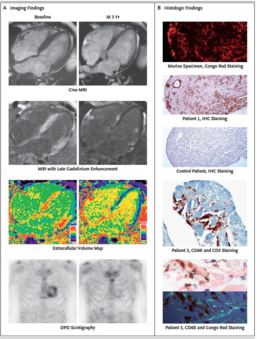
That's pretty striking
Reversal of amyloidosis cardiomyopathy in 3 individuals who had developed anti-ATTR amyloid antibodies
https://t.co/jtOucqp18T @NEJM@MariannaFonta11
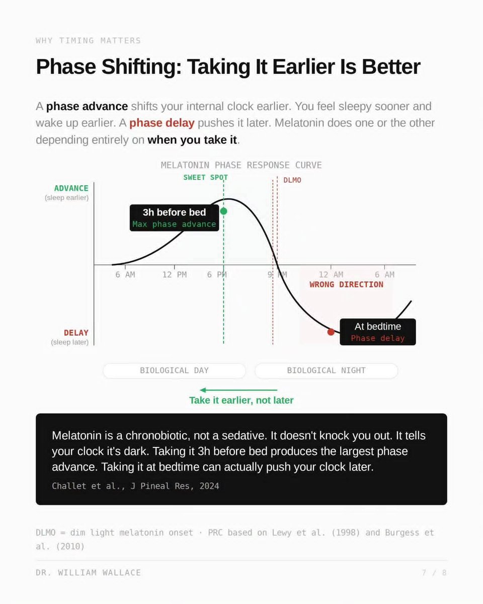
Most people take melatonin right before bed.
That might be one of the worst times to take it.
Melatonin isn't a sedative, it's a chronobiotic. It doesn't knock you out. It signals to your circadian clock that it's dark. And the effect it has on your sleep timing depends entirely on WHEN you take it.
There's a concept called the Phase Response Curve. It shows that melatonin taken ~3 hours before your usual bedtime produces the largest phase advance, meaning it shifts your internal clock earlier, so you fall asleep sooner and wake up easier.
But take it AT bedtime? You can actually push your clock in the wrong direction. That's a phase delay. You end up falling asleep later over time, not earlier.
The sweet spot sits just before dim light melatonin onset, the point when your brain would naturally start releasing melatonin. For most people, that's roughly 3 hours before habitual bedtime.
Timing > dose.
Lewy et al. (1998), Burgess et al. (2010), and Challet et al., J Pineal Res (2024).
Vitamin B12 is absorbed through two pathways. The first is intrinsic factor, a protein produced by parietal cells in the stomach. IF binds B12 in the small intestine and carries it across the gut wall via a receptor called cubilin in the distal ileum. This pathway is efficient but has a hard ceiling: it saturates at roughly 1.5 µg per dose. No matter how much B12 you swallow beyond that, IF cannot carry any more.
The second pathway is passive diffusion. About 1 to 2% of any oral dose diffuses across the intestinal lining without IF, and this occurs along the entire length of the gut. At dietary doses, this pathway is negligible. At supplement doses, it becomes the primary route of absorption.
Adams et al. (1971, Scand J Gastroenterol) measured whole body retention of radiolabeled cyanocobalamin at different doses. At 1 µg, roughly 50% was retained. At 5 µg, about 20%. At 25 µg, just over 5%. The NIH Office of Dietary Supplements reports approximately 2% absorption at 500 µg and 1.3% at 1,000 µg.
The fraction drops dramatically. But the total amount absorbed keeps rising. At 1 µg you absorb about 0.5 µg. At 1,000 µg you absorb roughly 13 µg total, of which approximately 10 µg comes from passive diffusion alone. The RDA is 2.4 µg. Even the backup pathway, working at 1% efficiency, delivers more than four times your daily requirement from a single pill.
This is the basis for high-dose oral B12 as an alternative to injections in patients who lack intrinsic factor. The NIH notes that high-dose oral supplementation "may be another treatment option" for pernicious anemia, though injections remain standard first-line therapy and the available randomized controlled trials comparing the two approaches are considered limited in quality.
One important nuance: absorbing B12 into your bloodstream is only the first step. After absorption, B12 must bind to a transport protein called transcobalamin to reach your cells. This complex, holotranscobalamin, is the biologically active fraction. It represents only about 20 to 30% of the total B12 circulating in your blood. The remaining 70 to 80% rides on a separate protein called haptocorrin, which does not deliver B12 to most tissues.
This is why serum B12 can be misleading as a status marker. A person can have a "normal" total serum B12 level while their holotranscobalamin, the fraction that actually delivers B12 to cells, is low. Methylmalonic acid is a more sensitive functional marker because it rises when cellular B12 is genuinely insufficient, regardless of what total serum B12 shows.
Absorption determines how much B12 enters your blood. Transport determines how much reaches your cells. Testing only total serum B12 measures neither of these processes accurately.
Adams et al., Scand J Gastroenterol, 1971
NIH Office of Dietary Supplements, 2024
Allen et al., J Nutr, 2018