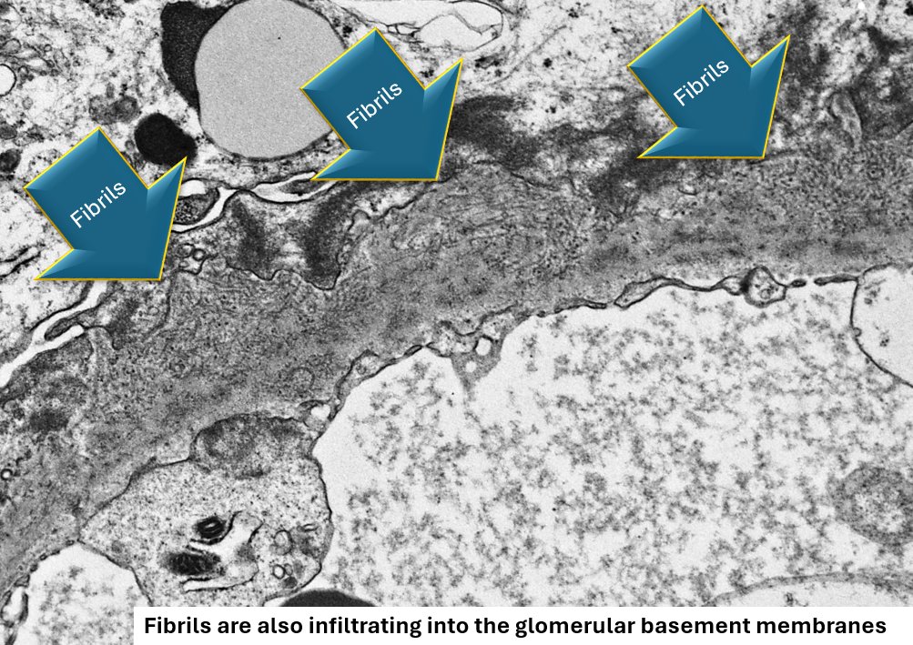
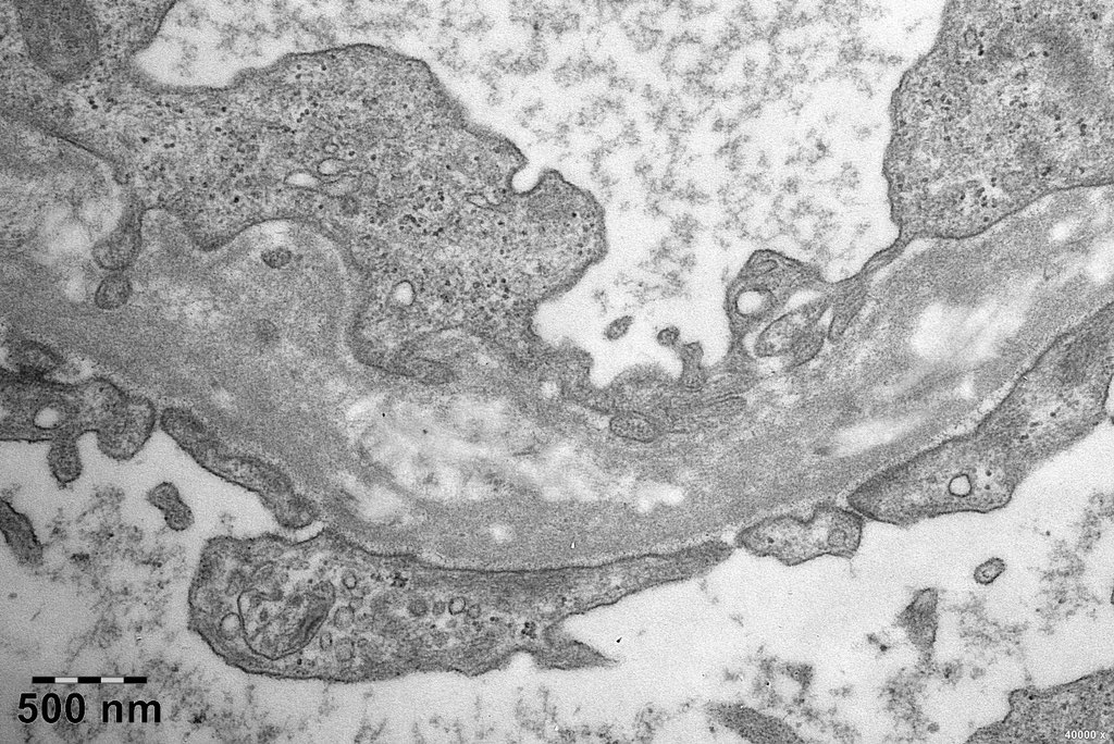
Case 4 (2026): A 20 year old presented with hematuria and mild proteinuria. The biopsy findings are shown. IF images of C3 stained sections are shown.
What’s the diagnosis?
Is there a new tissue marker for this entity?
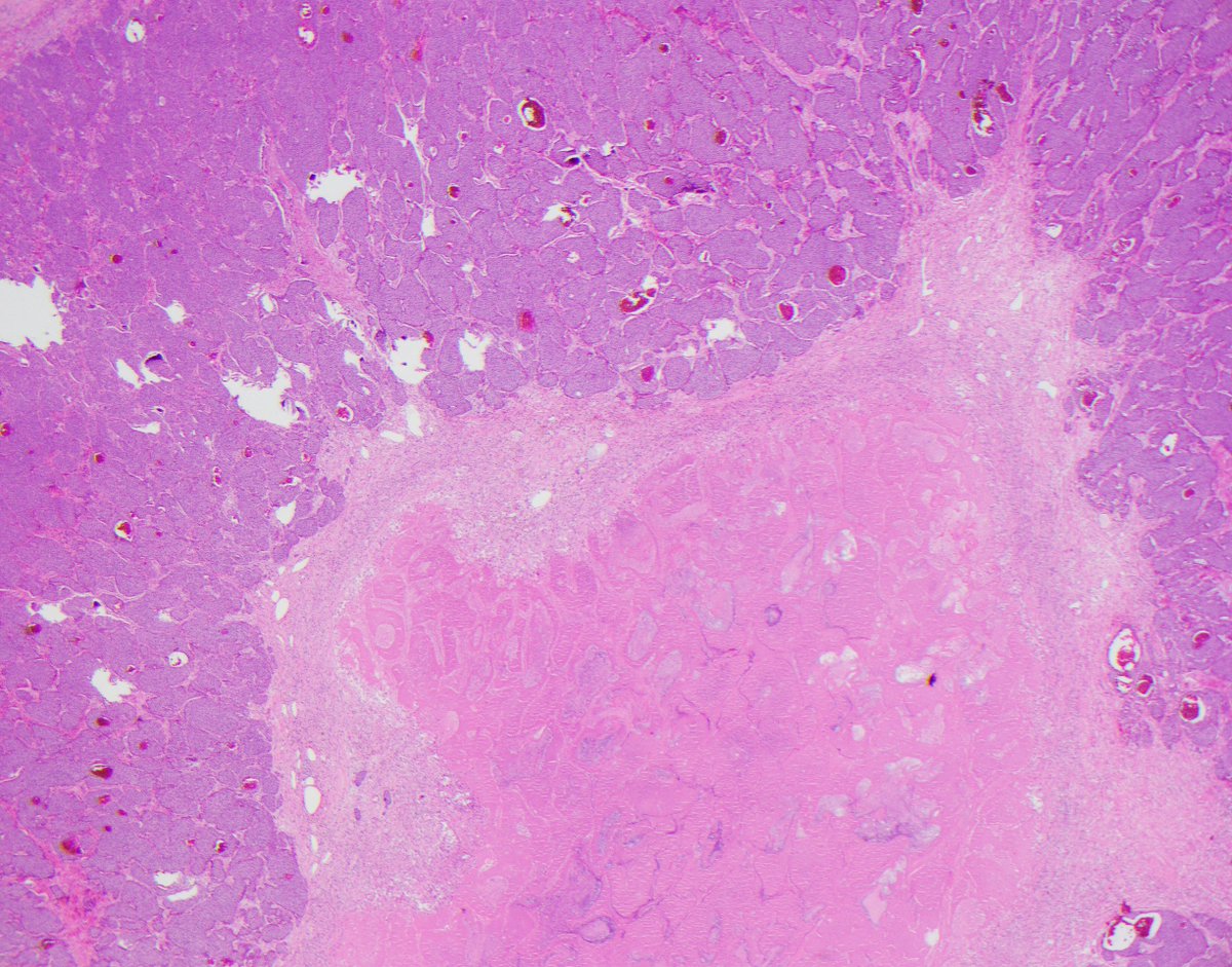
This adrenal cortical carcinoma presented as a liver mass. The CK8/18 highlights a tiny rim of compressed liver at the periphery. On a prior needle biopsy it was a mimicker of hepatocellular carcinoma but we figured it out thanks to modern immunohistochemistry.
#openaccess for 50 days to our article! Click on this link before February 25, 2026 to read and/or download! #gupath
@Shounak_Gupta @buraktekinmd@LiangChengMD
👉🏼 https://t.co/uLpr1eyv3e
When was the last time you saw this rare serous borderline tumor of the testis? Can you recognize a similar spectrum of associated histologic changes as those seen in ovarian serous borderline tumors? How do you distinguish these rare lesions from other Müllerian and non-Müllerian epithelial neoplasms?
You may want to take a look at this recent article published in the American Journal of Surgical Pathology @JLHornick - a beautiful study led by Prof. Christopher Przybycin. I am honored to be part of this work.
Full article is available at the following link: https://t.co/tnwhXRW0cZ
#renalpath Myelin and zebra bodies in LMX1B associated nephropathy. Misdiagnosed as fabry disease initially. Only found few moth-eaten appearance in GBM. No special drug used.
A membranous pattern is occasionally present in Fibrillary GN
Notice randomly arranged fibrils in the subepithelial regions. DNAJB9-positivity is seen in a similar distribution by IHC. Can be mistaken for membranous nephropathy: https://t.co/T8VO6Gc1Lm.
#renalpath#PathTwitter
New in #HumPathol: Unlocking Diagnostic Potential: A Retrospective Analysis of GPNMB Immunohistochemistry in Nearly 1000 Surgical Pathology Specimens. https://t.co/XKPEJXydcF #pathology#PathTwitter#PathX#GUpath#IHCpath
Read our latest #research in @Human_Pathology
Evaluation of 3,606 renal cell tumors for TFE3 rearrangements and TFEB alterations via FISH, NGS, and GPNMB IHC
Free access until July 12, 2025.
https://t.co/joB94wuum4
#GUPath
Infarct necrosis important to separate from true tumour necrosis in NETs.2 show true necrosis(abrupt transition, karyorrhectic debris, comedo-like)& 2 show infarct(gradual transition, zonal granulation tissue/myobroblasts)
If you think easy -> retweet
If you think hard -> like
This hepatic solitary fibrous tumors is infiltrative and has resulted in obstructed ducts - the appearance is not in line with that of mesenchymal hamartoma, which has more stellate mesenchymal cells. #UMiamiPath@ModernPathology
Listen to this podcast on renaming grade group 1 (GG1) prostate cancer as non-cancer. This is a very important discussion in which #pathologist, #urologist and men with prostate cancer need to actively participate to understand its future implications!
https://t.co/3jrgUIS56m
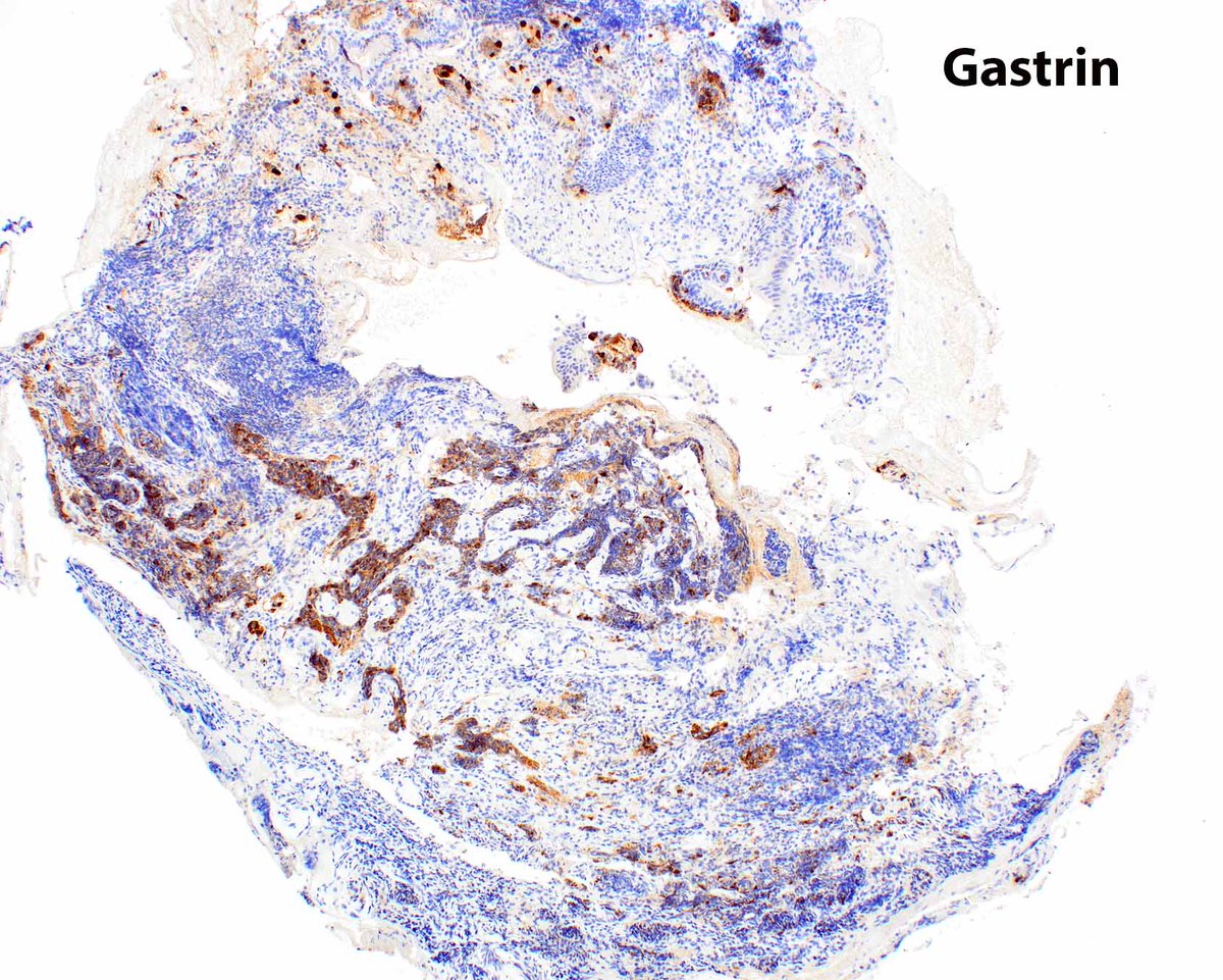
Duodenal neuroendocrine tumors can be mistaken for inflammation. The gastrin could indicate that this is a gastrinoma if the patient has elevated serum gastrin. The tumor is in the "gastrinoma triangle". The gastrinoma triangle image source: https://t.co/0N2HQuQ3Ws #UMiamiPath
chromophobe RCC to me is akin to testicular yolk sac tumor:
can show SO many different faces (morphologic patterns)!🤡
for a FANtabulous review of chromophobe RCCs, ✔️this paper - one of my all-time #GUpath fav's🥰