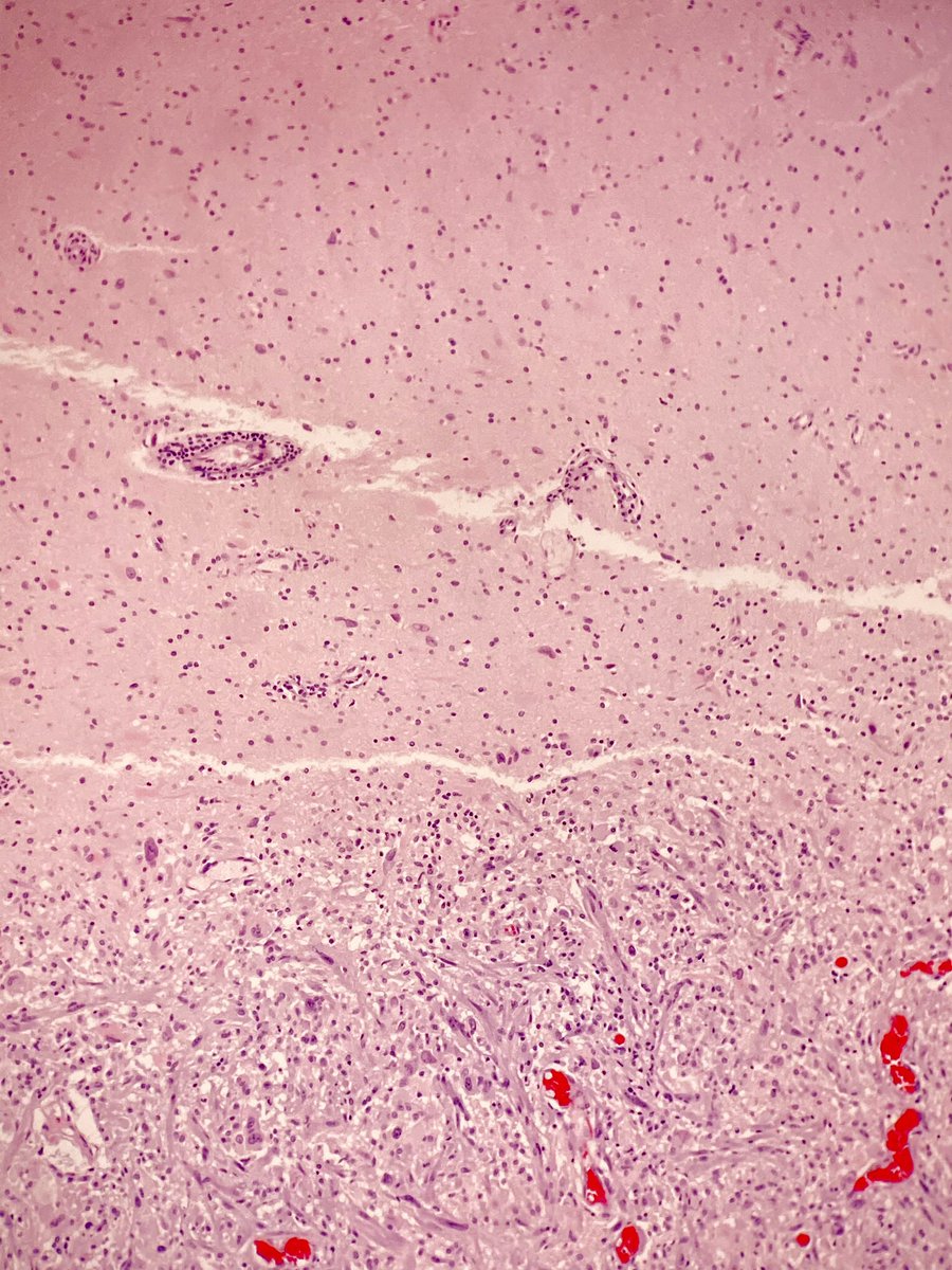
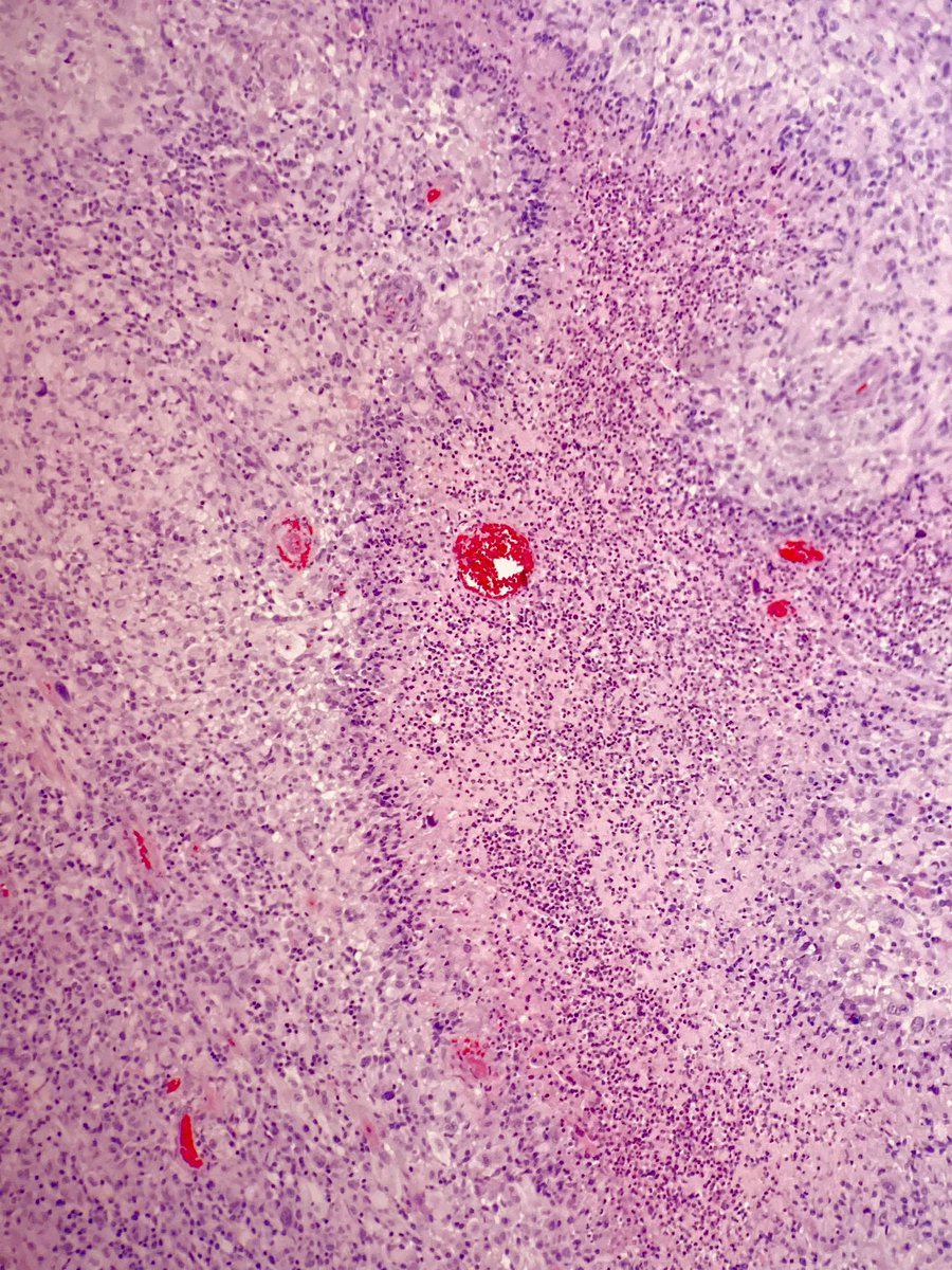
Gliosarcoma- Biphasic variant of glioblastoma with alternating glial and mesenchymal differentiation. Tumors have traditional features of GBM including palisading necrosis, microvascular proliferation, and high mitotic rate with additional sarcomatoid features. WHO grade IV.
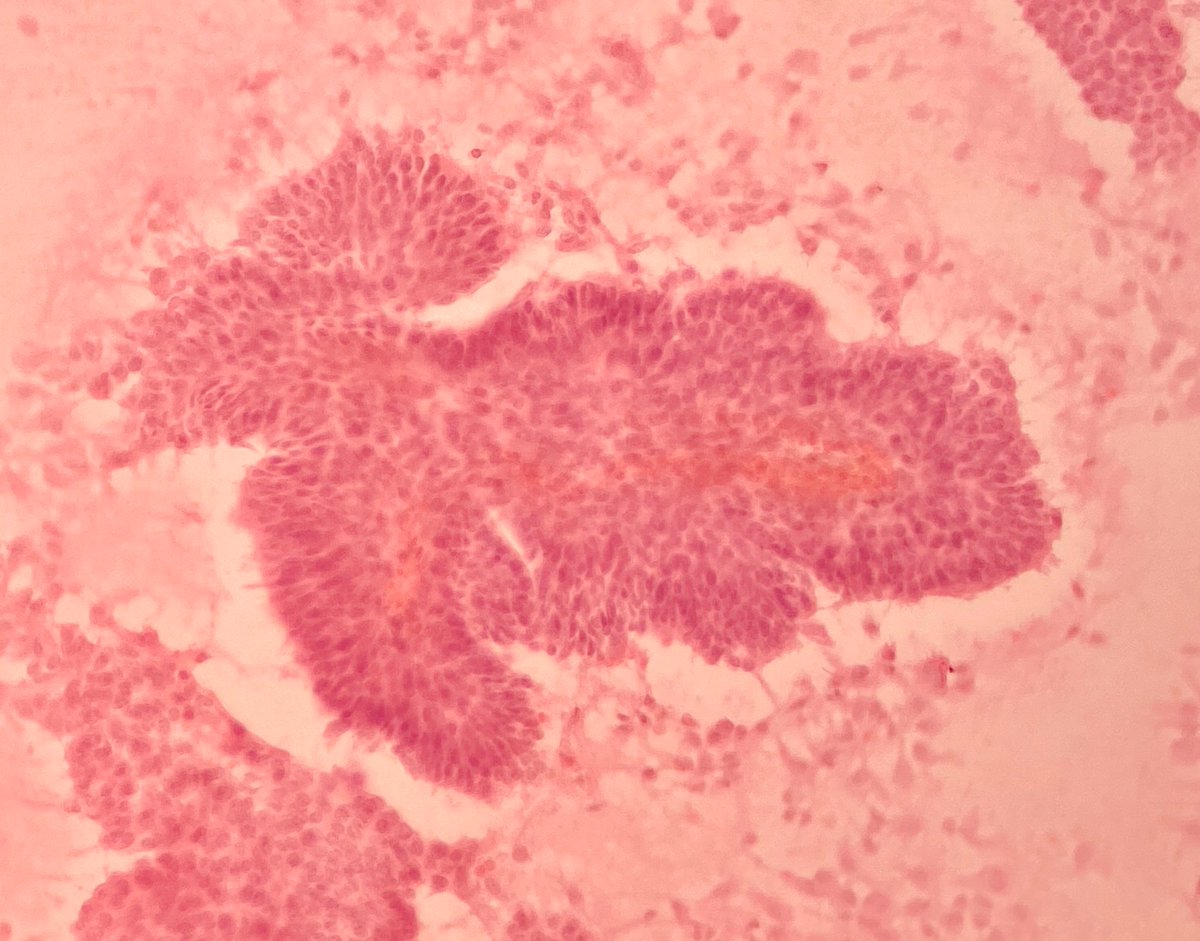
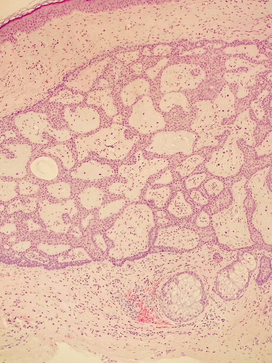
Firbroepithelioma of Pinkus: premalignant lesion composed of interconnected and branching strands of basaloid cells embedded in a loose fibrovascular stroma. Extends from epidermis to dermis.
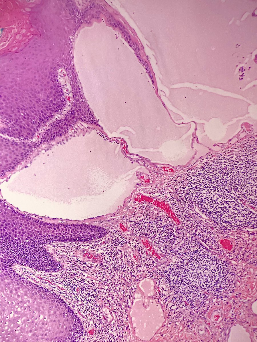
Lymphangioma: large lymphatic channels in loose connective tissue with associated lymphoid aggregates. Usually well circumscribed with focal cystic formation. May be due to lymphovascular malformation. CD31/CD34/D2-40 (+).
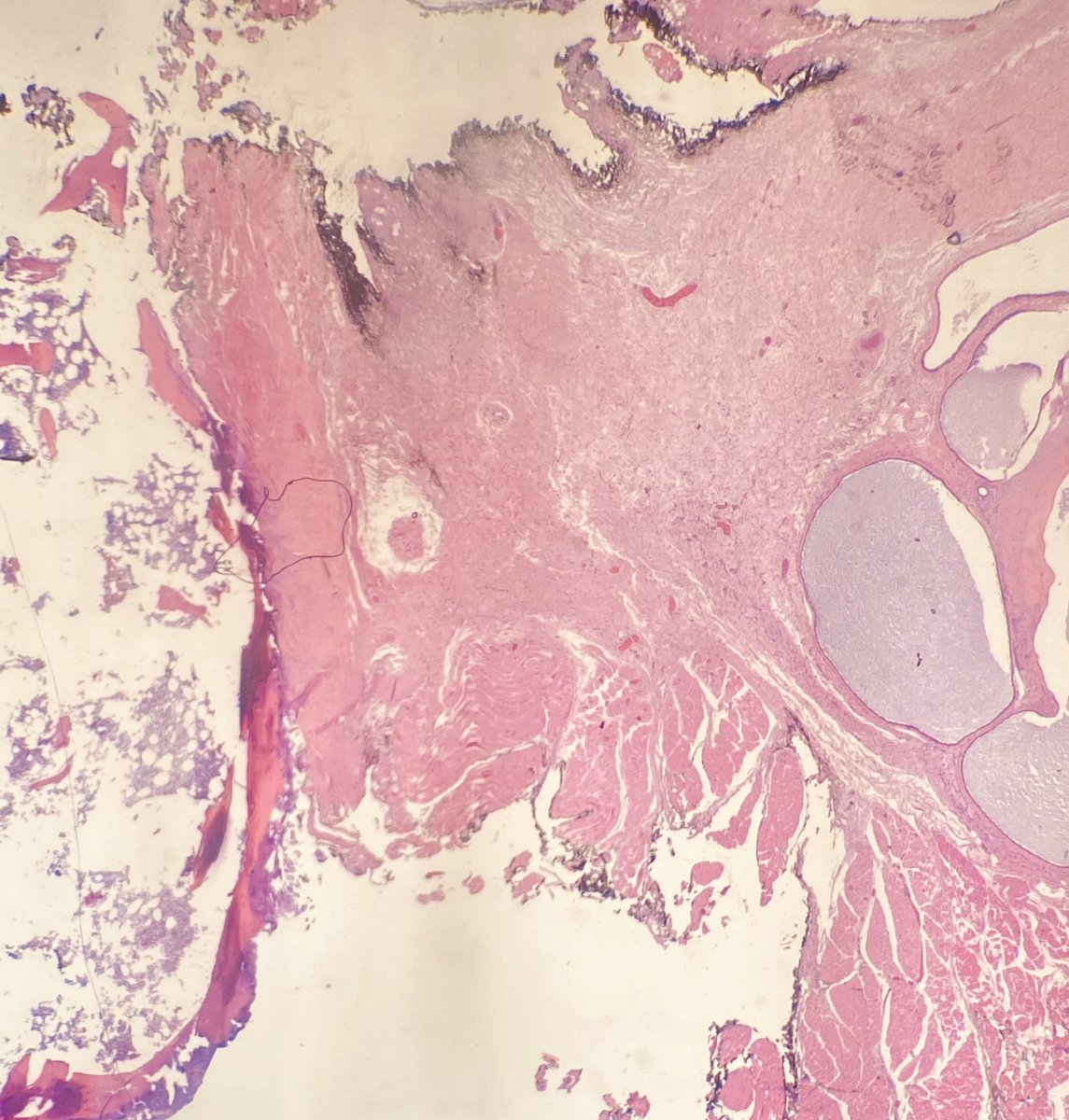
Cool to see hematopoiesis in the hyoid bone sent with this specimen! This entity may also have squamous lining or adjacent mucous type salivary glands.
Excision of a thyroglossal duct cyst attached to hyoid bone. Micro showed multilocular cyst lined by respiratory epithelium with adjacent thyroid follicular tissue and inflammation.
Post ET Myelofibrosis: This BMBX shows extreme hypercellularity with increased staghorn megakaryocytes in clusters. Reticulin showed MF3 fibrosis and diffuse collagen deposition.
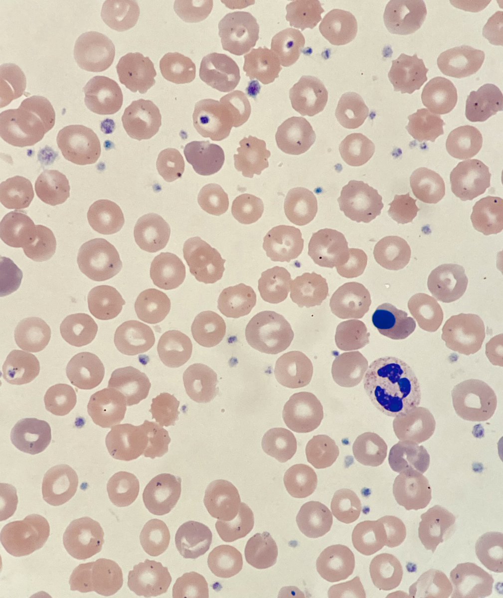
Blood smear from a patient with pyruvate kinase deficiency. Notable for echinocytes, polychromasia, and nRBCs. What is one way this patient was treated?
Use of Immunohistochemical Markers (HNF-1β, Napsin A, ER, CTH, and ASS1) to Distinguish Endometrial Clear Cell Carcinoma From Its Morphologic Mimics Including Arias-Stella Reaction by Dr. Jennifer X. Ji, et al. #GYNPath@ISGynP https://t.co/QDWso2ANk7
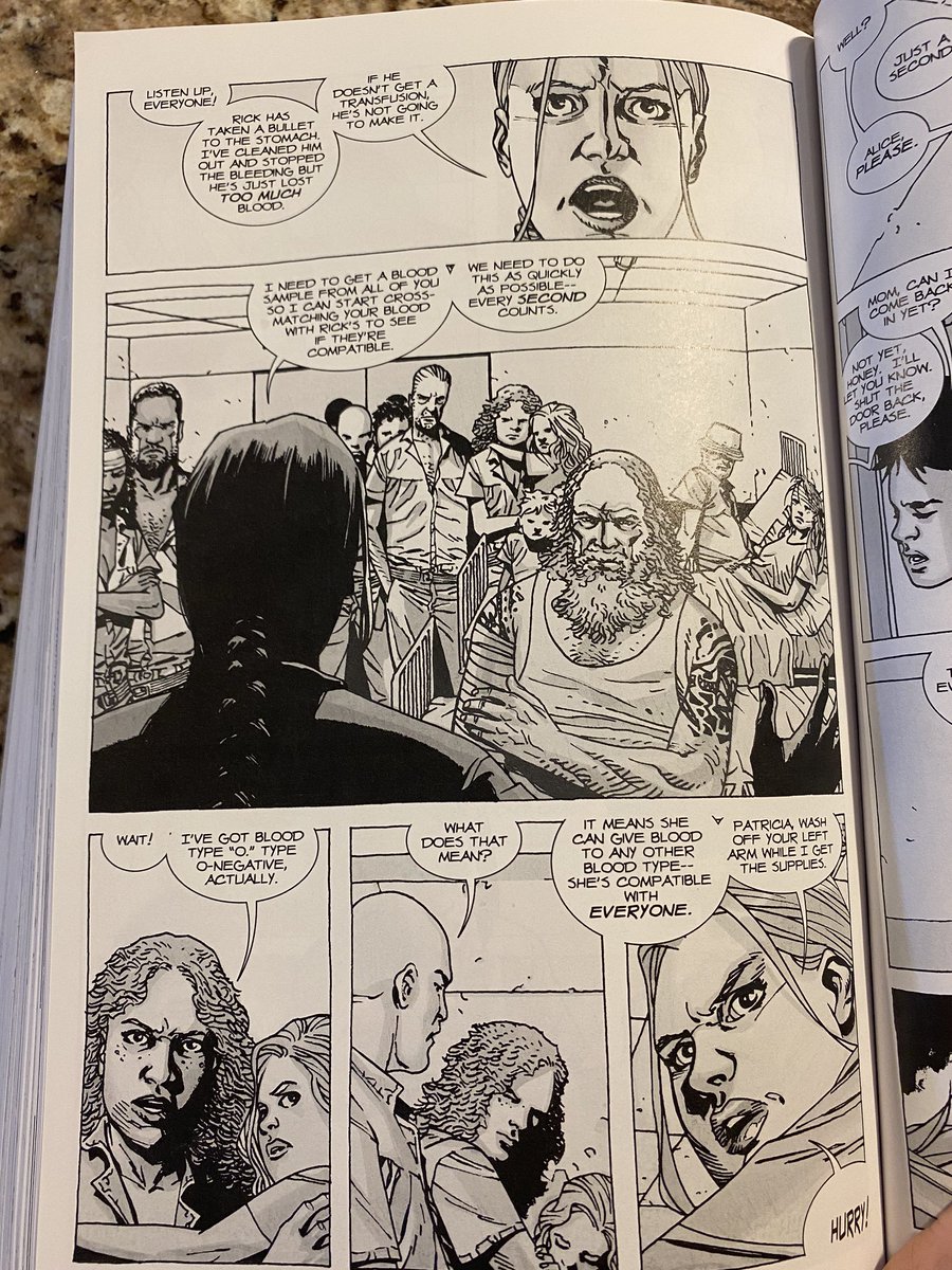
@TeamCaptainJohn@AABB I would have to look into that! I think a cross match would cover them in this hypothetical situation. And spoilers, but the transfusion went well haha.
An adult female presented with a 4th ventricle mass and hydrocephalus. A biopsy was done with IOC (Pic 1/2). Sections showed a neoplasm with papillary architecture and fibrovascular cores lined by flat surface epithelium (Pic 3/4).
A diagnosis of choroid plexus papilloma was rendered. This tumor is a WHO Grade 1 tumor most commonly in the ventricular system that may cause hydrocephalus (d/t obstruction or CSF overproduction), developmental delay, or epilepsy in children.