@VioletaCatala@JHernandez_RX@_uroima@ESUR_JN@residentesSERAM Excellent, findings are those of PI-RADS 2 as Dr @JHernandez_RX mentioned. Here is a summary of the findings 👏 if you have DCE dont forget to take a quick peak to late phases as inflammatory changes or necrosis will be more conspicuous 💡 and will help you solve the case!
Its been cloudy for some months now. While we still wait for clear skies here's a picture Id like to share from a camping trip last september. Took it with a small refractor telescope under very dark skies. The Veil Nebula a supernova remnant, wish you all a happy new year!
Estamos seguros que han adquirido habilidades de lectura de esta técnica tan útil como compleja y que harán un gran aporte, desde un enfoque interdisciplinario, en beneficio de los pacientes.
¡Los echaremos a faltar!! Equipo Uroima
@trigeminy_henry@ElhefnawiYara You are right, they totally do. We got some of them, all work fine and they sound like this or even more depending on the settings you choose
🚨🚨🚨
XXV Jornadas SERME
I Jornada SERME-SERPE
¡ENVÍA YA TU COMUNICACIÓN!
¡Últimos días para enviar tu comunicación!
Fecha límite 25 de septiembre de 2023
👇🏻👇🏻👇🏻
https://t.co/fdIqHer0oj
@teachplaygrub Very nice! Gotta love some of Tyson’s quotes hehe. I was looking forward for a cute guardsman fracture drawing. Nevertheless it was entertaining and useful, thank you 🙏🏽
@teachplaygrub Swi is such a wonderful sequence, not just to asses for bleeding (usually best to check recent bleed on flair than swi), but to asses collaterals and even the presence of a clot in the arterial structures, with very good correlation to MRA sequences. Thx for the post!
@docskalski@lovettdesign@GEHealthcare I bet Matt has the support of the entire radiology community. No matter the country we’re from and especially colleagues formed in the last decade, we’ve all grown up with his illustrations. #FOAMrad
@inigoiri Genial 👌🏽 en el formulario cuando pregunta nivel deseado significa que dará acceso a solo 1 de los 2 módulos? Ambos son atractivos a cualquier nivel
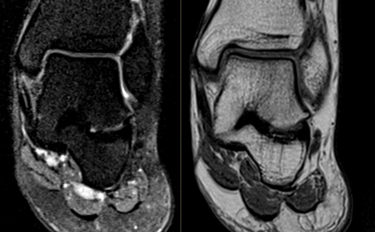
Talocalcaneal coalition type 4 (osseus) due to the lack of fibrous component as seen in the picture. Middle facet type being the most common and usually associated with pes planus (both depicted in this case). Also keep in mind the sinus tarsi, effaced with low fibrous signal 🧐
28YO female with chronic foot pain. Can you tell whats going on? how would you classify it? any known associations? Gotta love diagnostics best seen on T1. @SermeMsk#radres