Harlequin Color Change (Harlequin Sign of the Newborn)
Key Features
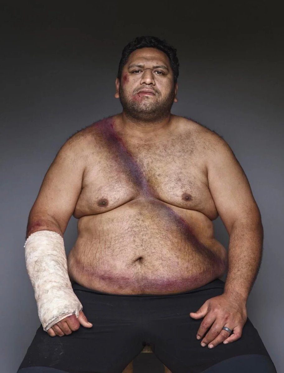
Classic Demarcation: A sharp, striking longitudinal midline demarcation dividing the body into a dependent hyperemic (deep red) side and an upper pale side.
Pathophysiology: It is a benign, transient vascular phenomenon caused by immature hypothalamic autonomic tone, leading to temporary imbalance in peripheral cutaneous vascular tone.
Triggering Factors: Typically seen when a healthy neonate (most commonly between 2 to 5 days of age) is placed on their side (lateral decubitus position). Gravity causes pooling of blood in the lower half of the body.
Prognosis & Management: Entirely benign and self-limiting. The color change typically fades or disappears within minutes to half an hour, or immediately when the infant changes position or cries/moves. No medical treatment or intervention is required.
Rare but classic.
This video shows a young woman with mucosal bleeding and a characteristic rash.
Labs: severe thrombocytopenia.
Can you name the clinical sign?
Acute Aortic Syndromes: Same emergency, different mechanism.
Although they share similar symptoms, acute aortic syndromes differ in how the aortic wall is injured. Recognizing these differences is essential for accurate diagnosis and treatment.
🟥 Aortic dissection An intimal tear allows blood to enter the aortic wall, splitting the media into a true lumen and a false lumen separated by an intimal flap.
🟥 Intramural hematoma (IMH) There is no visible intimal tear. Instead, bleeding occurs within the aortic wall, creating a hematoma that thickens the wall while the aortic lumen often appears relatively normal.
🟥 Penetrating atherosclerotic ulcer (PAU) An atherosclerotic plaque erodes through the intima into the media. Blood enters the aortic wall but is usually confined by surrounding scar tissue, often leading to a localized dissection or pseudoaneurysm.
These three conditions are closely related and can progress into one another if left untreated. Prompt recognition with CT angiography and urgent management can be lifesaving.
Reference: Isselbacher EM, Preventza O, Black JH III, et al. 2022 ACC/AHA Guideline for the Diagnosis and Management of Aortic Disease. Circulation. 2022;146:e334-e482.
🩺 أول ما بدأت شغلي في العناية المركزة في فترة الامتياز...
كنت أشوف المحاليل كلها نفس الشي، أي مريض يحتاج سوائل؟ خلاص علّق له محلول ملح أو رينجر لاكتيت وانتهينا! 😅
لين جاء يوم الاستشاري سألني في الجولة:
"ليش كتبت لمريض الكبد رينجر لاكتيت؟"
وقتها وقفت ساكت... وحسيت إني دخلت اختبار وأنا ما ذاكرت له. 😂
بعدها مع كثرة الحالات، اكتشفت إن المحاليل مو مجرد أكياس سوائل... كل واحد له شخصية، وله وقت يناسبه، ووقت ممكن يضر فيه المريض.
خلونا نبسطها بحيث حتى يفهمها غير الأطباء
Answer:
The structure shown is the Circle of Willis
The pathology is a ruptured berry (saccular) aneurysm arising from the middle cerebral artery.
Bonus: Although this specimen shows a middle cerebral artery berry aneurysm, the anterior communicating artery (ACom) is the most commonly involved artery in berry (saccular) aneurysms overall.
Heart's trabeculae carneae remind us that efficiency in biology often hides behind apparent disorder.
🌟 The trabeculae carneae are irregular muscular ridges that project from the inner surfaces of the heart’s ventricles, creating a characteristic spongy appearance. They are continuous with the ventricular myocardium and covered by endocardium.
1⃣ Three types can be distinguished:
▶️ Fixed ridges that reinforce the wall
▶️ Bridges that connect papillary muscles to chordae tendineae,
▶️ Free trabeculae, such as the moderator band in the right ventricle, which carries Purkinje fibres to the anterior papillary muscle and contributes to coordinated contraction.
🚩 Physiologically, these structures are essential to the mechanical and electrical performance of the heart:
2⃣ During systole, when intraventricular pressure peaks and the chambers contract powerfully, the trabeculae carneae prevent the smooth ventricular walls from collapsing inward or adhering to each other.
👁️Let me dive a little deeper...
In a perfectly smooth-walled chamber, such as a hypothetical ventricle without trabeculae carneae, strong contraction during systole would press the opposing endocardial surfaces together. Because the heart is filled with fluid, the expulsion of blood reduces internal volume sharply, and the negative pressure created when the walls approximate could cause local adherence—much like two wet glass plates or suction cups sticking together. The thin endocardium, coated by a film of plasma, would create a surface tension effect if pressed too closely, making it energetically costly for the walls to separate again during diastole.
So...
▶️Their uneven topography maintains small channels of blood between the ridges, preserving the internal geometry of the ventricle and ensuring that re-expansion during diastole occurs smoothly and without suction effects.
▶️This is particularly important in the left ventricle, where high pressures could otherwise flatten the endocardial surfaces.
3⃣ The trabeculae also contribute to the even distribution of wall stress, preventing localised overstrain and promoting efficient force transmission.
4⃣ Their pattern reduces the formation of large vortices that would increase turbulence and energy loss during ejection, while their subtle curvature promotes micro-vortices that facilitate rapid ventricular filling during diastole.
5⃣ Some trabeculae contain Purkinje fibres, enabling rapid conduction of electrical impulses and synchronising the contraction of the ventricular myocardium.
6⃣ Their tightening also stabilises the chordae tendineae and atrioventricular valves, ensuring unidirectional blood flow.
🔎 In sum, the trabeculae carneae are not mere structural ornamentation but a functional masterpiece: they strengthen contraction, optimise blood flow, and integrate electrical and mechanical harmony within each heartbeat.
#Physiology_RS #Cardio_RS #Physiology #Heart #Cardiology