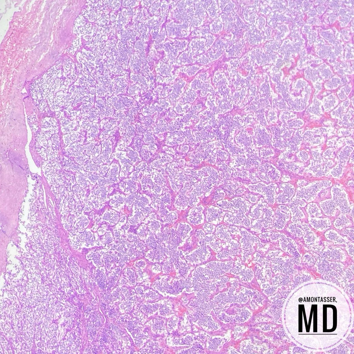
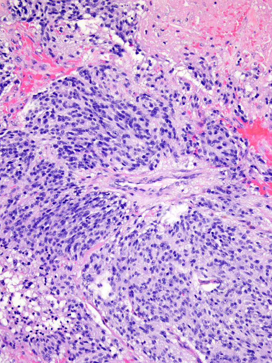
✅ Paraganglioma 🎯
• Neuroendocrine neoplasm of neuronal subtype showing classic Zellballen morphology.
• Characteristically negative for keratins and positive for GATA3.
• Strongly associated with germline mutations in SDH genes or VHL.
• All cases are considered to have a lifelong risk of metastasis.
The "zellballen pattern" is a characteristic histological feature, where tumor cells form small, rounded nests or "balls of cells" (zellballen in German) within a vascularized fibrous stroma, and is pathognomonic for paragangliomas.
https://t.co/OAMIQsD9mC
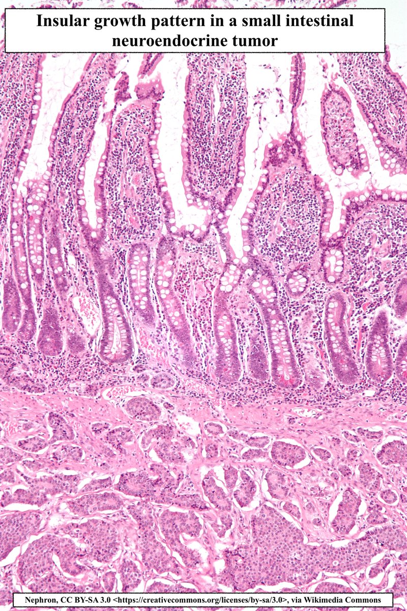
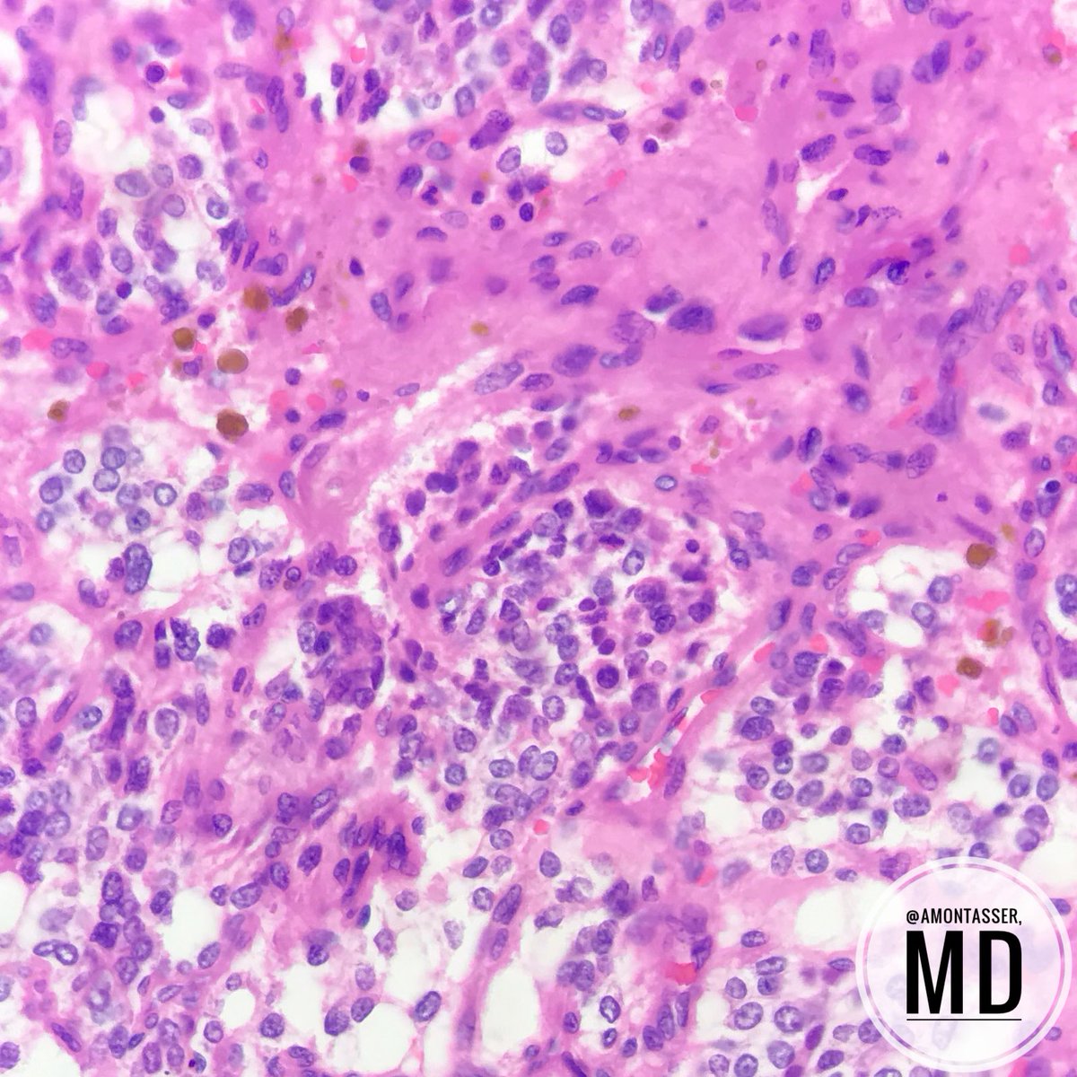
Insular Architecture
•Growth pattern comprised of tightly packed islands or nests
•May be seen in Neuroendocrine tumors and carcinomas, Poorly differentiated+medullary thyroid carcinoma
• Not to be confused w Zellballen pattern (has s100+ sustentacular cells)
#pathagonia
Adenoid Cystic Carcinoma (ACC)
Salivary Gland Tumor that is composed of both epithelial and myoepithelial cells.
Accounts for ~25% of primary salivary gland carcinomas
Can have a t(6;9) MYB::NFIB fusion.
Here’s ACC in a hard palate
Credit: Kelly Hall,MD
#pathagonia#pathx
Just a picture to appreciate classic morphology of Adenoid cystic carcinoma. Sometimes I forget to look at the finer details in more 'commonly' seen entities
#headandneckpath#PathX#Pathresidents#PathTwitter
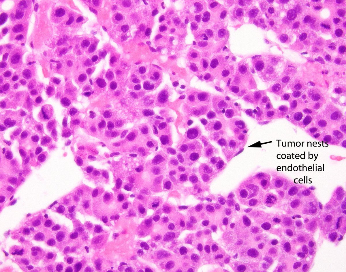
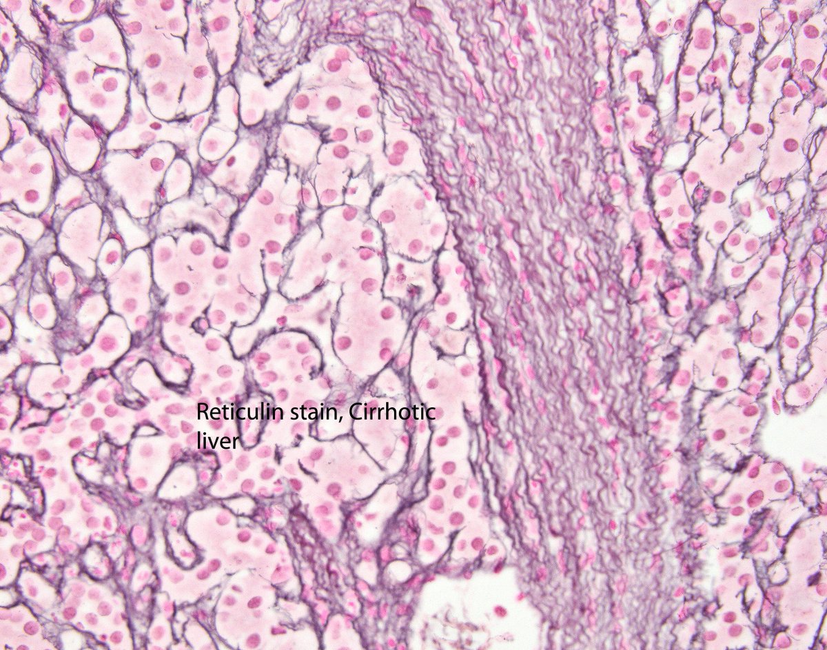
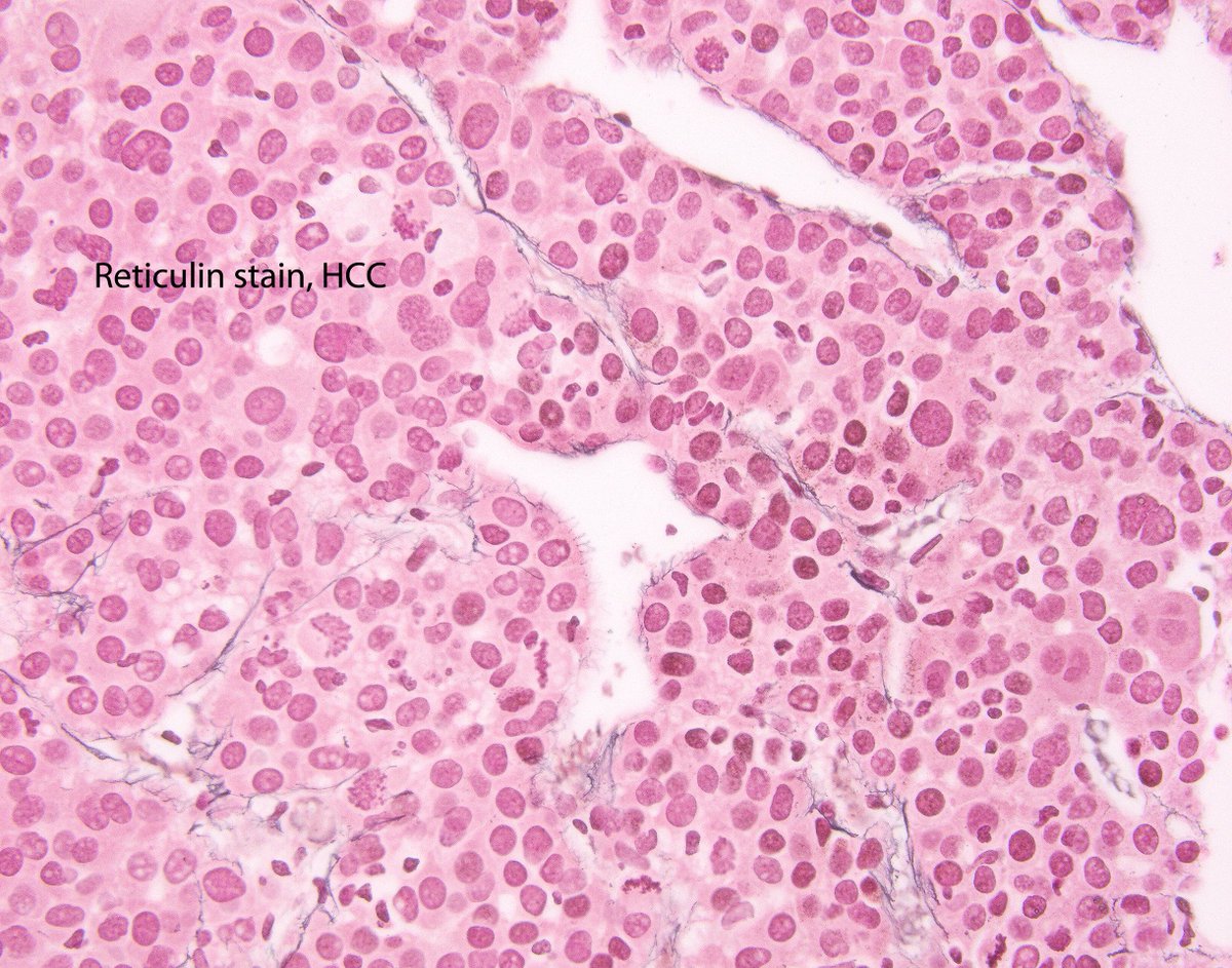
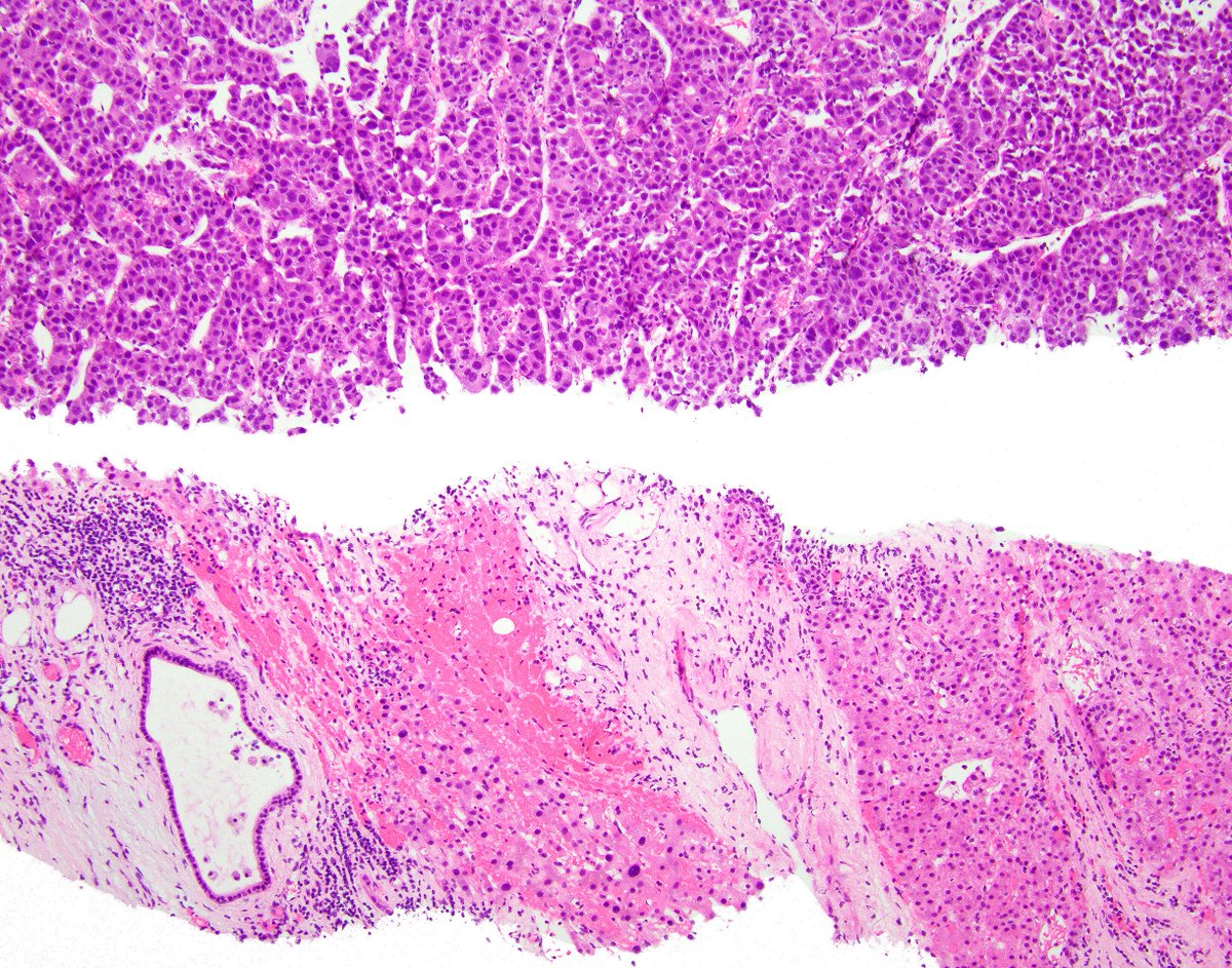
This is a nice example of hepatocellular carcinoma in a cirrhotic liver that shows the pattern of endothelial cells wrapped around tumor cell groups and the beautiful reticulin distribution in the tumor and the adjoining cirrhotic liver.
La biopsie confirme le diagnostic d'hépatite alcoolique aigue, en mettant en évidence la présence d'hépatocytes ballonisés, clarifiés avec corps de Mallory, de stéatose et d'un infiltrat polymorphe
Reminder: In children, the liver cells are arranged in two-cell-thick plates. The presence of two-cell-thick plates and rosette formation in adults indicates hepatocyte regeneration.
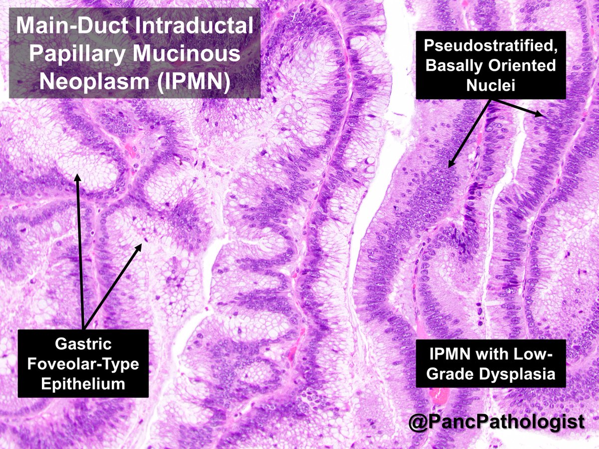
Odze and Goldblum Surgical Pathology of the GI Tract, Liver, Biliary Tract and Pancreas, 3rd Ed.