🧵 1/9
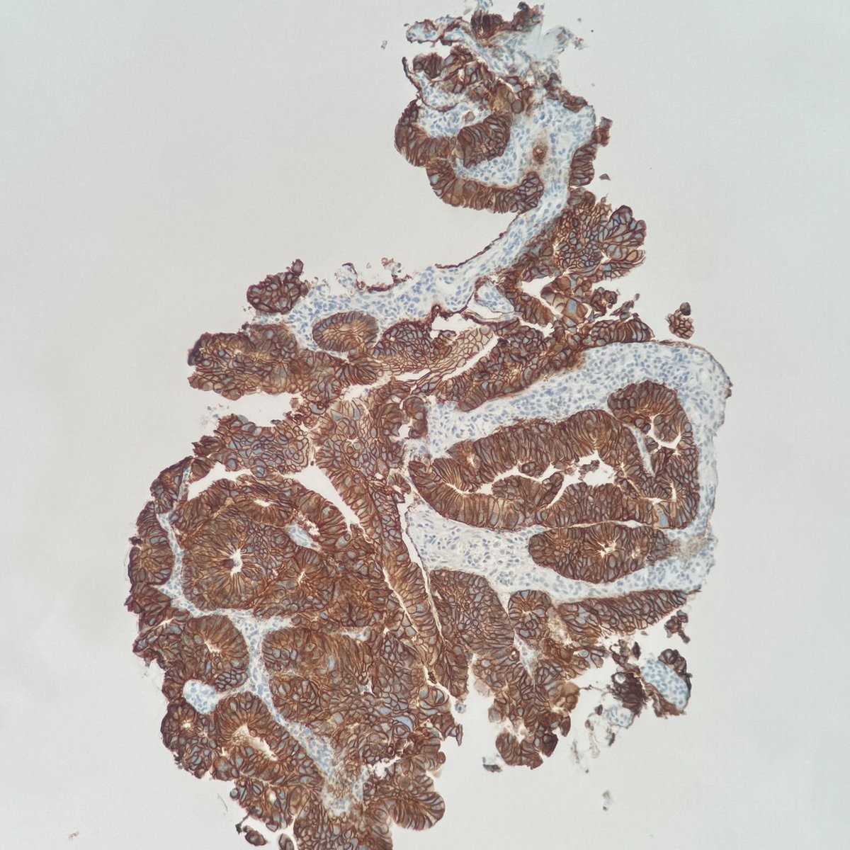
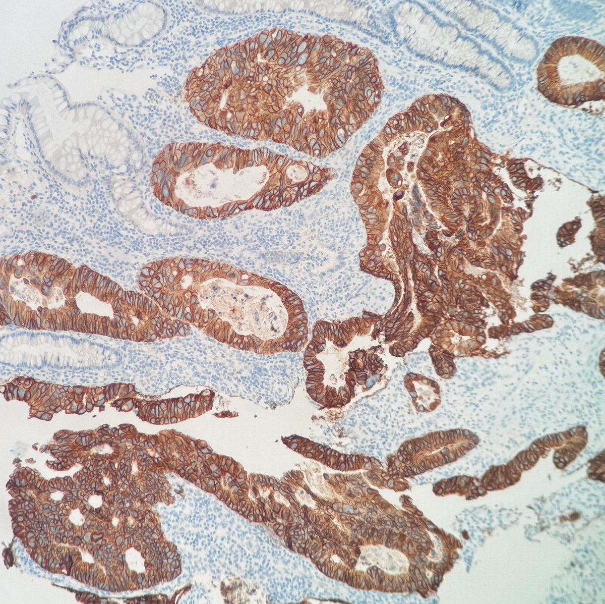
Elderly male. Metastatic colorectal cancer with liver mets. RAS wild-type — looks straightforward.
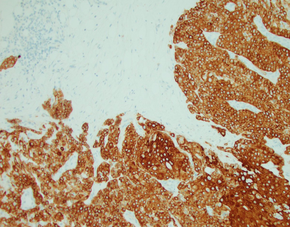
Then the molecular profile comes back: HER2 IHC 3+.
This changes the entire treatment plan. Here's why 👇
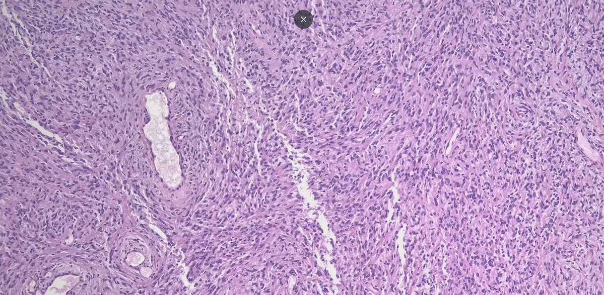
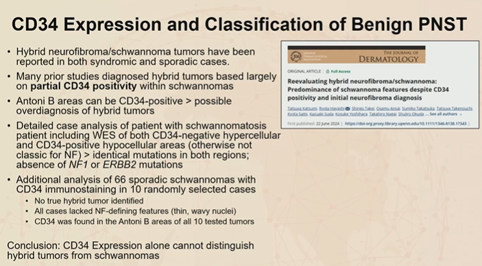
Pitfall ⚠️ Peripheral Nerve Sheath Tumors
Schwannomas can show CD34 positivity, especially in Antoni B regions.
This staining pattern may lead to overdiagnosis of hybrid nerve sheath tumors.
Dr. Dehner #USCAP2026#pathology#PathX#PathTwitter
🔬Tall cell carcinoma with reversed polarity (TCCRP),
emphasis: diagnostic pitfalls in papillary breast lesions🔬
TCCRP is a rare subtype of invasive breast carcinoma, usually small, low‑grade and clinically indolent, but easily confused with other benign and malignant papillary lesions and even with metastatic papillary thyroid carcinoma.
Definition 📚
Invasive breast carcinoma composed of solid or solid‑papillary nests of tall columnar cells with eosinophilic cytoplasm and reversed nuclear polarity (nuclei oriented toward the lumen), frequently associated with IDH2 p.R172 and PIK3CA hotspot mutations.
Epidemiology 👵
Occurs almost exclusively in women, typically postmenopausal, median age around 60–64 years.
Very low incidence; described mainly in small series and case reports.
No clear predilection for breast side or quadrant.
Sites / Clinical Features 🩻
Single, small breast nodule (mean ~1–1.7 cm; most are T1), detected on screening or as a palpable mass.
Imaging (mammography/US): solid hypoechoic mass, BI‑RADS 4–5, sometimes well circumscribed, sometimes irregular with posterior shadowing.
Axillary nodal metastasis is uncommon; distant metastasis is exceptional.
Pathogenesis / Molecular 🔬
Key driver: IDH2 p.R172 (R172S, R172G, R172W, R172T) mutations in the majority of tested cases.
PIK3CA mutations in roughly two‑thirds of tumors, often co‑occurring with IDH2 alterations.
Lack of BRAF mutations and RET/PTC rearrangements, which helps to exclude thyroid origin.
Diagnosis 🧪
Core biopsy can be tricky: solid/papillary architecture mimics papilloma, solid papillary carcinoma, or secretory carcinoma.
Tip: carefully search for reversed nuclear polarity at the periphery of nests and papillae, especially at the tumor–stroma interface.
IDH2 testing (IHC or NGS) is very useful in equivocal triple‑negative papillary lesions.
Histopathology 🧫
Architecture:
Expansile, circumscribed nests in fibrotic or hyalinized stroma.
Solid, solid‑papillary, and follicular/microcystic patterns with colloid‑like luminal material.
Fibrovascular cores often packed with foamy histiocytes.
Cytology:
Tall columnar or cuboidal cells, eosinophilic mitochondria‑rich cytoplasm.
Oval/elongated nuclei with grooves and occasional pseudo‑inclusions; minimal pleomorphism, rare mitoses.
Hallmark: reversed nuclear polarity (apical nuclei away from the basement membrane).
No continuous myoepithelial cell layer around invasive nests (p63, SMMHC, CK14 negative).
Immunohistochemistry 🎯
Overall profile:
Most cases are triple‑negative (ER−, PR−, HER2−) with low Ki‑67 (~1–10%).
Positive markers:
CK7 diffuse, CK5/6 in most cases.
GATA3, GCDFP‑15, mammaglobin (confirm mammary origin).
Calretinin often positive; S‑100/SOX10 may be focally positive.
IDH2 R172 IHC positive in most molecularly mutated tumors.
Negative / helpful in DDx:
TTF‑1/NKX2‑1, PAX8, thyroglobulin – argue against metastatic thyroid carcinoma.
Chromogranin, synaptophysin, INSM1 – usually negative, unlike classic solid papillary carcinoma.
Differential Diagnosis 🧩
⚠️Intraductal papilloma with usual ductal hyperplasia:
Preserved myoepithelium (p63+, SMA+), no diffuse reversed polarity.
⚠️Solid papillary carcinoma:
Strong ER positivity, neuroendocrine markers (CgA/Syn)+, CK5/6−, lacks reversed polarity.
⚠️Encapsulated papillary carcinoma:
Fibrous capsule, ER+, no characteristic follicular/colloid‑like areas of TCCRP.
⚠️Secretory carcinoma:
ETV6‑NTRK3 fusion, PAS‑positive secretion, strong S‑100; IDH2 wild‑type.
⚠️Metastatic papillary thyroid carcinoma (tall cell variant):
TG/TTF‑1/BRAF/RET+, GATA3−, no IDH2 R172 signature.
Prognosis 🧾
Very low recurrence risk; compiled series show ~2% recurrence with overall survival near 100% at medium‑term follow‑up.
Rare cases show late axillary recurrence or isolated bone metastasis.
Treatment 💊
Breast‑conserving surgery + sentinel lymph node biopsy is the most common and appropriate approach for small T1 tumors.
Chemotherapy and radiotherapy are often omitted or used selectively, given the indolent biology.
Future perspective: potential candidacy for IDH2 or PI3K/AKT/mTOR‑targeted therapies in rare aggressive or recurrent cases.
Take-Home Messages NotasPatologia ✅
📌TCCRP is rare but important: invasive, usually triple‑negative, yet clinically indolent.
📌Suspect TCCRP in solid/solid‑papillary, triple‑negative breast lesions with foamy histiocytes and reversed nuclear polarity.
📌IDH2 R172 + PIK3CA form its molecular “signature”; IDH2 IHC is an excellent shortcut in challenging cases.
📌A structured DDx with papilloma, solid papillary carcinoma, secretory carcinoma, and metastatic PTC is mandatory.
📌Correct recognition of TCCRP prevents overtreatment compared with conventional high‑grade triple‑negative breast cancer.
📚Selected References📚
WHO Classification of Tumours of the Breast, 5th ed.
Lu Y et al. Tall Cell Carcinoma with Reversed Polarity of the breast. The Breast. 2025.
Lei Z et al. Case report: Tall cell carcinoma with reversed polarity of the breast. Frontiers in Oncology. 2024.
Zhang X et al. Tall cell carcinoma of the breast with reverse polarity. Gland Surgery. 2021.
Wang H et al. Tall cell carcinoma with reversed polarity: case report and review of the literature. Discover Oncology. 2026.
🚨Disclaimer
This text is an educational summary for healthcare professionals and students. It does not replace full pathology reports, local guidelines, or individualized clinical decision-making.
Hashtags
#pathology #PathTwitter #BreastPathology #NotasDePatologia #MolecularPathology
WHO 2021 Thymoma Classification in a Glance! 🔬
Master Type A, B (B1-B3), AB, and Metaplastic variants with our simple memory tricks. Essential for surgical pathology!
Follow us for more daily high-yield tips! 👇
#Thymoma#WHO2021#Pathology#Oncology#MedEd#SurgicalPathology
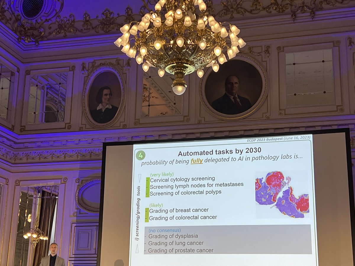
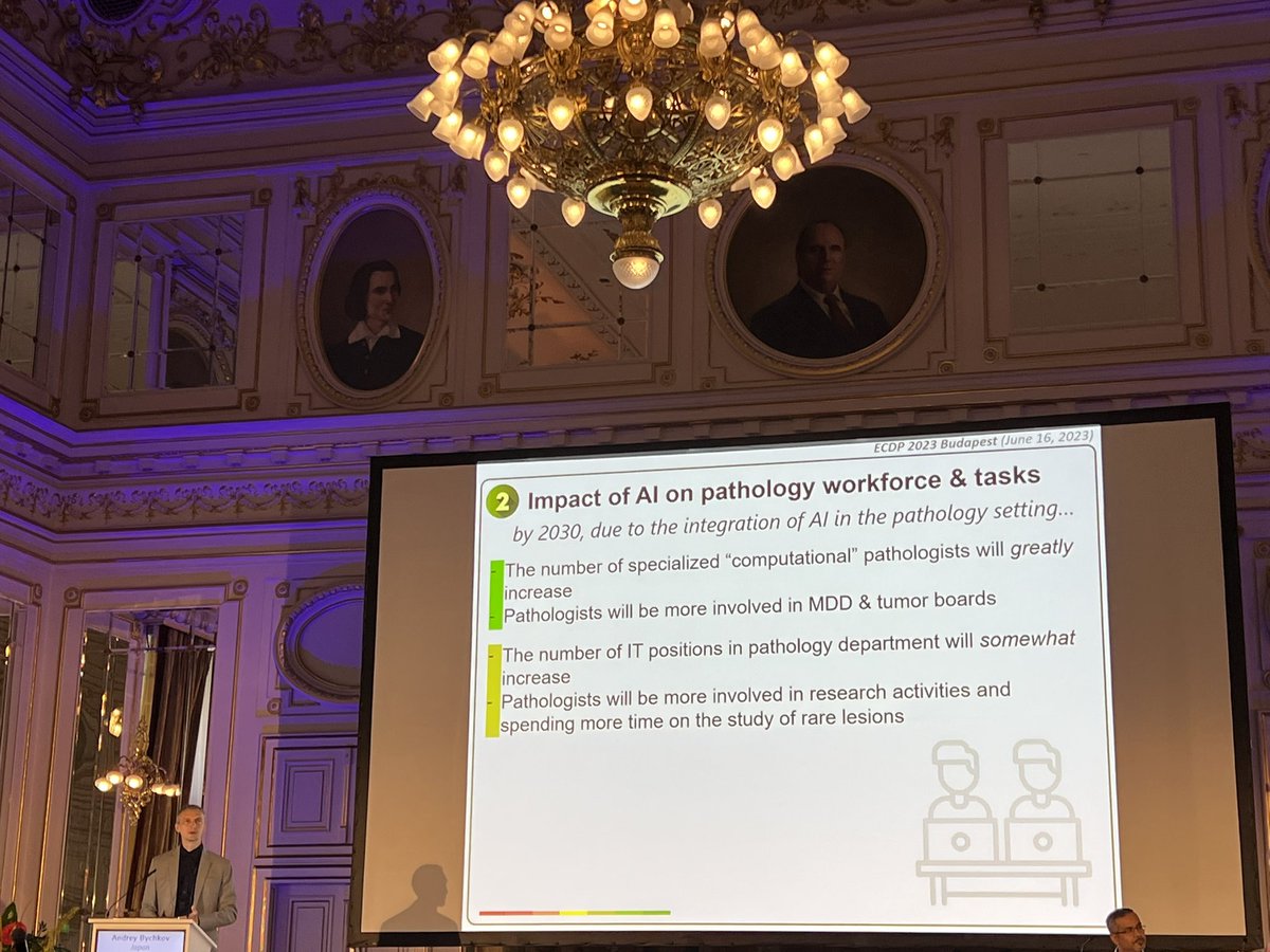
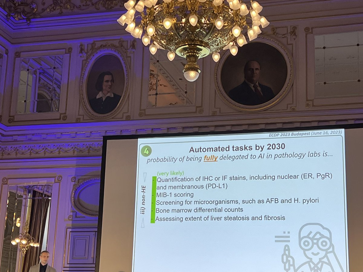
A stunning afternoon lecture by Andrey Bychkov at #ECDP2023 about the near future of #ArtificialIntelligence applied to #pathology with a prediction made by a group of experts in the field through a Delphi Study
Please read the excellent full article: https://t.co/p0IBtQd4Xo
One in a million Monday.
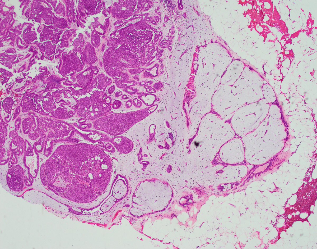
Unwittingly saved by the clinician. This case was not a consult, it was just a good old biopsy from a satellite hospital for whose patients we provide pathology diagnostic services. In that hospital, breast surgeons coordinate breast patient care, perform biopsies, discuss treatment and surgical plans with patients, and eventually refer to oncology as needed. Unlike our radiologists and oncologists here at WashU, the surgeons at that hospital do not want to get a tumor diagnosis then get an addendum with the biomarkers. They like to see a complete report all at once. I can't say I dislike that practice, and after this particular case, I like it even more, because it saved me from a major (albeit exceptionally rare) misdiagnosis.
The images below depict a tumor I was squarely going to call invasive solid papillary carcinoma because, to my eyes, that's exactly what it looks like. Except that's not what it is. And because I waited for the biomarkers to sign it out, I was fortunate to find out it was triple negative, which would be extremely unusual for solid papillary carcinoma. So, I contacted the surgeon and dug into her chart, and sure enough, the patient had a history of metastatic mucoepidermoid carcinoma of the minor salivary glands. That's when I started seeing a hint of squamous differentiation and possible mucous cells. CK5/6 and p63 were done and were confirmatory. MAML2 FISH was also positive and sealed the deal. I did not find a similar case in the literature.
Lessons from this monumental near miss.
1. Clinician preferences did save me this time, but the clinician's omission of pertinent medical history is a common and persistent problem.
2. Always remember that breast biomarkers are not only predicitve and prognostic, but also diagnostic. If their pattern does not fit with the tumor's morphology, take a deep breath and dive into those muddy diagnostic waters.
3. Again and again, be vigilant, look for clinical information, don't make assumptions. Easier said than done, of course, and I do not want to encourage paranoia because I am not the paranoid type, but sharp alertness is one absolute must, and not allowing hasty explanations for things that don't quite fit is another.
4. Morphology is key, but nature loves mimicry. Don't be a hammer. Everything is not a nail.
@washupathedu @wusm_pathology #PathTwitter #Breastpath #ENTpath
Friday cuteness. Some cases are just visually appealing aside from being diagnostically challenging. This is an example of a fibroadenoma involved by an expansile and somewhat uniform pattern of usual ductal hyperplasia, the most florid example I have seen within a fibroadenoma. Fortunately, CK5/6 accentuated the mixed nature of the proliferation and saved the day :) @wusm_pathology @washupathedu #PathTwitter #BreastPath