Research is to see what everybody else has seen and to think what nobody else has thought.
--Albert Szent-Gyorgyi
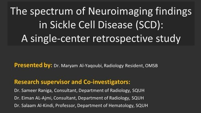
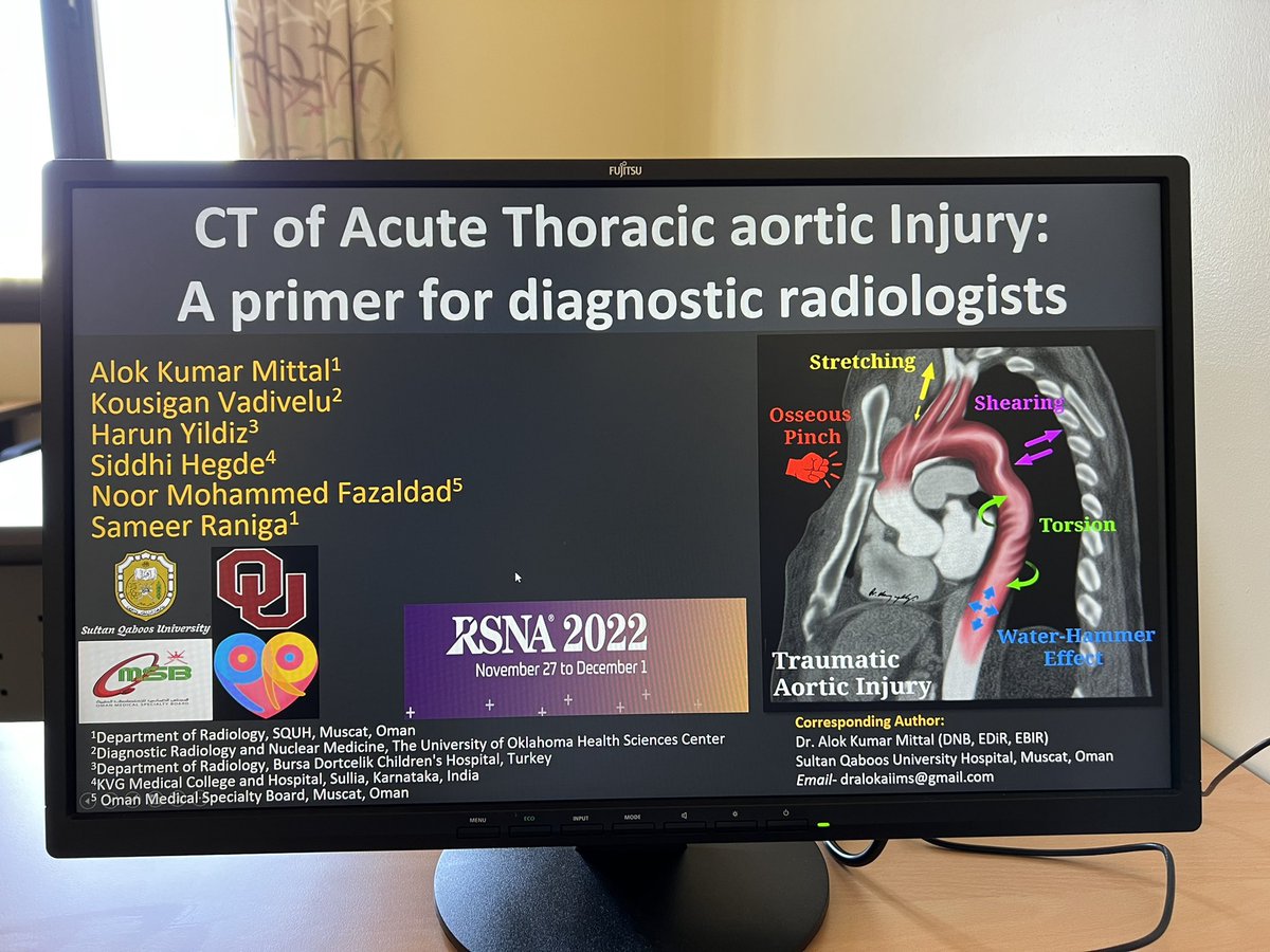
I supervised two trainees who presented their research at the Radiology Training Program's Research Day.
Congratulations to Dr. Noor for winning the second prize!
@OMSB_OM
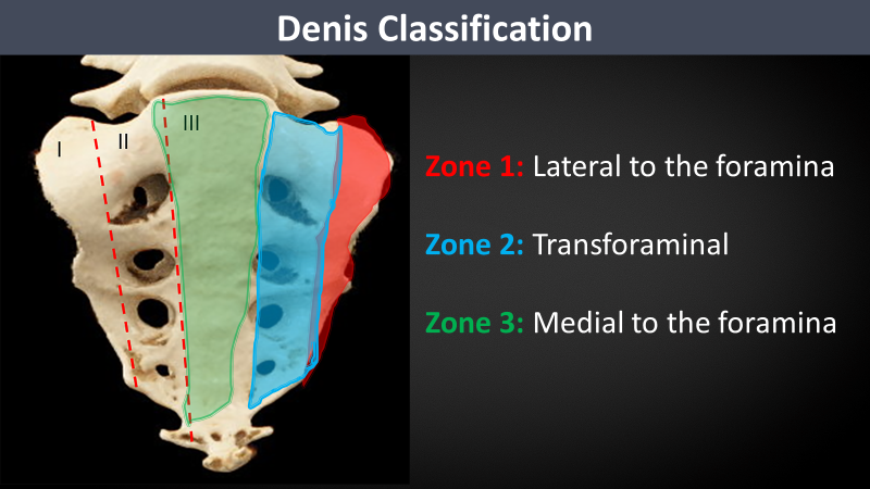
Denis et al. classified sacral fracture patterns into three zones based on the relationship of # to the neural foramina. Zone 1 and 2 are longitudinal, while zone 3 is classically reserved for sacral fractures with a transverse component, such as U- or H-shaped fractures.
In patients with sacral fracture, which imaging features suggest traumatic spinopelvic dissociation?
Follow this link: https://t.co/arLSWBdeGS
#RGphx @cookyscan #tweetorial
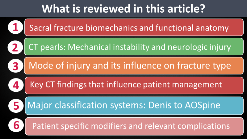
How much do you know about different sacral fracture patterns, classification systems, and their influence on patient management?
In @RadioGraphics, Dreizin et al. of discuss this important topic.
Link: https://t.co/arLSWBdeGS
#RGphx @cookyscan @RSNA
Academic writing is a superpower. It's a key to publishing, getting a new job or tenure, and becoming known in the field
These 10 AI-powered websites will help you with EVERY part of your manuscript and will save you 100+ hours:
#openscience#AcademicTwitter#phdvoice#phdchat
Thanks for coming along on the journey! Check out the incredible full-length @RadioGraphics article by Dr. Alexander, @ErbenYoung, @DrOverfieldMD, and @mpcaserta with plenty of tips and useful information, and videos.
Link: https://t.co/cPuhfiGwiv
#RGphx @cookyscan #tweetorial
CEUS evaluation can be suggested for patients who cannot undergo CTA or for problem-solving after an inconclusive CTA. It can precisely classify the endoleak type including origin, the direction of flow, and contrast material timing.
#RGphx
15/15
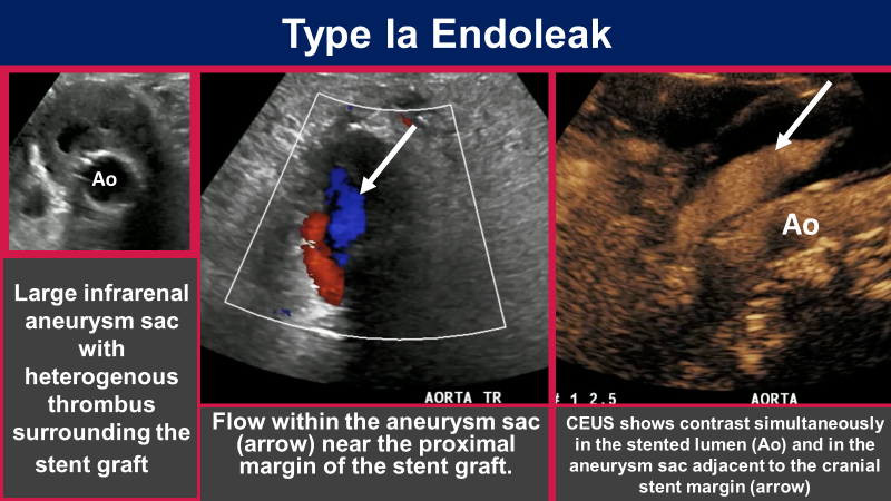
Type I endoleak occurs due to insufficient attachment of the stent graft limb to the native arterial wall and may be proximal (type IA) or distal (type IB), with resultant sac expansion from antegrade blood flow.
#RGPhx
10/15
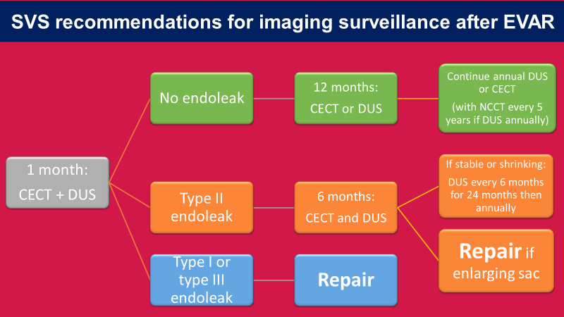
Multiphasic CT is the investigation of choice for imaging surveillance after EVAR. The authors summarize the 2018 Society of Vascular Surgery (SVS) guidelines for CECT and Doppler ultrasound.
#RGPhx
6/15
This review in @RadioGraphics monograph by Dr. Alexander, @ErbenYoung, @DrOverfieldMD, and @mpcaserta focuses on the role of CEUS in diagnosing endoleaks after endovascular repair (EVAR) of abdominal aortic aneurysms (AAA).
Link: https://t.co/hI32oNnY94
#RGphx@cookyscan1
1/15
Elective repair of Abdominal aortic aneurysms (AAA) with endovascular stent grafts (EVAR) requires lifetime imaging surveillance for potential complications, most commonly endoleaks.
What is your approach to post-EVAR imaging?
Stay tuned for a Tweetorial...
@RadioGraphics
This is what you do if you want to be a top educator.
Prepare well in advance- edit-revise- practice- repeat.
Respect your audience. Ensure that your teaching is worth their time and efforts.
Learning from @drmankad#RCRDubai
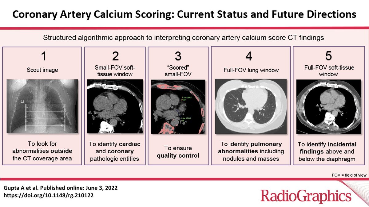
Calcium score is an indespensible tool for stratifying cardiovascular risk, predicting patient outcomes, and guiding preventive therapy. This article focuses on scan protocol, methods of calculating a score, and MESA percentile scores. https://t.co/s5idANQGwV
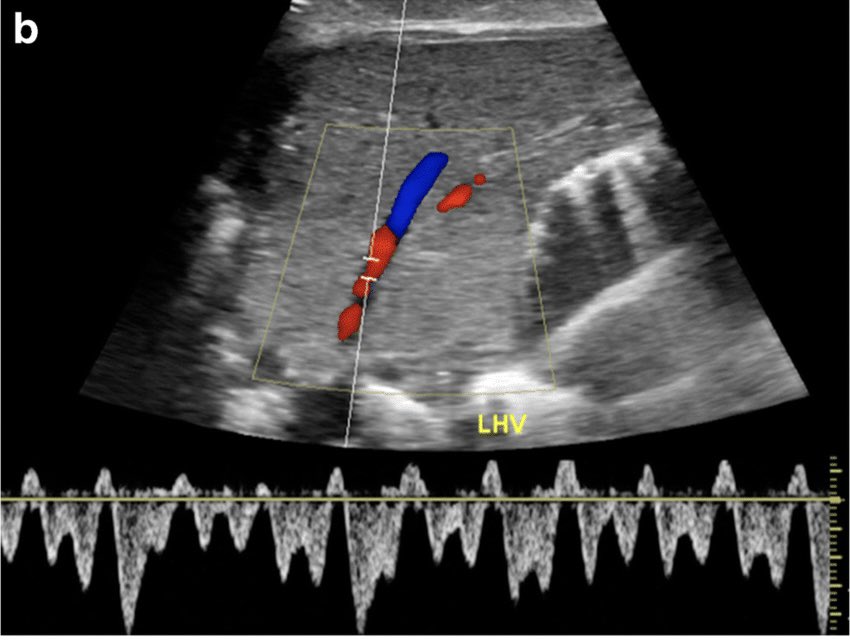
Normal hepatic vein:
1. Triphasic waveform owing to right atrial contractions.
Loss of phasicity can occur in early post op period due to graft edema.
@samrad77
Few key points about different vascular anastomoses in liver transplant and normal postoperative sonographic findings.
Typically 3 vascular anastomoses are done namely:
1. Hepatic arterial
2. Caval/hepatic venous
3. Portal venous.
@samrad77
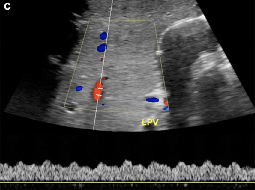
Normal portal vein:
1. Monophasic
2. Continuous hepatopetal flow
3. Mild cardiac & respiratory phasicity
4. Mean velocity 15-18 cm/s
5. Diameter: 13-16 mm
Caliber change of 5 mm at the anastomotic site is insignificant.
Vel >50cm/s is common due to post op edema/size mismatch