Our colleague Eva Yi Xie, a Princeton neuroscience PhD student, was detained by ICE at Reagan National Airport on her way home from SfN, where she’d just presented her poster. She’s being held in Chantilly, VA. Please help us bring her home. 📷
Recent TMS trials have shown that fMRI-guided TMS can outperform conventional scalp-based targeting for depression.
In our new Brain Stimulation commentary, we explore some of the exciting questions these advances open up.
https://t.co/dE1Xpb26cO
Heading to #SfN2026? 🧠 We’re presenting a new method that substantially reduces scan time for reliable individual brain network estimation, including recently identified intereffector regions and intLANG from just 14 min of fMRI.
Come chat! Nov. 18, 8-9 AM, Poster LBP077.04.
Excited to share my first journal paper as first author!
Using fPET-FDG, we identified a robust superior-inferior pattern in metabolic connectivity driven by minute-scale slow dynamics.
Grateful to my advisor Dr. Jingyuan Chen and collaborators!
https://t.co/Jeagxc7VQk
📣 New preprint: Multiple Nested Distributed Language Networks in the Human Brain. 📣
Brain regions specialized for language have been extensively described. But are they organized into one network, or multiple? Our new preprint provides evidence for the latter:
https://t.co/GCD49Jly69
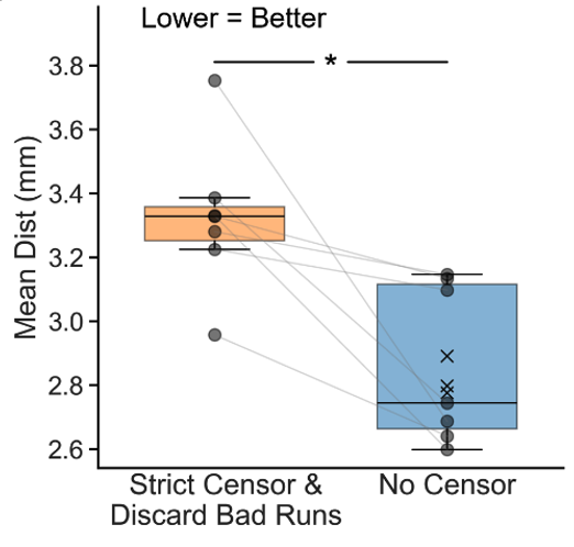
Do you censor high motion frames in fMRI? In two preprints by @twktan@mandyfmejia, we find that we may be censoring too much!
https://t.co/kSTADNYy93
https://t.co/TaQtTipAm3
In personalized TMS, strict censoring leads to worse targets than no censoring even with high motion!
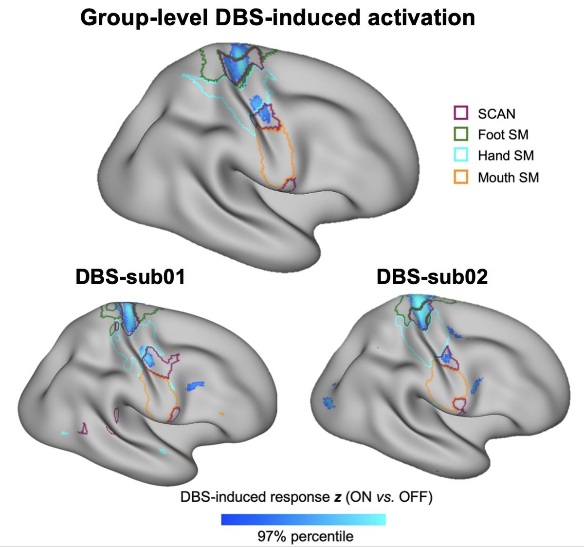
Thrilled to announce our new paper in @NatureNeuro !
We managed the impossible: precision functional mapping during #DBS, with 11.7h fMRI/patient.
Selective DBS-induced deactivations in the SCAN, building on our recent @Nature paper. @hesheng3@iamzhangvv
https://t.co/d8xOuNI31w
It’s time to rethink Parkinson’s disease. Our work reframes PD as a disorder of the somato-cognitive action network (SCAN), and shows that normalization of SCAN connectivity represents a shared mechanism across diverse effective therapies.
https://t.co/OuqOQcOcYq
@ndosenbach@DavidRen555@iamzhangvv@bttyeo@foxmdphd
Schizophrenia’s Unique Brain Network Identified
Researchers have identified a unified brain network linking regions of atrophy associated with schizophrenia, providing a clearer understanding of its neuroanatomy.
By analyzing data from over 90 studies with more than 8,000 participants, they found a consistent connectivity pattern involving regions like the insula, hippocampus, and fusiform cortex.
This network is distinct from those associated with other psychiatric or neurological conditions and remains stable across different stages and symptoms of schizophrenia.
The findings suggest that atrophy in high-risk individuals shares similarities with those who progress to clinical schizophrenia, offering predictive insights.
This research also supports the development of personalized treatments using brain stimulation techniques like transcranial magnetic stimulation.
A clinical trial targeting this network is planned, aiming to refine therapies for schizophrenia.