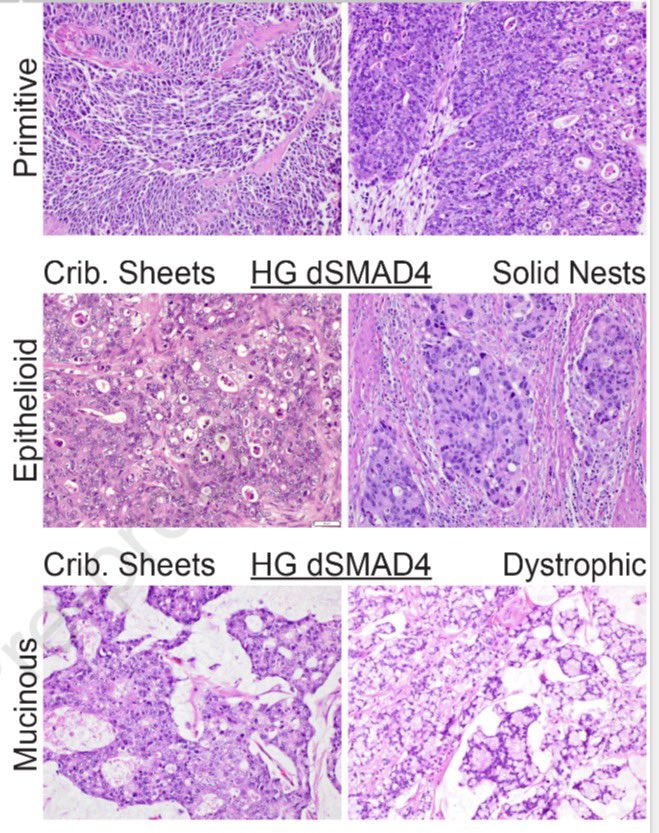
I’m thrilled to share that our study on SMAD4-deficient colorectal adenocarcinoma has just been published in Modern Pathology.
Grateful to my amazing mentor Dr. Oliver G. McDonald for his tremendous support 🙏🏻
@ModernPathology@LizMontgomeryMD@UMiamiPathology#GIPath#PathX
Let’s enjoy this cutie which pops up often on RISE and board exams, but not so frequently under the microscope! Once you see it, you’ll never forget it!
A beautiful case of Sex Cord Tumor with Annular Tubules (SCTAT):
• Positive for Calretinin, Inhibin, and WT-1
• Approximately one-third of cases are associated with Peutz-Jeghers syndrome (STK11 mutation)
#PathX #GYNpath @UMiamiPathology
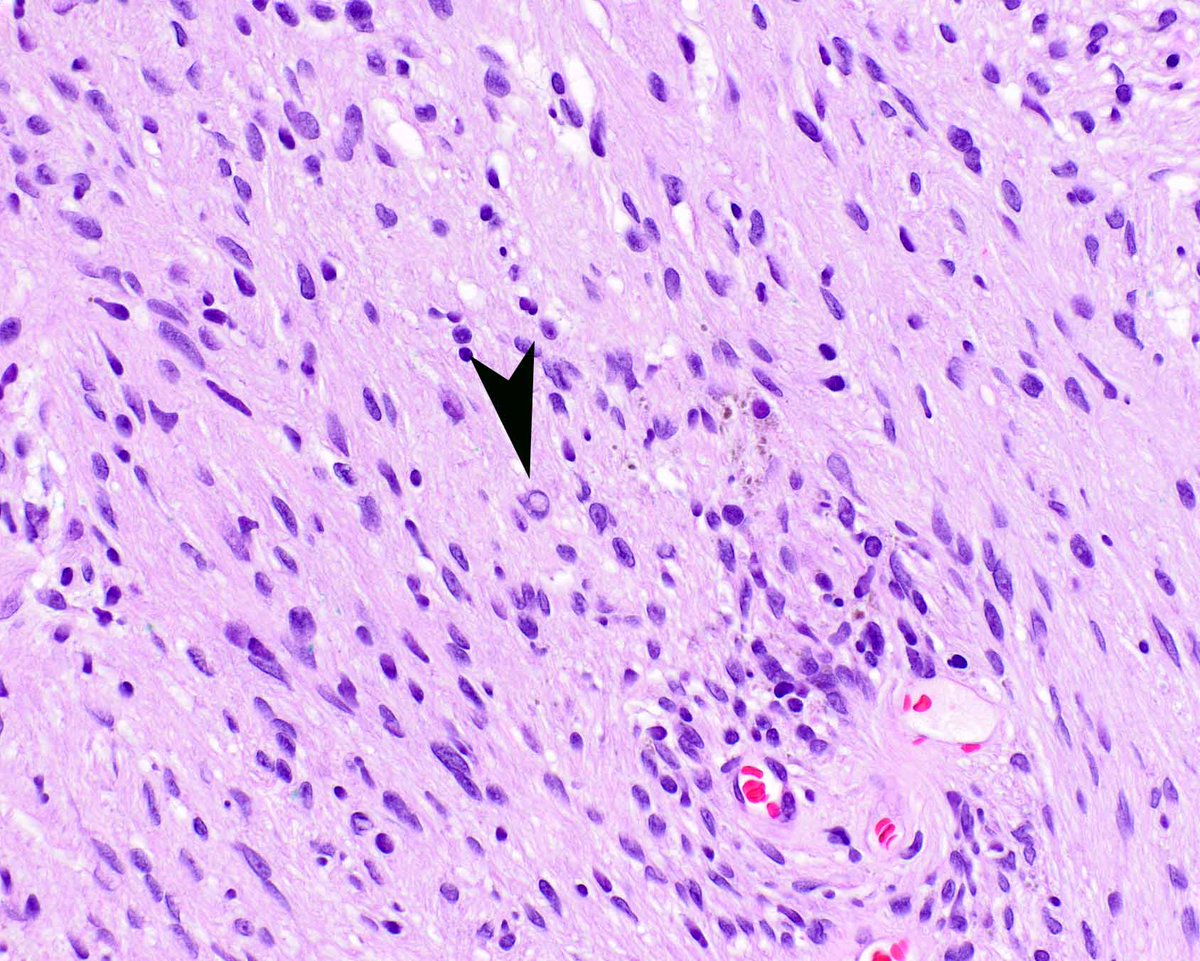
Plexiform schwannomas can be associated with central neurofibromatosis (NF2); finding this lesion may be a clue to screen the patient for central nervous system schwannomas. Note the intranuclear cytoplasmic invagination (arrow). #UMiamiPath@ModernPathology@science_press
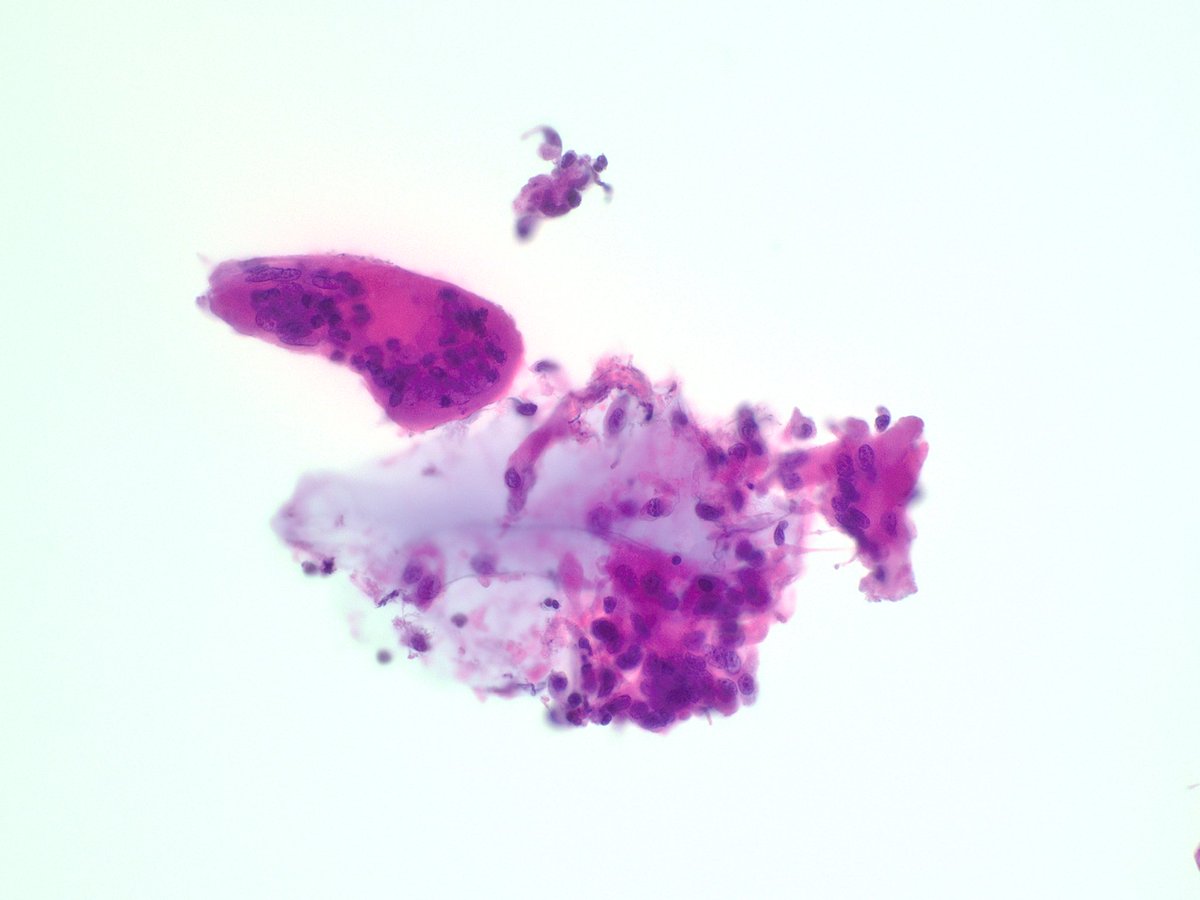
Uncommon complication? Not in Miami! 🔬
This cytology slide shows a granulomatous reaction to hyaluronic acid fillers—clusters of epithelioid histiocytes and giant cells fighting the filler, forming granulomas. This is a rare side effect of fillers… but in Miami, where cosmetic procedures are a beauty staple, we see it more often than expected. Keep it on your radar to make the diagnosis! 💡 #PathologySpotlight #MiamiBeauty #FillerFacts
🙌This month’s member spotlight, we are featuring Katherine Drews-Elger, MD, PhD➡️ To read the complete member profile and to catch up on FSP news and updates, visit https://t.co/SrWHpgK2EK. As a member of FSP, you could be our next member spotlight of the month!
Brilliant aspiring pathologist Valentina delivered an exceptional teaching session on spindle cell neoplasms of the breast — a challenging topic. She flawlessly integrated it with the UM Jeopardy game spirit. Chocolate provided.
Granular cell ameloblastoma
rare histological subtype of ameloblastoma accounting for less than 5% of all cases.
The characteristic microscopic features of granular cells are attributed to the increased presence of lysosomes in the cytoplasm of the tumour cells. The acquisition of granular cell phenotype has been attributed to an aging or degenerative change in long-standing lesions
Immunohistochemical studies proved that the granular cells are (+) for cytokeratin, CD68, lysozyme and alpha-1-antichymotrypsin, but (-) for vimentin, desmin, S-100 protein, neuron-specific enolase and CD15, indicating epithelial origin and lysosomal aggregation
🔬 Join the @FLPathologists Precision Medicine Academy on March 12, 2025, at 12:00 PM ET for a session on Molecular & Genomic Biomarker Testing in Hematopathology!
📢 Featuring guest speaker @drruizcor
✅ Earn 1 CME Credit
Register now: https://t.co/HkZWE9h7s2