I’m thrilled to finally share our discovery of a pathway enabling mtDNA release from mitochondria and activation of the innate immune cGAS-STING pathway, an epic collaboration between Shadelab and @manorlaboratory! Read on for details!
Combining #b3d and #Eevee with #JupyterNotebooks and #MolecularNodes, you can interactively style and render proteins from inside a live notebook.
Actually works a lot better than I thought it might. #GeometryNodes inside of python notebooks is a cool combo
#b3d#blender#geometrynodes
I revised and simplified my algorithm of inertia.
combining it with collision deformer it can give this kind of puffy soft body like result in real time.
Excited to release our latest work, led by @hbozhao in collaboration with our friend Andrej Košmrlj!
In this preprint (https://t.co/uJNIj2zaYb), we study how chemotactic active matter phase separates—and find many cool surprises. 🦠🚀🔬
Tweetorial follows... [1/8]
🚨Crowded scenes are notoriously difficult for #ComputerVision models.
In our new work lead by @zhoumu53 & @LStoffl we developed a novel approach called BUCTD that is state-of-the-art on crowded 🐒🐠&🕺human pose estimation benchmarks!🔥
https://t.co/pPI8DlxR8Q
#CVPR2023 workshop
Excellent new fine-grained tracking from DeepMind:
TAPIR: Tracking Any Point with per-frame Initialization and temporal Refinement
arxiv: https://t.co/ffkLzkSnem
project: https://t.co/RRqsvfQ4SA
tldr: TapNet for localization then PIPs-style refinement; outperforms everything!
Identify cell types in your electron microscopy (EM) data using MorphoFeatures.
Developed by @embl researchers, MorphoFeatures is a novel unsupervised deep learning method for extracting cellular morphology directly from 3D EM images.
https://t.co/HI5JmbgNyt
Our paper on #tardigrade walking (with @Duranigrade, Deborah Johnston, and @DJCohenEtAl ) is out now in
@PNASNews : https://t.co/XDzBurTwTq
Follow this little guy, strolling on his way to an explainer thread 🧵[1/n]
Fibroblasts are cool!
Mammary fibroblasts are good at inducing organoid branching, but how do they do it? Let’s find out in our new preprint. A thread.🧵🧵🧵
paper: https://t.co/1n9xgwOdS7
Group of @KoledovaZuzana@Science_MEDMUNI
collaboration with @slvfre@institut_curie
1/12
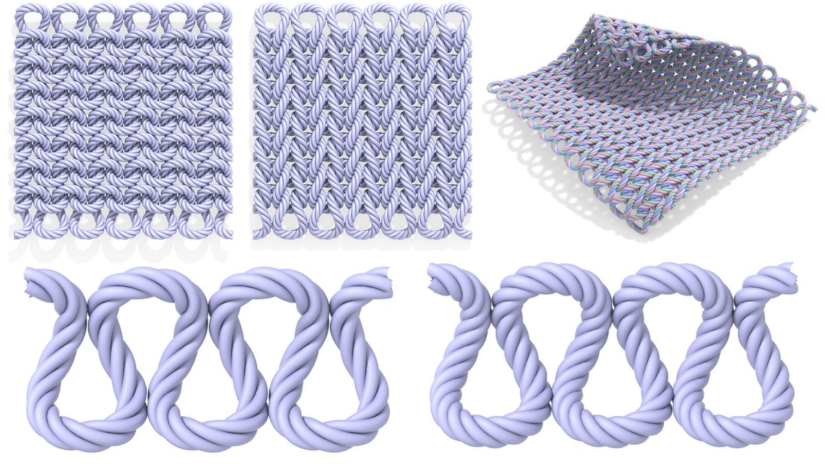
For fun (and because I couldn't find any!) I wrote down some closed-form parametric equations for plain-knit yarns and the twisted fibers running around them:
https://t.co/vYV8EIPJWX
Also includes C code to generate curves, and displacement/alpha maps for making tiled patterns.
Pretty awesome discovery: a single shape that tiles the infinite plane without repetition.
If you're staring straight down at a checkerboard, there's no way to tell where you are: every part looks the same.
But here, the relative arrangement of tiles encodes your location.
napari-skimage-regionprops allows you to:
- interactively get objects size, shape✅
"But I want to relate to objects from other channels!"
You can write python code to do that.
OR...
I am introducing multichannel summary statistics to https://t.co/Sjli3itVgH🚀🙂give it a try!
I'm creating 3D tours of open single cell RNA-seq data in UMAP space. Here, using data from 50k brain cells by the @AllenInstitute . (https://t.co/aOSpYfve70). The UMAP position and class of every cell is read into Blender, realized as a stylized, class-specific model I sculpted
@foodskop@NeedhiBhalla@born2raisecell@BementLab ohhhhh yes we've definitely chatted about this stuff : ) the parallels how the wave generators for native cortical actin dynamics his group sees are controlled and this system point to some universal themes for patterning https://t.co/qXUAdfE2Ed
Our first preprint is out! Led by Rohith Rajasekaran (@born2raisecell): “A programmable reaction-diffusion system for spatiotemporal cell signaling circuit design”
A visually stunning toolkit to build cellular radios that probe or control cell biology.
https://t.co/RA18asTEBQ
Are you looking for ready-to-use widgets and type annotations for your @napari_imaging plugins? I made a lot of napari-specific, very useful stuffs here 👇👇
https://t.co/k99JZ8tdH0