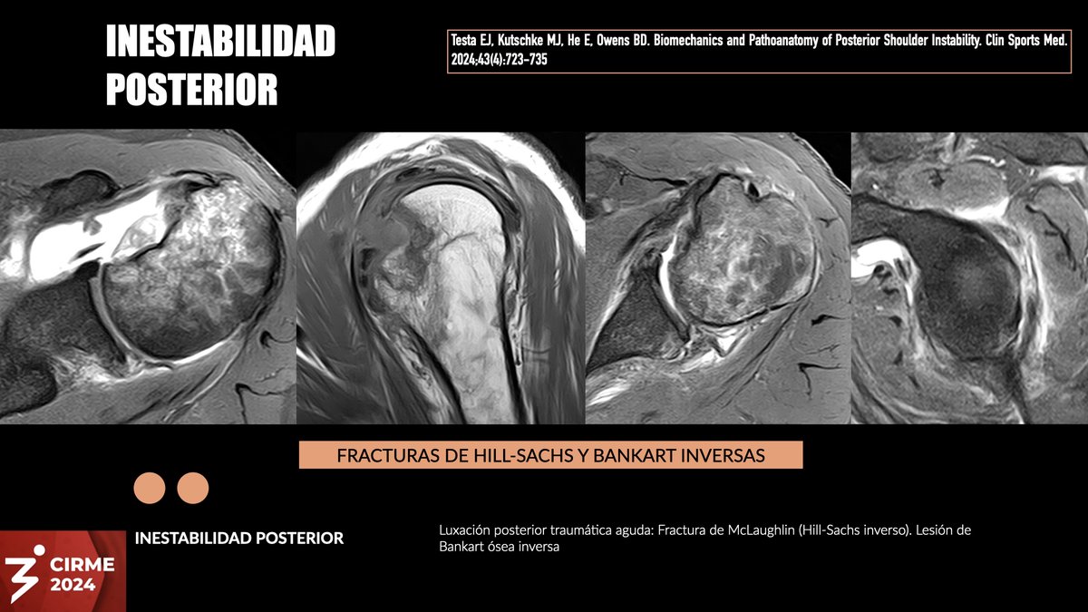
Luxación traumática aguda reducida en un jugador de rugby. Defecto óseo bipolar con lesión de Hill-Sachs inversa (fractura de McLaughlin) y lesión de Bankart ósea inversa menor.
Un día para inicio de CIRME 2024 en BsAs. Abierta modalidad online.
https://t.co/osGcbwQc1a
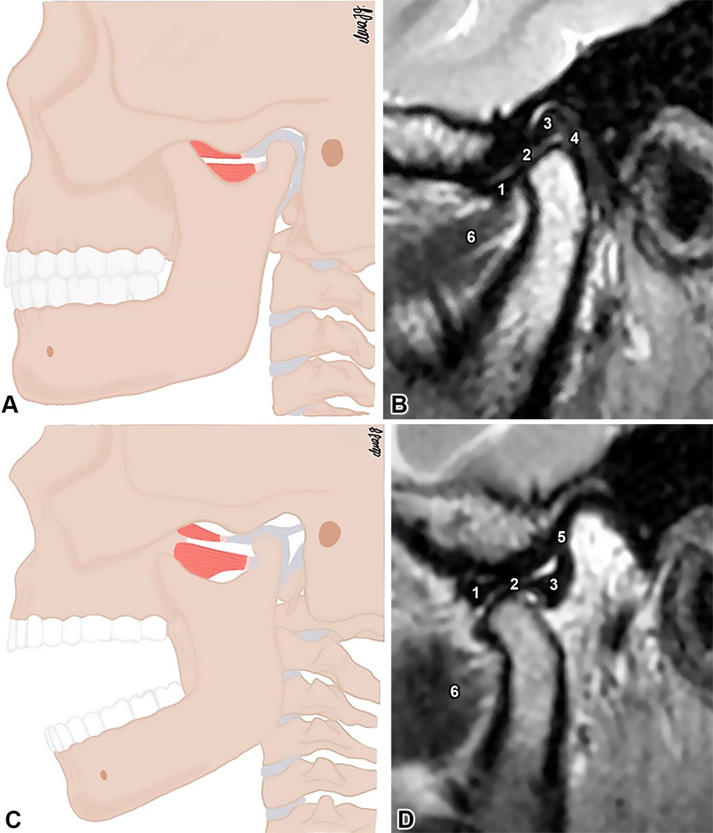
Ever curious about conditions linked to temporomandibular joint derangement? This article dives into TMJ anatomy, its biomechanics, and normal & abnormal imaging findings, with arthroscopic correlation! https://t.co/RRIJxK6xEA
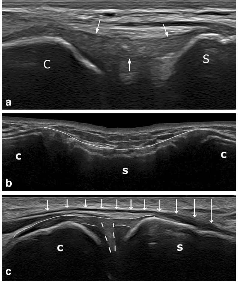
The usefulness of the fibrillar pattern and pennation angle in the ultrasound diagnosis of muscle injury
A 17-year-old patient who, while braking while playing football, feels a sharp pain in the anterolateral region of the proximal third of the thigh that prevents him from continuing to play sports.
Typically, the axial plane (the short axis of the muscle) is used to study fibrillar tears, with a high diagnostic sensitivity. However, on many occasions, it can be easier to scan through the long axis of the muscle to visualize the different muscle fascicles and the pennation angle in their entirety, being able to make the diagnosis by observing the interruption and loss of tension of the muscle fascicles associated with the loss of the normal pennation angle.
In the present case, the normal fibrillar pattern is initially visualized in the medial region of the rectus femoris muscle until the presence of a fibrillar tear in the posterolateral myofascial junction of its proximal third, where there is a sudden interruption of the fibrillar pattern due to the presence of an intramuscular hematoma.
Fantastic arricle - for those who infiltrate and scan the sc joint.
Anatomical study of the sternoclavicular
joint using high-frequency ultrasound
FREE PDF
https://t.co/Xsm6Xmsq6h
PECTORALIS MAJOR TENDON:
2 layers:
•ANTERIOR: Clavicular & superior Sternal segments
•POSTERIOR: remaining Sternal segments
Slides from my last lecture at @RadiologiaChile congress
iliacus muscle injury: MRI diagnosis
A 16-year-old patient who, while playing soccer, felt inguinal pain when changing direction when defending possession and, three days later, a snapping sensation when flexing and extending.
The physical examination showed no pain when flexing against resistance but discomfort when externally rotating against resistance.
An MRI was performed, which showed a significant fibrillar tear in the myotendinous junction of the iliac muscle, with a hematoma with clots at the level of the muscle injury.
Due to greater access to ultrasound and MRI, pelvic muscle injuries (flexors, rotators, and hip stabilizers) are increasingly frequently diagnosed. In my personal statistics, they are the third most frequent group after quadriceps and hamstrings.
Despite being less symptomatic injuries, the importance of an adequate diagnosis and treatment lies in the fact that if they are not correctly managed, there is a risk of secondary overload of other muscle groups that cause a fibrillar tear.
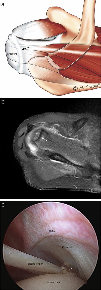
A fantastic article on rotator cuff anatomy and tear patterns. Relevant to both ultrasound and MRI.
Rotator cuff tear patterns: MRI appearance and its surgical relevance
https://t.co/ovhv7cjCoj
Young patient from today's consultation, referred with a question about gout in the little toe. No risk factors for gout. Recently had bilateral carpal tunnel surgery - without effect. What is the most likely diagnosis or underlying disease?
Utility of the fascicular anatomy of the Achilles tendon in the diagnosis and treatment of partial tear
A 44-year-old amateur soccer player, upon starting a sprint, experienced debilitating pain in the posterior region of the ankle and heard a pop, which made it impossible for him to continue physical activity. Upon physical examination, he presented significant pain in the distal end of the Achilles tendon, with a negative Thompson test.
An MRI was performed, which showed a partial tear of the Achilles tendon, with complete distal interruption of the fibers dependent on the medial gastrocnemius muscle.
The Achilles tendon is a common tendon of the gastrocnemius and soleus muscles. The fibers rotate depending on each muscle until inserted into the calcaneus. Multiple cadaver studies serially analyzed the location of these fibers in cross-sections.
In this patient's example, the tendon involvement corresponds to the fibers dependent on the medial gastrocnemius muscle, which could be useful for assessing conservative or surgical treatment.
The variability of the APL muscle can range from supernumerary tendons to variable sites of insertion, most frequently on the first metacarpal bone, the APB muscle and the trapezium
https://t.co/nqpQb6ZSXJ
What ist the main reason of APL tenosynovitis and enthesopathy at the insertion? Hint: it's not De Quervain Tenosynovitis
Adhesive capsulitis of the hip: diagnosis
A 55-year-old patient with gluteal pain of 6 months' duration associated with decreased range of motion of the hip joint.
An AMRI is requested to rule out a tear of the acetabular labrum. The study shows an increase in thickness of the lower and upper bands of the iliofemoral ligament (measuring 13 and 15 mm, respectively) associated with pericapsular edema, consistent with adhesive capsulitis.
An ultrasound-guided intra-articular infiltration of 1 cc of Trigon and 1 cc of 2% mepivacaine is performed, and the pain is entirely resolved after five days. Treatment is associated with physiotherapy and rehabilitation, and the range of joint mobility recovers after three weeks.
In the article “Capsular ligaments of the hip: anatomic, histologic, and positional study in cadaveric specimens with MR arthrography” (doi: 10.1148/radiol.12111320), the average thickness of the capsular ligaments of the hip joint is described.