High-Risk Indicators for Fontan Completion — #2 Anatomic & Surgical History:
Prior pulmonary artery or pulmonary venous interventions after the Glenn stage often indicate underlying anatomic or hemodynamic complexity, increasing the risk of suboptimal Fontan physiology.
Fontan Pathway Diameter — Size, Flow Reserve, and Exercise Physiology🏃🌊
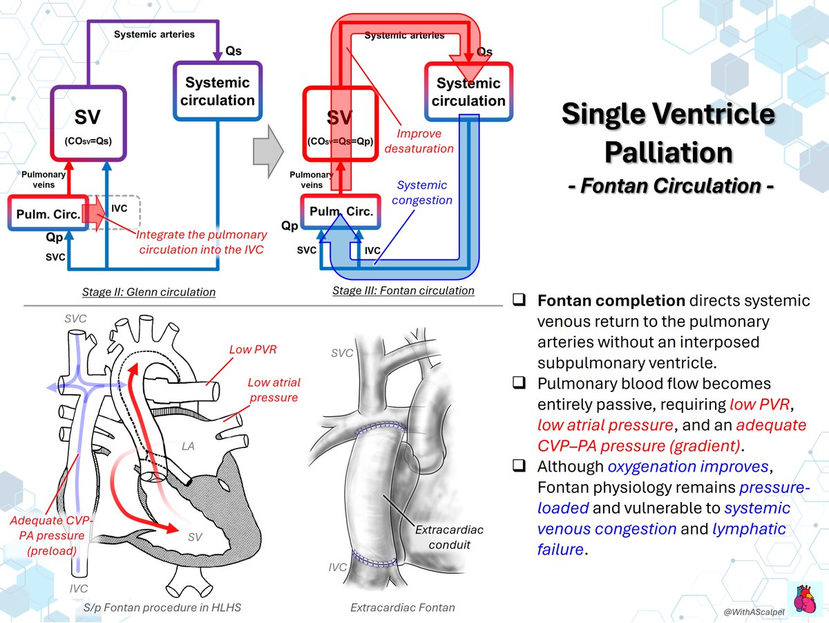
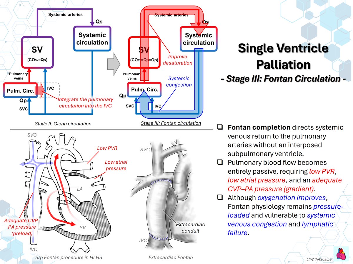
👉Fontan circulation directs systemic venous return to the PAs without a subpulmonary ventricle, making flow dependent on low PVR and an adequate CVP–PAP gradient.
👉The single ventricle supports systemic output through a venous pathway, where congestion and limited flow reserve become key constraints.
👉In 67 adults at exercise cath (mean age 30.4±7.9 yr), larger diameter correlated with less reserve: Qp −2.3%/mm, Qs −2.2%/mm, peak VO₂ −0.8%/mm.
(https://t.co/9JptVF39bg)
@MayoClinicCVS #JTCVS
Fontan Procedure—Lateral Tunnel vs Extracardiac Conduit💡
👉The lateral tunnel directs IVC flow through an intra-atrial pathway, offering some growth potential but exposing the atrium to dilation, arrhythmia substrate, and potential leaks.
👉The extracardiac conduit routes IVC flow outside the atrium, providing a smooth venous pathway but with no growth potential and risks of thrombosis and conduit-related obstruction.
👉Regardless of staging strategy, successful Fontan circulation depends on a low-resistance, unobstructed systemic venous pathway to the pulmonary arteries.
Fontan Procedure: Circulation and Surgical Pathways
👉Fontan circulation directs systemic venous return to the PAs without a subpulmonary ventricle, relying on low PVR and a sufficient CVP–PAP gradient.
👉Extracardiac Fontan divides the IVC at the RA junction and connects it to the PA using a PTFE conduit, avoiding the atrium but lacking growth.
👉Lateral tunnel Fontan routes IVC flow through an intra-atrial patch pathway, preserving some growth potential but adding arrhythmia and leak risks.
Single-Ventricle Circulation: From Parallel Flow to Fontan Series Circulation
👉In biventricular circulation, the RV drives pulmonary flow and fills the LV, creating a true series circuit.
👉In single-ventricle physiology, one ventricle supplies systemic and pulmonary beds, causing volume overload and flow competition.
👉Glenn unloads the ventricle with partial oxygenation; Fontan completion creates series flow without a subpulmonary ventricle.
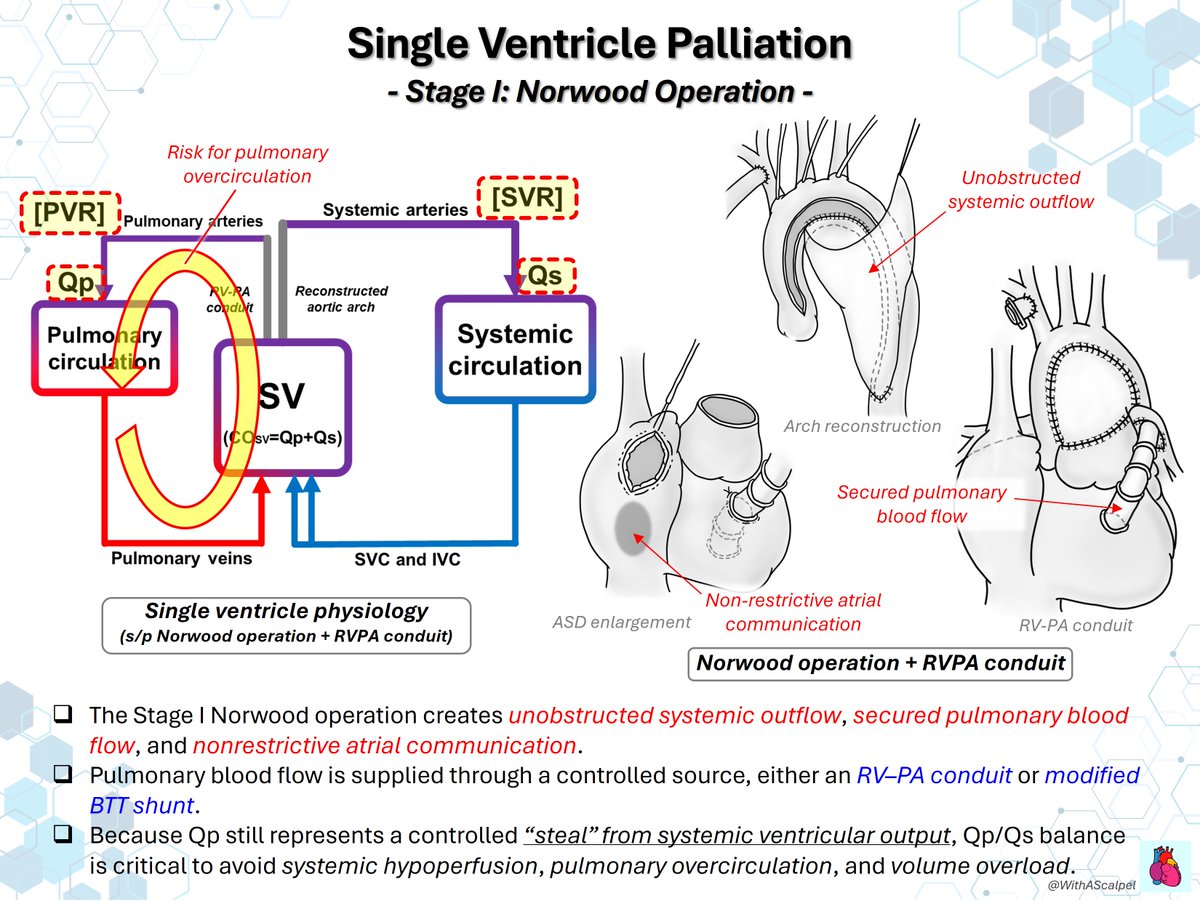
Single Ventricle Palliation #2: Norwood → Glenn → Fontan
👉Stage I Norwood secures systemic outflow, controlled pulmonary blood flow, and nonrestrictive atrial communication.
👉Stage II Glenn diverts SVC flow to the PAs, making pulmonary flow passive while reducing single-ventricle volume load.
👉Stage III Fontan directs systemic venous return to the PAs; low PVR and low atrial pressure are essential for flow.
Fenestrated Fontan #1: Concept and Physiology
👉A fenestration creates a controlled right-to-left shunt from the Fontan pathway to the atrium, functioning as a pop-off.
👉By lowering Fontan pressure, it preserves ventricular preload and cardiac output when PVR is elevated or filling is fragile.
👉This may ease early venous congestion and pleural effusions, but the trade-off is lower systemic arterial saturation.
VSD Patch Closure for Perimembranous VSD with Inlet Extension🧐
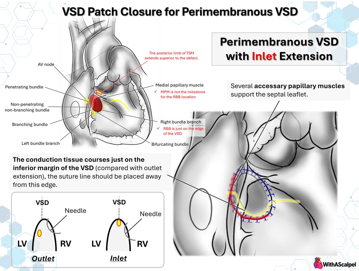
👉In inlet-extended perimembranous VSD, the conduction axis courses along the inferior margin, so sutures should be placed away from this edge.
👉Accessory papillary muscles and septal leaflet attachments can obscure the true rim; medial papillary muscle is not a reliable landmark for the right bundle branch.
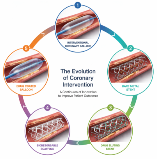
Back to the future - Lessons from past management of atherosclerotic coronary vascular disease that can guide contemporary PCI practice
📺 & 📰https://t.co/6CCrmMoLpU
Reflecting on historical perspectives, this session replay imparts valuable lessons to guide PCI practice today:
🟣 best practices in vessel preparation
🟣the significance of DES
🟣a pathologist’s insight into mechanisms of PCI failure to improve patient outcomes.
Physician team: @twj1974 , Patrick W. Serruys, @mc_morice, Bruno Scheller, @Antocol17, @AlokeFinn, Joanna Wykrzykowska
#interventionalcardiology #EuroPCR
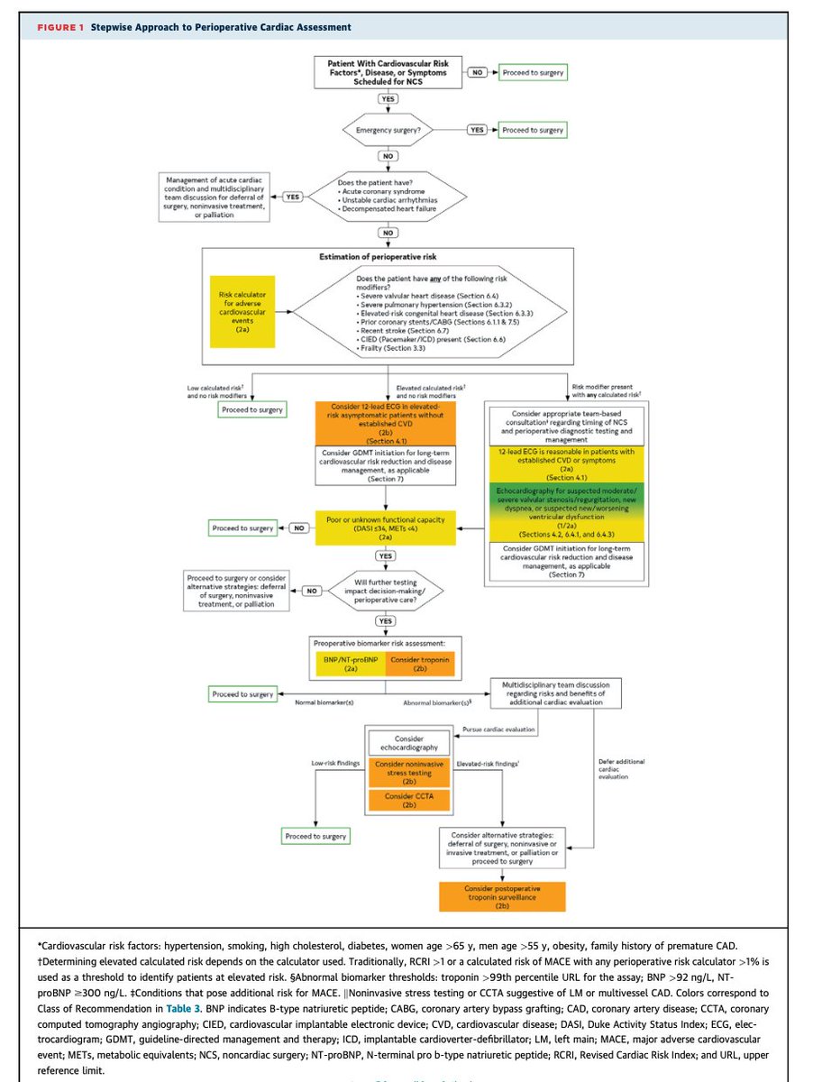
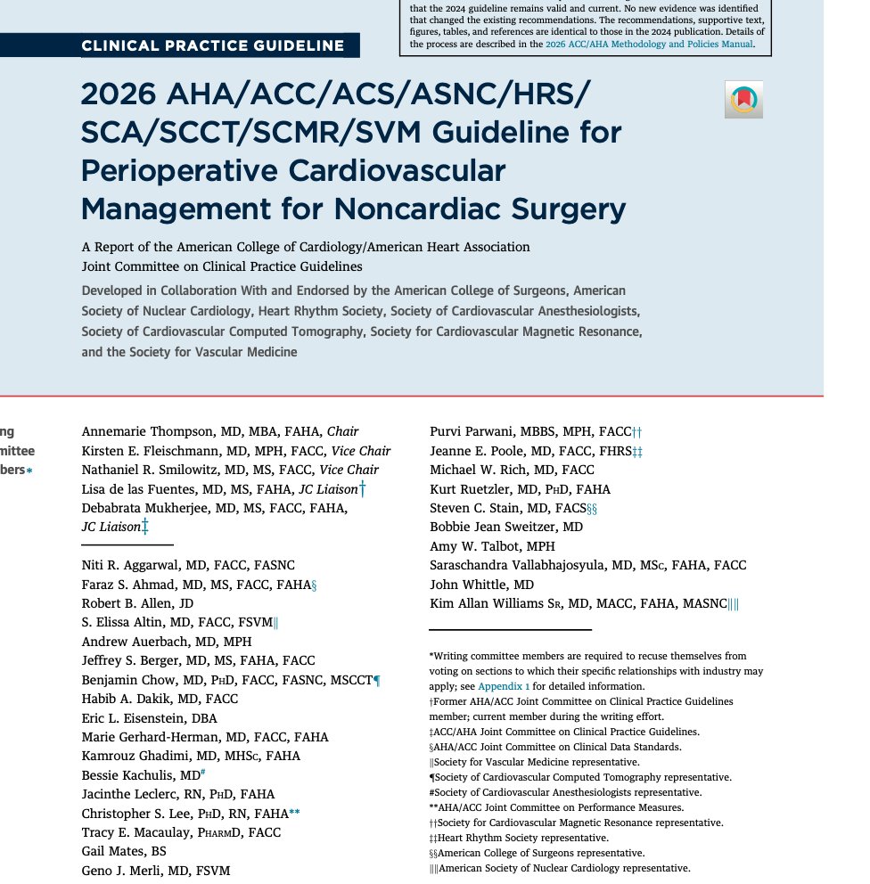
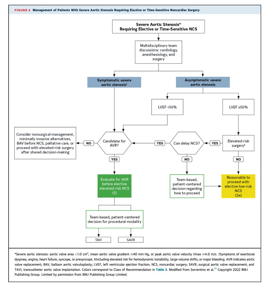
Guías 2026 de la AHA de manejo preoperatorio en cirugía no cardíaca (alias valoración preoperatoria). Recién salida del horno ayer. Puntos importantes:
🔴 Los iSGLT2 se suspenden 3 días antes por riesgo de cetoacidosis euglucémica (nota: la evidencia nueva apunta a que tal vez no se requiera).
🔴 Los aGLP-1 semanales se suspenden más de 1 semana antes (por broncoaspiración). Metformina: continuar.
🔴 Betabloqueador: continuar. Si apenas lo vas a iniciar: empezar 7 días antes.
🔴 IECA/ARA-II pueden omitirse 24 horas en hipertensión controlada + cirugía de alto riesgo, pero se continúan en insuficiencia cardíaca.
🔴 Prueba de estrés y angioTC coronaria: solo con riesgo alto (como <4 METs por DASI y otra indicación. CAC de 0 en los últimos 2 años: pasa a cirugía sin más.
🔴 El ECG en asintomáticos con cirugía de bajo riesgo: no aporta, no es necesario.
🔴 En cirugía de alto riesgo, >65 años, o sintomáticos: considera medir BNP o NT-proBNP). En general no se necesitan seriados.
🔴 Anticoagulación oral (con ACOD) sin puente en general, sin heparina. Puente solo si: AVK y riesgo trombótico muy alto: válvula mecánica mitral, EVC/AIT en los últimos 3 meses, trombofilia o CHA₂DS₂-VASc ≥7.
🔴 Tiempos e infarto. Después de un stent farmacoactivo: esperar 6-12 meses si cirugía electiva. Después de un EVC/AIT se esperan ≥3 meses.
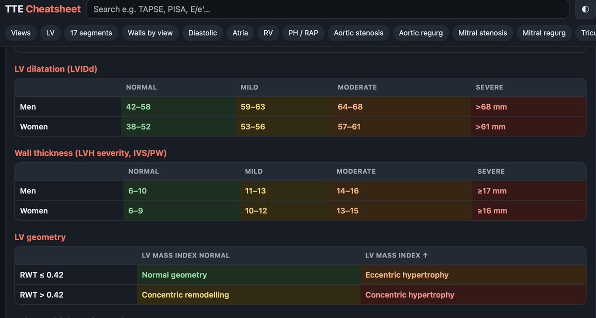
🫀 TTE cheatsheet update, now at https://t.co/5rQ5TyJadK, still free.
• Updated based on feedbacks
• Drop a screenshot of measurements→ values auto-filled, cross-checked & interpreted
Runs in your browser, nothing uploaded/downloaded @ASE360#EchoFirst#CardioTwitter
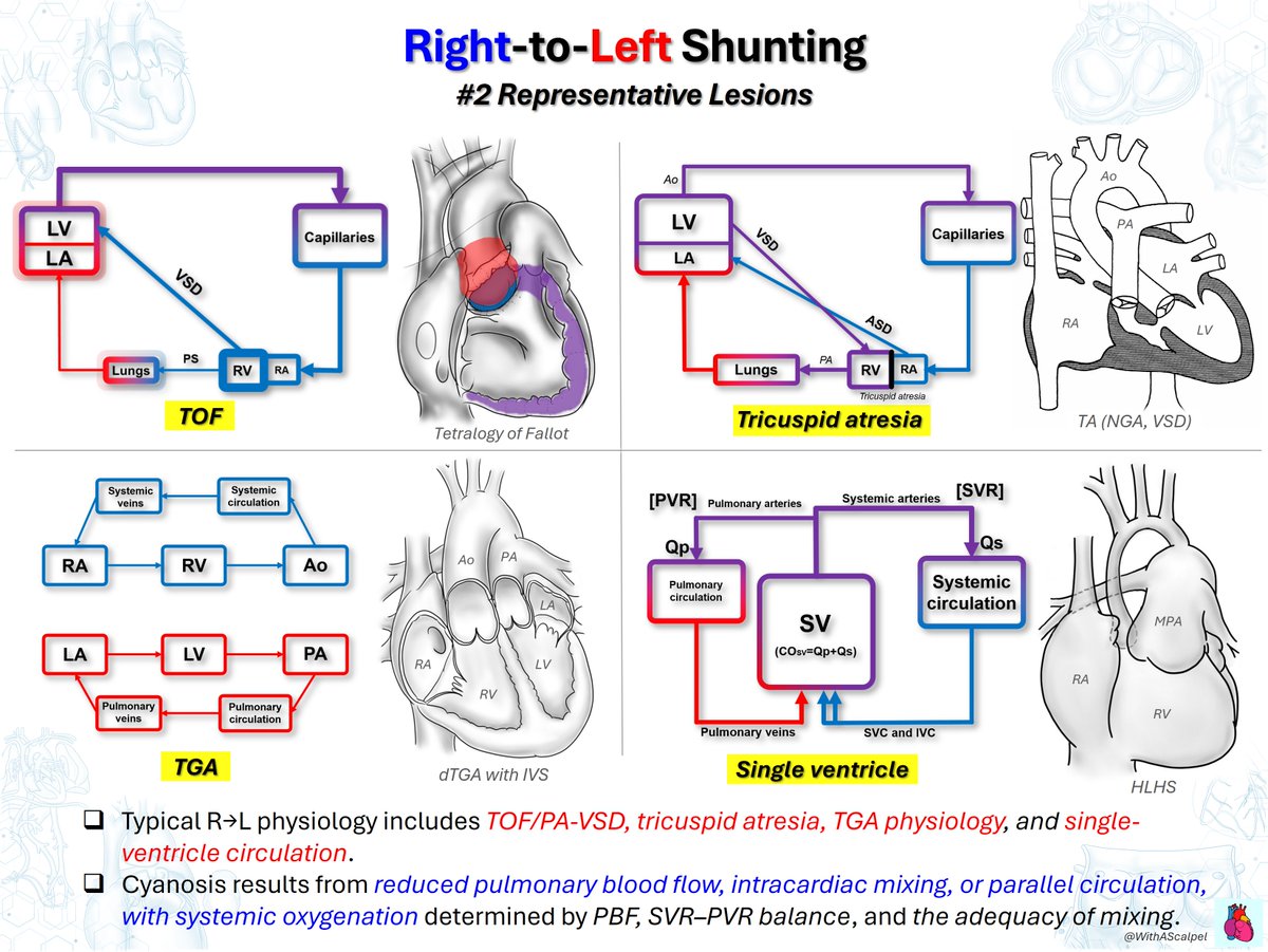
Right-to-Left Shunting: Physiology and Representative Lesions
👉Right-to-left shunting allows systemic venous blood to bypass effective pulmonary oxygenation, causing arterial desaturation and cyanosis.
👉In TOF/PA-VSD and tricuspid atresia, systemic oxygenation depends largely on pulmonary blood flow and the degree of intracardiac mixing.
👉TGA creates parallel circulations, while single-ventricle physiology depends on effective mixing and the balance between Qp and Qs.
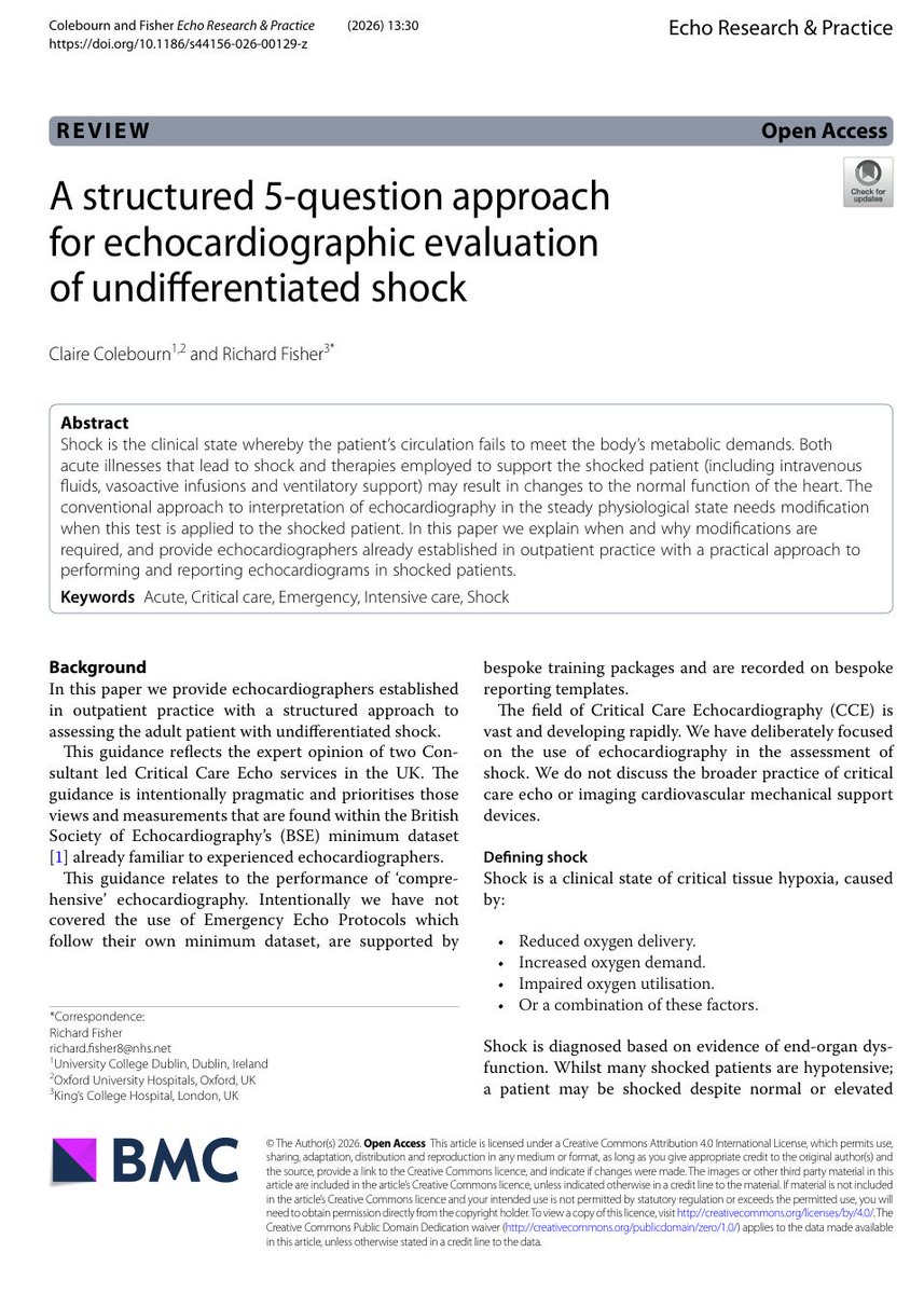
❤️Echocardiography in shock: stop asking only “What is the EF?”
In undifferentiated shock, echocardiography is most useful when it answers physiological questions, not when it simply describes cardiac anatomy.
Colebourn and Fisher propose a pragmatic 5 question framework for approaching the shocked patient:
1. What are the stroke volume and cardiac output?
Start with flow. Importantly, cardiac index is not a binary marker of shock. A patient may maintain cardiac output through tachycardia despite a low stroke volume, while another may have low output because bradycardia prevents compensation. SVI, CI, heart rate and rhythm therefore need to be interpreted together.
2. If output is abnormal, why?
LV failure? RV failure? Low preload? Valve disease? Mechanical complication? Obstruction?
And do not forget dynamic LVOTO. Critical illness can combine low LV volume with hypercontractility, and escalating vasoactive support without recognising LVOTO may paradoxically worsen shock.
This is also where LVEF can mislead us.
A “normal” EF does not guarantee adequate forward flow. EF depends on LV volume and afterload and does not distinguish forward from regurgitant stroke volume. Vasoplegia may even make LV systolic function appear reassuring until vascular tone is restored.
3. Is left atrial pressure elevated?
E/e′ >14 may support elevated LAP in general ICU populations, but performs less reliably in cardiogenic shock and should not be interpreted alone. A normal LA size does not exclude an acute rise in LAP.
4. Are pulmonary pressure and PVR elevated?
Now the assessment moves to the RV pulmonary circulation interaction, where pressure, afterload and RV adaptation matter.
5. How is the situation evolving?
Perhaps the most important question.
The echo we see is not the patient's fixed cardiovascular phenotype. It is the heart under the current preload, afterload, vasoactive therapy and ventilatory conditions.
Noradrenaline can increase BP while decreasing SV in one patient, yet increase both BP and SV in another.
This is why critical care echocardiography should increasingly be thought of as:
Flow → Mechanism → Filling pressures → RV pulmonary circulation → Response over time
Not a photograph.
A dynamic physiological assessment.
This paper represents pragmatic expert guidance rather than a validated diagnostic algorithm.
Reference����
Colebourn C, Fisher R. A structured 5-question approach for echocardiographic evaluation of undifferentiated shock. Echo Res Pract. 2026;13:30. https://t.co/z8VVd7Am2K
#CriticalCare #Echocardiography #POCUS #Shock #Hemodynamics #Cardiology #IntensiveCare
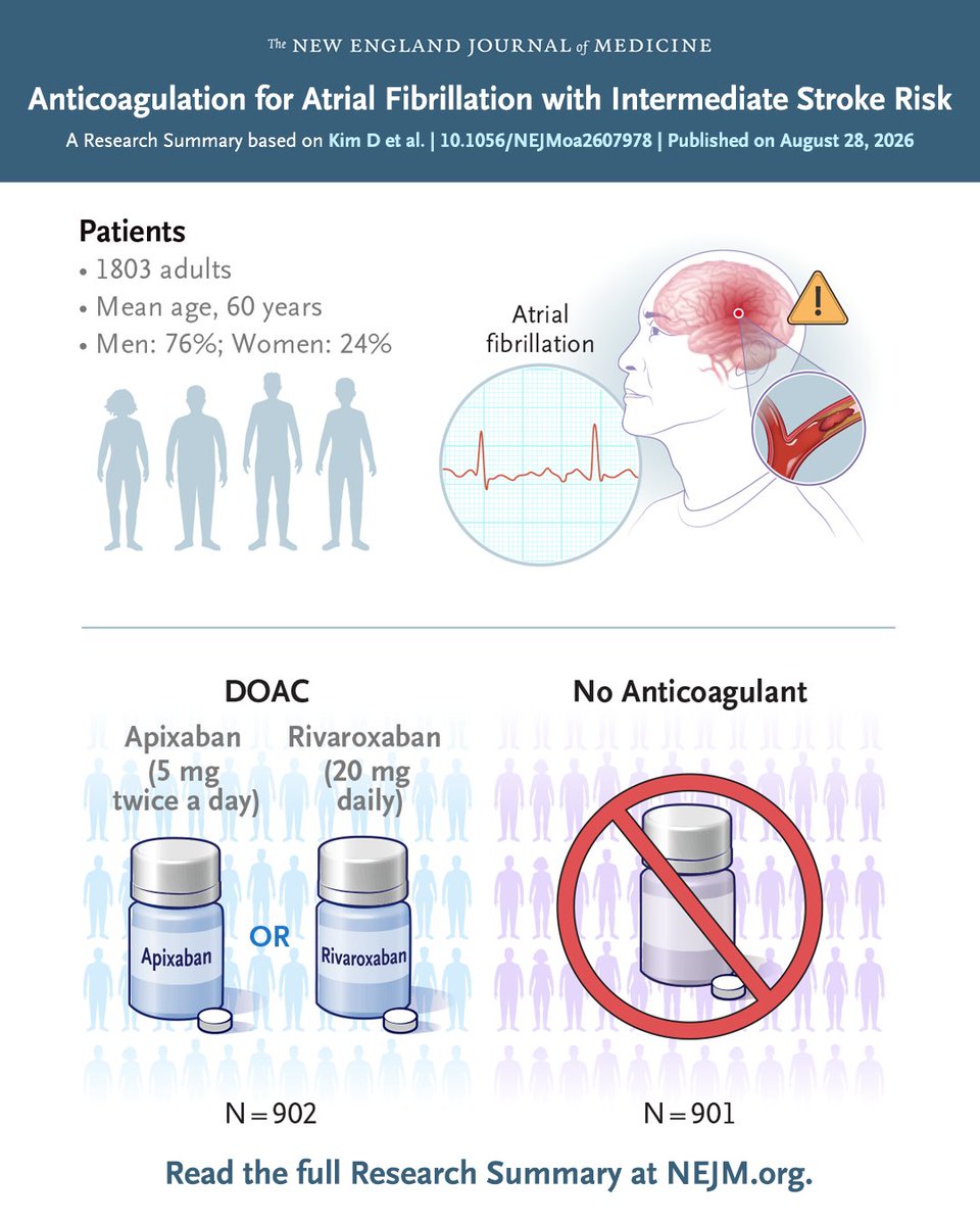
In patients with #AFib and intermediate stroke risk, direct oral anticoagulation led to a lower risk of stroke, systemic embolism, major bleeding, or death from cardiovascular causes than no anticoagulation. Full SINGLE-AF trial results and Research Summary: https://t.co/rISG8pcl17
#ESCCongress
Dear cardiology fellows 🫀
I built a free TTE cheatsheet with @claudeai I keep open while reading echos: guideline cutoffs, 17-segment walls by view, valve grading and 15 built-in calculators (AVA, SV/CO).
Check it out 👇
https://t.co/MZzWwYgeMe
#EchoFirst#CardioTwitter
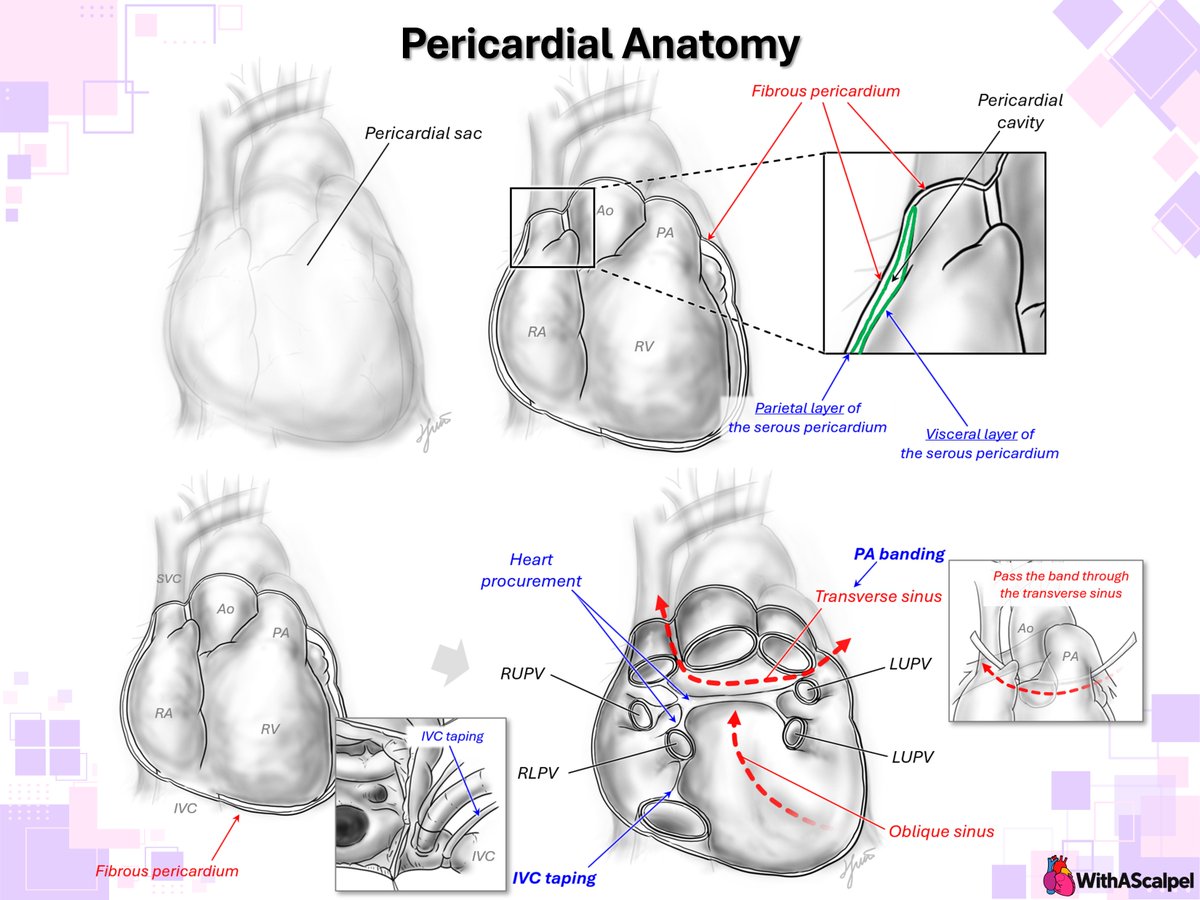
Pericardial Anatomy — Series: #1–2
👉The pericardium consists of fibrous and serous layers surrounding the heart, with a potential pericardial cavity between the parietal and visceral serous layers.
👉Posterior pericardial reflections form the transverse and oblique sinuses and define important surgical pathways around the great vessels and pulmonary veins.
↓ Full series below
Q. Which loop diuretic carries the highest risk of causing ototoxicity when given in high intravenous doses?
A) Spironolactone.
B) Furosemide.
C) Hydrochlorothiazide.
D) Acetazolamide.